NEURODIAGNOSTIC EVALUATION p1
1/120
There's no tags or description
Looks like no tags are added yet.
Name | Mastery | Learn | Test | Matching | Spaced | Call with Kai |
|---|
No analytics yet
Send a link to your students to track their progress
121 Terms
Based on image reconstruction from sets of quantitative x-ray measurements
Computed Tomography (CT or CAT Scan)
contains x-ray source and detectors; tilts from axial to coronal
Gantry
Differential absorption of x-ray beam by different tissues produce varied levels of density in the image which are measured in?
Hounsfield units (HU)
These tissues are considered HYPODENSE
Air, Fat, Water/CSF
Hounsfield units of:
Air
Fat
Water/CSF
-1000
-120
0
The following are considered HYPERDENSE:
Hemorrhage, Calcification, Bone
Hounsfield units of:
Hemorrhage
Calcification
Bone
70-80
80-100
+400
This tissue is between hyperdense and hypodense
White matter
Hounsfield units of white matter
22-32
Tissue/s considered black on the CT gray scale
Air, CSF, Fat
Tissue/s considered white on the CT gray scale
Extravasated blood, Calcium
Tissue/s considered dark gray on the CT grayscale
Inflammation (contrast enhancing), Edema
Tissue/s considered gray on the CT grayscale
Brain, Tumor (can be white and contrast enhancing)
Tissue/s considered very white on the CT grayscale
Bone
Type of CT scan used for identifying acute hemorrhage (gold standard for acute neurotrauma)
non-contrast CT (NCCT)
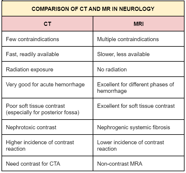
Advantages: Fast - identifies blood - shows bony structures
Sign/s associated with loss of insular cortex
Insular ribbon sign
Sign/s associated with hyperattenuating vessel filled with acute thrombus
dense MCA and dot sign
During a stroke, there is a loss of gray-white matter __________ in the NCCT image
differentiation
Sign/s associated with decreased density of basal ganglia
disappearing basal ganglia sign
This CT scan type is used to enhance differences in tissue density, demonstrate vasculature and vascular pathology and detects areas of BBB breakdown
Contrast Enhanced CT (CECT)
IV iodinated water-soluble contrast is given through IV line
Contraindications or limiting factors of CECT
Allergic reaction
Contrast induced nephropathy (increased creatinine)
CT scans are more reliable for ______ brain parenchymal or _______ hemorrhage especially _______ (aneurysm that popped)
acute
extra axial
SAH
Indications for emergency CT:
Acute or chronic FND
Head or facial trauma
Headache
Abrupt or worsening
SAH traumatic vs. non-traumatic
Change in mental status
New-onset seizure
Limitations of CT scan
Imaging of posterior fossa → linear artifacts (not as clear)
Ionizing radiation → pregnant
Advantages of CT-scan
Speed
Cost
Availability
Repeating scan during a bolus of IV contrast that produces a dynamic set of images that are real time 4D images of blood flow through the intracranial vessels
CTA
CT scan that produces functional images of brain parenchymal blood flow
CTP
True or False:
Catheter Angiography is more widely available, less specialized are required and there is no risk of dissection or stroke.
False:
CTA to boi
True or False:
Catheter angiography is more time consuming than CTA
False:
False, CTA has a time-consuming process requiring to edit and generate rendering
CTP allows quantitative measures of the following:
cerebral blood volume
cerebral mean transmit time
time to peak
cerebral blood flow
CTP can be used as a quick screening method to assess _______ and for differentiating ________ and __________
Acute cerebral ischemia
Infarct
Penumbra
Infarct signs:
_____ MTT
Decreased _____ and _____
Infarct signs:
prolonged MTT
Decreased CBV and CBF
Ischemic penumbra signs:
prolonged _____ and _____
normal or increased ______
mildly reduced _____
____________
Salvageable
Ischemic penumbra signs:
prolonged MTT and TTP
normal or increased CBV
mildly reduced CBF
compensatory vasodilation
Salvageable
This imaging tech has the ability to distinguish between pathologic soft tissues and identify pathologic abnormalities
MRI
Origin of MRI signal:
hydrogen nuclei
consist of a single proton that is constantly spinning
Most precise and sensitive imaging for detecting CNS tissue pathology
MRI agen
MRI demonstrates a ____________ contrast between different tissues compared to CT or UTZ
Significantly higher
Limitations of MRI:
____ (broke boi boi boi)
Lengthy (________ min for cranial CT)
Distortion of images by ________
_________ and level of cooperation of patient
_________ (i.e. pacemakers, infusion pumps, cochlear implants, aneurysm clips)
_________ (i.e. ventilators)
________ → possibility of developing _______ of fetuses of animal (not 100%)
Limitations of MRI:
Cost
Lengthy (30-40 min for cranial CT)
Distortion of images by artifacts
Claustrophobia and level of cooperation of patient
Implanted devices (i.e. pacemakers, infusion pumps, cochlear implants, aneurysm clips)
Machines (i.e. ventilators)
Pregnant → possibility of developing cataracts of fetuses of animal (not 100%)
Safety Concerns of MRI:
Powerful magnetic field (its in the name bozo)
Gadolinium chelates
Gadolinium chelates can cause what in patients with renal dysfunctions?
Nephrogenic Systemic Fibrosis
Basic sequences of MRI:
Displays brain and spinal cord anatomy
Evaluates subacute hemorrhage, lipids, paramagnetic metals, or proteinaceous composition of lesions
Baseline for comparison of CE images
T1 WEIGHTED
What color is the CSF in T1 weighted MRI
Dark
T2-Weighted:
Display brain and spinal cord ________
Emphasize long T2 relaxation times = _____
Emphasize short T2 relaxation times = ______
T2-Weighted:
Display brain and spinal cord pathology
Emphasize long T2 relaxation times = bright
Emphasize short T2 relaxation times = dark
Brain:
CT GRAYSCALE
MRI T1 SIGNAL
MRI T2 SIGNAL
All gray
Air:
CT GRAYSCALE
MRI T1 SIGNAL
MRI T2 SIGNAL
All black
CSF:
CT GRAYSCALE
MRI T1 SIGNAL
MRI T2 SIGNAL
CSF:
Black
Black
White
Fat:
CT GRAYSCALE
MRI T1 SIGNAL
MRI T2 SIGNAL
Fat:
Black
White
Less white
Calcium:
CT GRAYSCALE
MRI T1 SIGNAL
MRI T2 SIGNAL
Calcium:
White
Black
Black
Bone:
CT GRAYSCALE
MRI T1 SIGNAL
MRI T2 SIGNAL
Bone:
VERY white
Black
Black
Extravasated blood:
CT GRAYSCALE
MRI T1 SIGNAL
MRI T2 SIGNAL
Extravasated blood:
White
White
Black
Inflammation:
CT GRAYSCALE
MRI T1 SIGNAL
MRI T2 SIGNAL
Inflammation:
Dark gray, contrast enhancing
Gray, gadolinium enhancing
White
Edema:
CT GRAYSCALE
MRI T1 SIGNAL
MRI T2 SIGNAL
Edema:
Dark gray
Gray
White
Tumor:
CT GRAYSCALE
MRI T1 SIGNAL
MRI T2 SIGNAL
Tumor:
Gray or white, contrast enhancing
Gray or white, gadolinium enhancing
White
MRI sequence used to eliminate signal from CSF
T2 sequenced (T2 flair)
T2 Flair highlights subtle brain pathology; specifically useful for _________ diseases and lesions that are near CSF compartments
White matter diseases
T2 flair demonstrates ________ tumor components as well as associated mass effect and edema
non-enhancing
Presents as a periventricular white matter lesion, radially oriented to bodies of lateral ventricles
Multiple Sclerosis
These lesions can be found periventricular, deep and subcortical/juxtacortical white matter and corpus callosum
Supratentorial lesions
Used to eliminate signal from fat
Useful in dx fat containing lesions like lipoma and dermoid cyst
Short Tau Inversion Recovery (STIR)
For detection of early ischemic brain injury
can help with identifying chronicity of infarct
Diffusion Weighted Imaging (DWI)
This measures the extent to which diffusivity of water is restricted from free diffusion, presumably due to structural barriers such as cell membranes or association of water with larger molecules that have lower diffusion coefficients
Interpreted along with DWI
Apparent Diffusion Coefficient (ADC)
A hyperintense DWI can mean?
Stroke
This MRI shows sensitivity to small amounts of blood and blood breakdown products (hypointense)
to detect bleeding
Gradient Recalled Echo (GRE)
Tissue components characterized in Gradient Recalled Echo (GRE):
calcification or iron content
MRI that signifies breakdown of BBB
Gadolinium Contrast Enhancement
Gadolinium Contrast Enhancement characterizes:
brain tumors
metastases
Infections
Inflammation
Takes pictures while blood is flowing, signal is related to flow phenomenon
Magnetic Resonance Angiography (MRA)
MRA is used to detect:
Stenosis
Thrombosis
Dissections
Aneurysms
evaluation following interventions for aneurysm cerebrovascular malformations (AVM)
CE
evaluate neck vasculature
CE + MRA
For viewing sinuses
Evaluate for patency of dural venous sinuses in venous sinus stenosis or thrombosis
Magnetic Resonance Venography (MRV)
Use of magnetic resonance for localization of cerebral activation
Used to obtain functional information by visualizing cortical activity
Detects subtle changes in blood flow or blood oxygenation in response to stimuli or actions
Functional MRI
Clinical applications of Functional MRI
Cortical mapping of known cognitive and motor functional units (researches)
Presurgical and pre-therapeutic planning
MRI technique that measures water molecule diffusion and its direction
Diffusion Tensor Imaging (DTI)
Clinical applications of Diffusion tensor imaging
Clinical applications:
Asses integrity of white matter tracts
Presurgical and pre therapeutic planning
map of neural connections
human connectome
Used to differentiate lesions (i.e Tumor recurrence (higher choline to creatine ratio) vs. radiation necrosis)
MR Spectroscopy (MRS)
Metabolite that signifies normal neuronal tissue / neuronal integrity
Decreases in destructive lesions and decreased neuron density
N-acetyl- aspartate (NAA)
Metabolite that signifies energy stores
Creatine
Metabolite that signifies components of the cell membrane and myelin/ membrane turnover
Increases in rapidly dividing tumors
Choline (Cho)
Metabolite that signifies anaerobic metabolism
Lactate
Marker of astrocytes/astrogliosis seen in Multiple Sclerosis
Inositol or Myoinositol
MRI that quantifies blood flow through biologic tissues
(cerebral blood volume, cerebral blood flow and mean transit time)
MR Perfusion Imaging
Clinical applications of MR Perfusion Imaging
Cerebrovascular disease
Brain tumors and metastasis
Memorize:
Molecular imaging used to better understand the biochemical processes that underlie disease
Positron Emission Tomography (PET) And Single-Photon Emission Computed Tomography (SPECT)
A radioactive compound, in trace amounts with a pharmaco- kinetic behavior that targets a molecular pathway related to the pathology of a certain disease
radioligand
Screening extracranial carotid and vertebral arteries for atherosclerosis
Real-time imaging of anatomy, physiology (hemodynamics) and pathophysiology of extracranial circulation
Extracranial Ultrasound (Duplex Ultrasound or Color Doppler)
True or False:
Extracranial ultrasound can be used for carotid imaging to detect carotid stenosis after surgery or stenting, it has a sensitivity of 87% and a specificity of 86%
False:
it has a sensitivity of 86% and a specificity of 87%
haha parang tanga oh
Extracranial ultrasound can also be used to determine carotid _______ size, morphology and carotid ________ thickness
Plaque
Intima media
If these are seen in extracranial ultrasound there is an inc risk of stroke or MI
Plaque surface irregularity
Echoluscent carotid plaque (Soft)
If these are seen in extracranial ultrasound there is an dec risk of stroke or MI
Echodense carotid plaque (calcified)
may be a marker of presence of active plaque in other vascular beds
True or False:
Vertebral arteries can also be examined using extracranial ultrasound but is limited d/t bony structures of the neck
True
Non-invasive US technology that monitors blood flow velocity and blood flow direction in large intracranial arteries
Intracranial Ultrasound (Transcranial Doppler or TCD)
Clinical applications of Intracranial US:
Stenosis or occlusion of a major intracranial artery in COW or VB system and monitoring of thrombolytic activity in acute stroke
Vasospasm in SAH → increased blood flow velocity
Brain death
Limitations of intracranial US:
____________ dependent
___________ rate of inability to perform TCD due to inadequate windows
Limited to ________
Operator dependent
10-15% rate of inability to perform TCD due to inadequate windows
Limited to large cerebral arteries
Transducer placed over open fontanelles or thin calvarium
Ultrasound in Babies
Ultrasound in babies is used to detect:
Intracerebral and subdural hemorrhages
mass lesions
congenital defects
High resolution images of extracranial and intracranial vasculature
Accessing femoral nerve and threading a catheter into the precerebral vessels
Gold standard for viewing cerebral blood vessels
Cerebral Angiography
This is the process where soft tissue is eliminated from image leaving only the contrast enhanced vasculature (DSA)
Digital subtraction process