2.2 Animal Tissues, Organs & Organ Systems
1/94
There's no tags or description
Looks like no tags are added yet.
Name | Mastery | Learn | Test | Matching | Spaced | Call with Kai |
|---|
No analytics yet
Send a link to your students to track their progress
95 Terms
Why does food need to be digested?
Large and insoluble
Broken down — can be absorbed by cells
What organs are the digestive system made up of?
Glands — salivary glands and pancreas
Stomach
Small intestine
Liver
Large intestine
Explain the function of the glands (salivary glands and pancreas) in the digestive system.
Produces digestive juices — contains enzymes
breaks down food
Explain the function of the stomach in the digestive system.
Produces HCl
kills bacteria
optimum pH for protease enzyme
Explain the function of the small intestine in the digestive system.
Soluble molecules are absorbed into blood
Explain the function of the liver in the digestive system.
Produces bile → stored in gall bladder
Helps digest lipids
Explain the function of the large intestine in the digestive system.
Absorbs water from undigested food → produces faeces
passes out of body through rectum and anus
Enzymes __________ specific reactions in living organisms due to the shape of their _________ _____.
catalyse
active site
What do digestive enzymes do?
Converts food → small soluble molecules
Can be absorbed into bloodstream
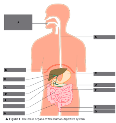
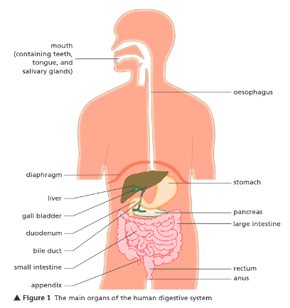
Label this diagram of the human digestive system.
A — mouth (teeth, tongue, and salivary glands)
B — oesophagus
C — stomach
D — pancreas
E — large intestine
F — rectum
G — anus
H — appendix
I — small intestine
J — bile duct
K — duodenum
L — gall bladder
M — liver
N — diaphragm
What are enzymes?
Biological catalysts
Increase rate of reactions in living organisms
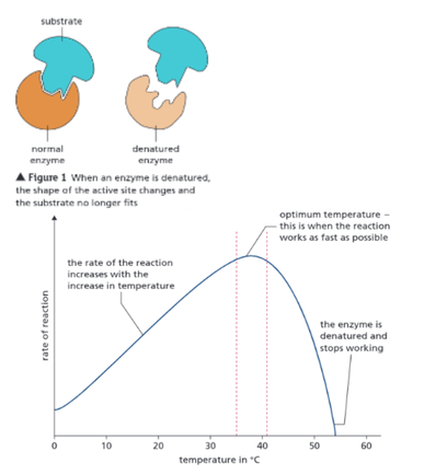
Why is the shape of an enzyme vital to its function?
Each enzyme has uniquely shaped active site — substrate binds to
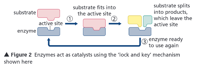
Explain the ‘lock and key’ theory.
Shape of substrate complementary to shape of active site
They bind → forms enzyme-substrate complex
Reaction takes place — products released from surface of enzyme

Why do enzymes require an optimum pH and temperature?
They are proteins
What is the optimum temperature for digestive enzymes?
37 °C

Explain the effect of temperature on enzyme action.
Increased temperature = increased rate of reaction (up to optimum)
Above optimum — rate rapidly decreases and reaction stops
Temperature = too hot → bonds in structure break
Shape of active site changes → substrate doesn’t fit
Enzyme is denatured and doesn’t work
What is the optimum pH for most enzymes?
7 — some in stomach have lower
Explain the effect of pH on enzyme action.
pH = too high/low → active site changes shape — substrate doesn’t fit
Enzyme is denatured and doesn’t work
What do carbohydrases do (give an example)?
Carbohydrates → simple sugars
Amylase — starch
Where is amylase produced? (3)
Salivary glands
Pancreas
Small intestine
What do proteases do (give an example)?
Proteins → amino acids
Pepsin
Where is pepsin produced?
Stomach
What do lipases do?
Lipids (fats) → glycerol and fatty acids
Where are lipases produced? (2)
Pancreas
Small intestine
What are the products of digestion used to do?
Build new carbohydrates, lipids, and proteins
Glucose → respiration
Explain the role of bile in the digestive system.
Alkaline — neutralises HCl made in stomach
Emulsifies fats → forms small droplets → increases SA
Alkaline conditions and large SA → increases rate of fat breakdown by lipase
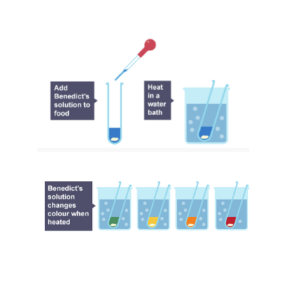
What is used to test for sugars?
Benedict’s solution
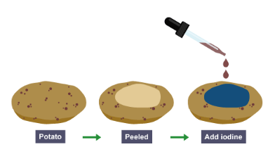
What is used to test for starch?
Iodine
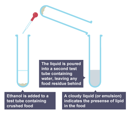
What is used to test for lipids?
Ethanol
What is used to test for proteins?
Biuret reagent
Describe how you test for sugars.
Put food sample in test tube
Add a few drops of Benedict’s solution
Place test tube in water bath for 5 mins
Results: blue → green → yellow → orange → red
green = less glucose
red = more glucose
Describe how you test for starch.
Put food sample in test tube
Add a few drops of iodine
Results: blue-black if starch is present
Describe how you test for lipids.
Put food sample in test tube
Add a few drops of distilled water
Add a few drops of ethanol
Shake solution gently
Results: goes cloudy if lipid is present
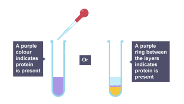
Describe the test for proteins.
Put food sample in test tube
Add 1 cm³ of biuret solution A and biuret solution B
Shake solution gently
Results: turns purple if protein is present

Required Practical 5 — Enzymes:
Describe a method to investigate the effect of pH on the rate of reaction of amylase enzyme.
Place one drop of iodine solution into each well of spotting tile
Get three test tubes:
2 cm³ — starch solution
2 cm³ — amylase solution
2 cm³ — buffer solution (pH 5)
Place test tubes in water bath (30°C) — leave for 10 mins → solutions reach correct temp
Combine solutions into one test tube — mix with stirring rod
Put in water bath — start stopwatch
After 30s — transfer one drop of solution to well in spotting tile
Iodine turns blue-black → starch is present
Take sample every 30s until iodine remains orange
Starch is no longer present — reaction is complete
Repeat whole experiment using different pH buffer solutions
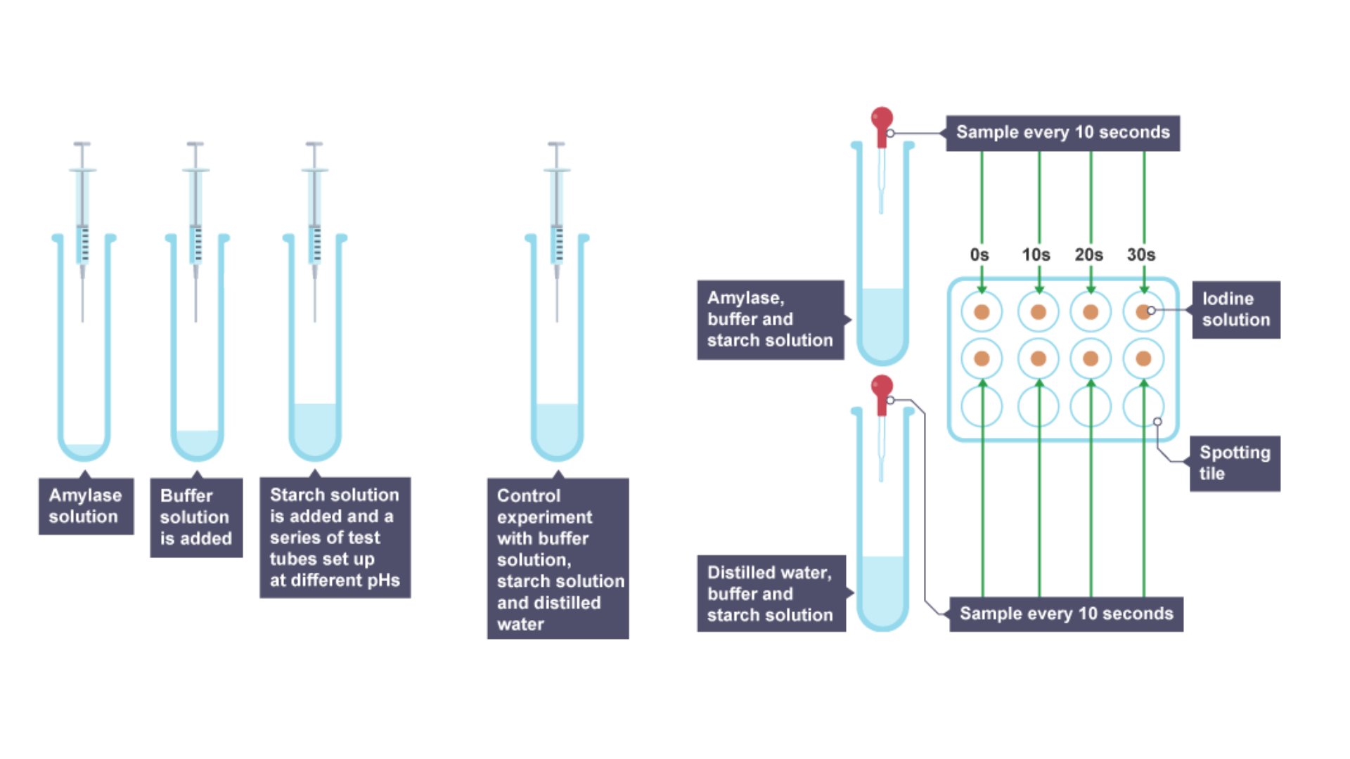
What are the problems wit RP 5 — Enzymes?
Taking samples every 30s → only have approx. time for complete reaction
take samples every 10s
Observation of colour change is subjective
What is the heart?
Organ that pumps blood around the body — double circulatory system
Where does the right ventricle pump blood?
Lungs → gas exchange
Where does the left ventricle pump blood?
Around the rest of body
What are the three types of blood vessel in the body?
Arteries
Veins
Capillaries
What is the natural resting heart rate controlled by?
Group of cells in right atrium — pacemaker
What are artificial pacemakers?
Electrical devices — correct irregularities in heart rate
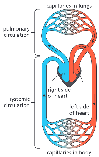
Explain a double circulatory system.
Deoxygenated blood → right atrium → right ventricle → lungs for gas exchange
Oxygenated blood → left atrium → left ventricle → O₂ blood around body


What do arteries do?
Carry blood (usually oxygenated) from heart → organs of body


Explain how the structure of arteries relates to their function.
Thick walls — muscle and elastic fibres
Stretch as blood is pumped into them under pressure from heart
Returns to original shape
Felt as a pulse

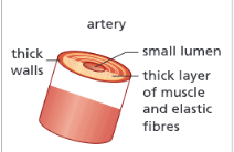
What happens if an artery is damaged?
Blood pumps out rapidly every time heart beats
It is under pressure


What do veins do?
Carry blood (low in O₂) from organs → heart
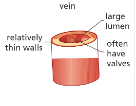

Why do veins have thinner walls than arteries?
Blood is not under pressure — no pulse

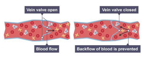
Why do veins have valves and what do they do?
Open as blood flows through them → heart
Closes if blood flows back → prevents backflow
Blood squeezed back → heart — skeletal muscles


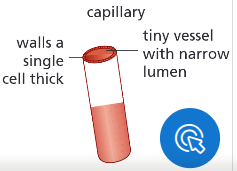
Explain the function of capillaries.
Allows blood to flow close to cells — enables substances to move between them


How does the structure of capillaries relate to their function?
One cell thick wall — short diffusion pathway
Permeable walls — substances can move across them
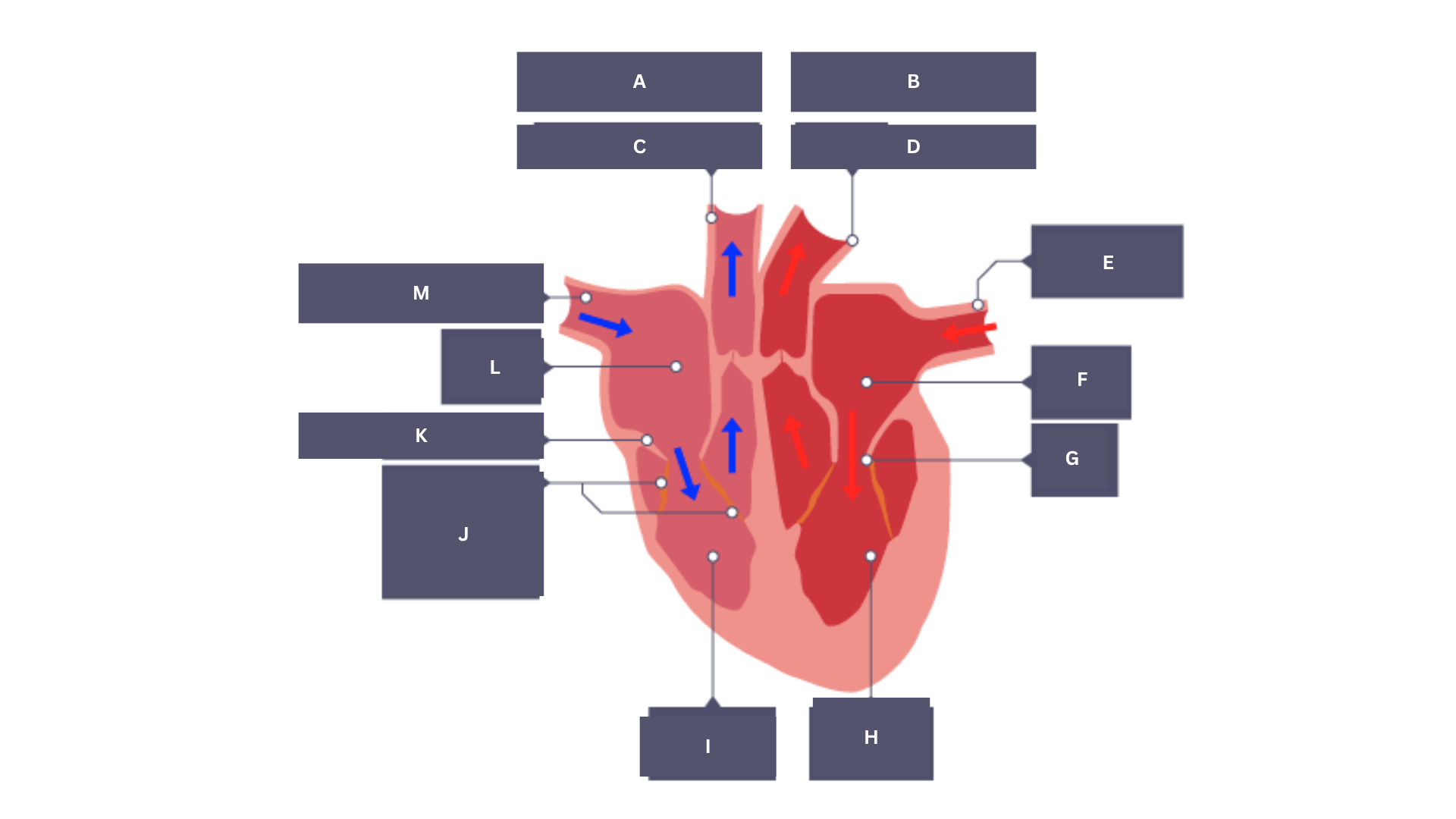
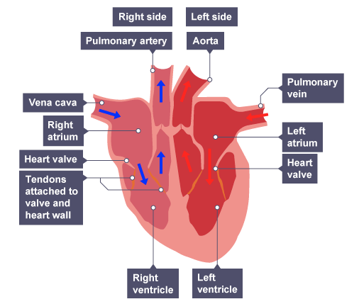
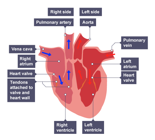
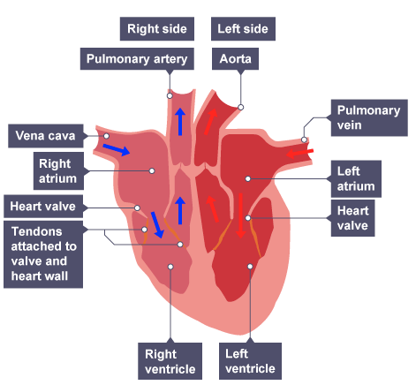
Label this diagram of the heart.
A — Right side
B — Left side
C — Pulmonary artery
D — Aorta
E — Pulmonary vein
F — Left atrium
G — Heart valve
H — Left ventricle
I — Right ventricle
J — Tendons — attached to valve and heart wall
K — Heart valve
L — Right atrium
M — Vena cava
Explain how the structure of the heart relates to its function.
Muscular walls — provides strong heartbeat
Muscular wall of left ventricle thicker — blood is pumped around body not just to lung
4 chambers — separates oxygenated and deoxygenated blood
Valves — prevents backflow of blood
Coronary arteries — gives heart oxygenated blood

Explain the process of blood circulation in the heart.
Blood flows to:
right atrium — through vena cava
left atrium — through pulmonary vein
Atria contract → forces blood into ventricles
Ventricles contract pushes blood in:
right ventricle → pulmonary artery → lungs
left ventricle → aorta → body
Valves close — prevents backflow of blood

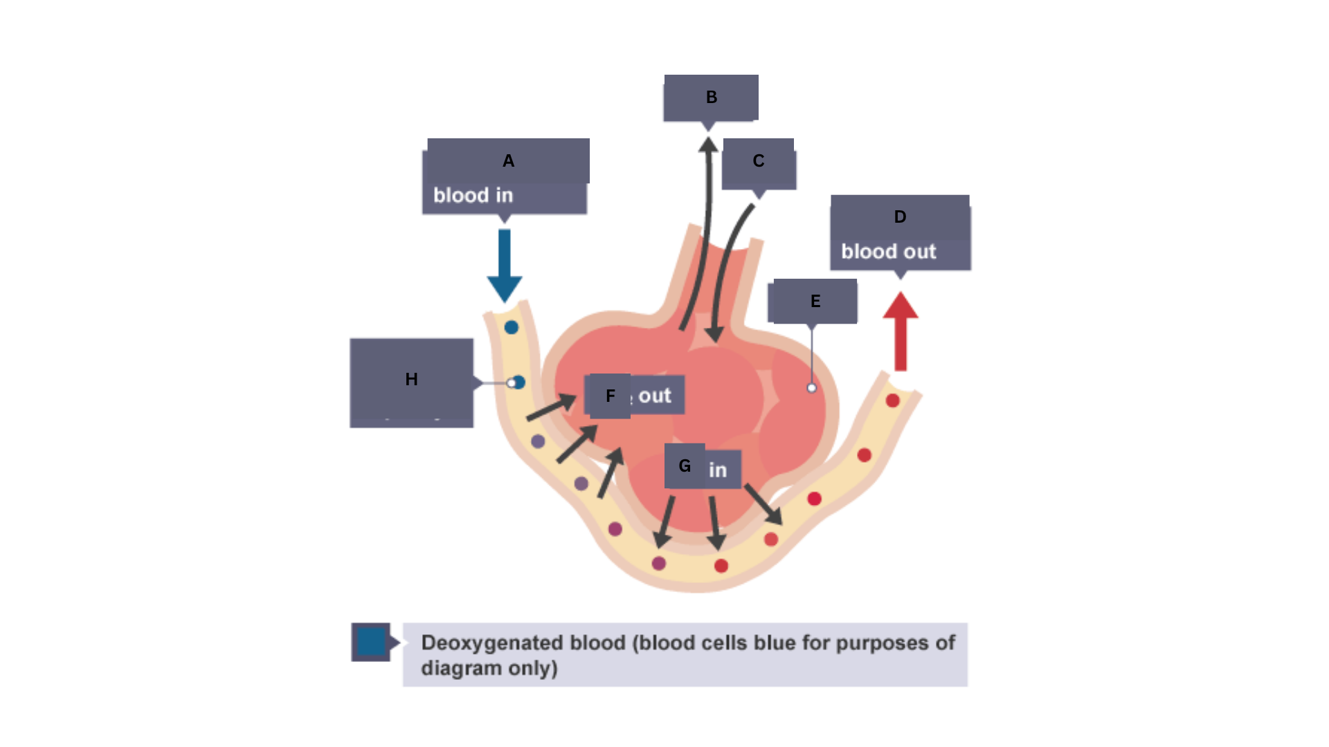
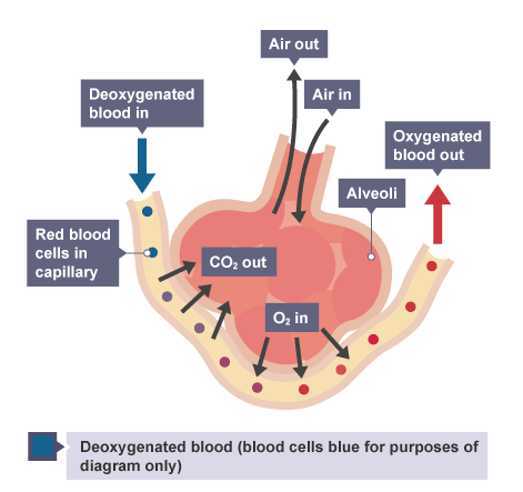
Label this diagram of the human gaseous exchange system.
A — Deoxygenated
B — Air out
C — Air in
D — Oxygenated
E — Aveoli
F — CO₂
G — O₂
H — Red blood cells in capillary
Explain the process of ventilation.
Ribcage moves up and out → diaphragm moves down — volume of chest increases
increased volume = lower pressure
Air drawn into chest as air moves from high pressure (environment) → low pressure (lungs)
Opposite when exhaling
Explain the process of gas exchange.
Inhalation → aveoli fill with O₂
Deoxygenated blood in capillaries surrounding aveoli — came from pulmonary artery
has lots of CO₂ — product of respiration
O₂ diffuses down concentration gradient → capillary bloodstream — has low O₂ concentration
CO₂ diffuses down concentration gradient from blood → aveoli
How are aveoli adapted for gas exchange?
Small and arranged in clusters — large surface area for diffusion
Capillaries provide large blood supply — maintains concentration gradient
Thin walls — short diffusion pathway
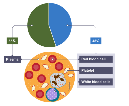
What is blood?
Tissue consisting of plasma and:
RBC — red blood cells
WBC — white blood cells
Platelets

What is plasma and what is its function?
Liquid that carries components in blood — e.g. RBC, WBC, platelets, amino acids, urea etc.
What are red blood cells and what is their function?
Carry O₂ molecules from lungs → cells in body
Biconcave disc — large surface area
No nucleus — more space for O₂
Contains haemoglobin — binds to oxygen
What are white blood cells and what is their function?
Part of immune system
body’s defence against pathogens
Have a nucleus
Different types:
produce antibodies
engulf and digest pathogens
produce antitoxins
neutralise toxins produced by microorganisms
What are platelets and what is their function?
Small fragments of cells + no nucleus
Clot blood at site of wound
clot dries and hardens → forms a scab → new skin grows
prevents microorganisms entering
No platelets = excessive cuts and bruising from cuts
Coronary heart disease is a non-communicable disease.
What is a non-communicable disease?
Cannot be spread between individuals
Explain what coronary heart disease is.
Fatty material builds up inside coronary arteries
Reduces blood flow
Lack of oxygen for heart muscle

What are stents and how do they help with coronary heart disease?
Metal tubes — keeps arteries open → blood can flow
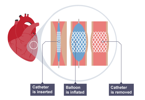

What are the advantages and disadvantages of stents?
Lowers risk of heart attack
Quick recovery time
Risk of heart attack during procedure
Risk of infection
Blood can clot around stent
What are statins and how do they help with coronary heart disease?
Drugs — decrease LDL cholesterol levels (cause coronary heart disease)
What are the advantages and disadvantages of statins?
Reduce risk of:
strokes
heart attacks
coronary heart disease
Increase levels of HDL cholesterol
Needs to be taken continuously
Side effects
May not have immediate effect
What are faulty valves and what problems can they cause?
Becomes stiff → cannot open
Damaged → leaks
Blood backflows → heart doesn’t work efficiently
What are the advantages and disadvantages of biological valves (e.g. pigs)?
No anti-clotting drugs
Readily available
Ethical/religious objections — use of animal tissue
Higher chance of rejection
Tissue can harden over time → less effective
Doesn’t last very long (12-15 years)
What are the advantages and disadvantages of mechanical valves?
Long lasting
Anti-clotting medication needed
Medication → excessive bleeding
What are the disadvantages of a heart transplant?
Requires donor — recently died
Not always available
Can be rejected by immune system
What are the advantages and disadvantages of artificial hearts?
Less likely to be rejected by immune system
Risk of infection
Mechanical parts could wear out
Blood clots → strokes
Medication to prevent this → thins blood → affects bleeding
What is the definition of health?
State of physical and mental well-being
Diseases, both ________________ and ________________, are major causes of ill health.
Other factors including ______, ______, and _______ situations may have a profound effect on both ___________ and ________ health.
communicable
non-communicable
diet
stress
life
physical
mental
How can different types of diseases interact (examples)?
Defects in immune system → more likely to suffer from infectious diseases
Viruses in cells → trigger cancers
Immune reactions caused by pathogen → trigger allergies, skin rashes, asthma
Severe physical ill health → depression
What can risk factors be?
Aspects of a person’s lifestyle
Substance in person’s body or environment
What are the causal mechanisms for certain risk factors? (6)
Diet, smoking, exercise → cardiovascular disease
Obesity → type 2 diabetes
Alcohol → liver and brain function
Smoking → lung disease and lung cancer
Smoking and alcohol → unborn babies
Carcinogens — e.g. ionising radiation → cancer
Explain the causal mechanisms for cardiovascular disease.
Diet — lots of LDL cholesterol = blocked arteries → increases blood pressure
Smoking — damages artery walls
Exercise — lowers blood pressure → reduces strain on heart
Explain the causal mechanisms for type 2 diabetes.
Obesity — affects metabolism → fat molecules released into blood → can affect cells uptake of sugar
Explain the causal mechanisms for liver and brain function.
Alcohol — fatty liver → liver failure
Alcohol — damage nerve cells in brain
Explain the causal mechanisms for lung disease and lung cancer.
Smoking — damages cells in lining of lungs
Many diseases are caused by the ___________ of a number of factors.
interaction
What is cancer the result of?
Mutations in cells → uncontrolled growth and division
What are benign tumours?
Growths of abnormal cells — contained in one area
Don’t invade other parts of body
What are malignant tumours?
Cancers
Invade neighbouring tissues — spread to different parts of the body in blood
Forms secondary tumours
Scientists have identified ___________ risk factors for various types of cancer.
There are also ___________ risk factors for some cancers.
lifestyle
genetic
What are some lifestyle risk factors for cancer?
Smoking
Obesity
UV light
Viral infection
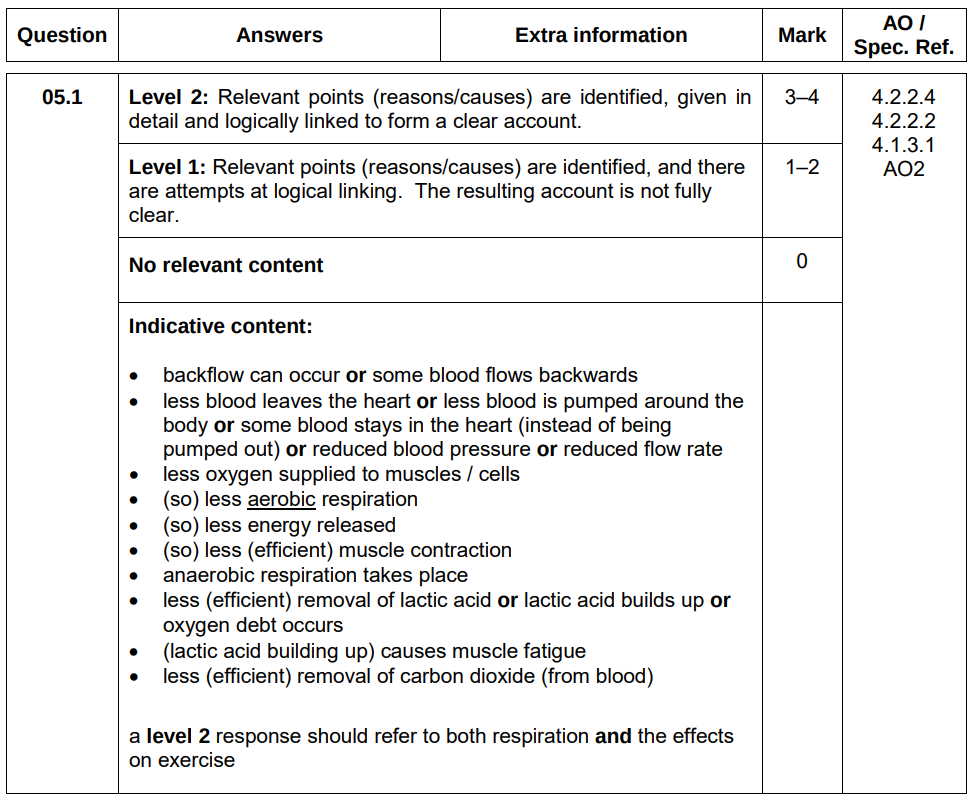
![<ul><li><p><strong>Figure 6</strong> shows the internal structure of the human heart.</p></li><li><p>One of the heart valves is labelled.</p></li><li><p>Sometimes a valve in the heart can start to leak.</p></li></ul><p>Explain why a person with a leaking heart valve has difficulty exercising. [4 marks]</p>](https://assets.knowt.com/user-attachments/ce969d28-0590-421c-bb07-ef57463e5a7b.png)
Figure 6 shows the internal structure of the human heart.
One of the heart valves is labelled.
Sometimes a valve in the heart can start to leak.
Explain why a person with a leaking heart valve has difficulty exercising. [4 marks]
Backflow of blood
Less blood pumped around body
Less oxygen supplied to muscles
Less aerobic respiration → less energy released
Anaerobic respiration → lactic acid build up → muscle fatigue
A patient with a leaking heart valve may have the valve replaced.
A study compared two different types of replacement heart valve:
mechanical valves
biological valves from pigs
The data used in the study was collected from female patients aged 50–69.
Table 4 shows the data.
Metric | Mechanical Valve | Biological Valve |
Number of patients given the valve | 2852 | 1754 |
Number of patients who died from heart-related problems after valve replacement | 180 | 178 |
Percentage of patients alive after 5 years | 91 | 89 |
Percentage of patients needing a second valve replacement within 6 years | 2.2 | 5.2 |
Percentage of patients who had a blood clot on the brain after surgery | 5.8 | 0.1 |
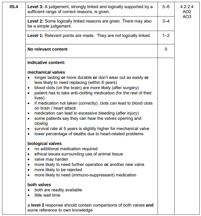
Evaluate the use of mechanical replacement heart valves and biological replacement heart valves. [6 marks]
Use information from Table 4 and your own knowledge.
Mechanical valves — longer lasting
Mechanical valves — blood clots more likely (5.8% > 0.1%)6
patient has to take anti-clotting medication
Medication for mechanical valves → excessive bleeding
Mechanical valves — lower percentage of deaths due to heart-related problems (6.3% < 10.1%)
Biological valves — more likely to be rejected
patient has to take immuno-suppressant medication
Both valves are readily available
Biological valves are better — low risk of blood clots and no anti-clotting medication needed → improves patent’s quality of life
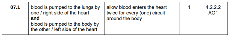
Define the term double circulatory system. [1 mark]
Blood enters the heart twice for every one circuit around the body
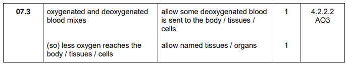
Explain why having only one ventricle makes the circulatory system less efficient than having two ventricles. [2 marks]
Oxygenated and deoxygenated blood mixes
Less oxygen reaches the tissues
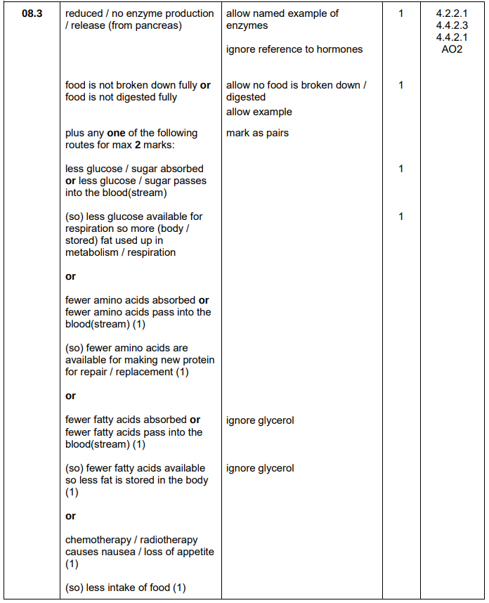
Pancreatic cancer develops when a malignant tumour grows inside the pancreas.
The pancreas produces digestive enzymes.
One symptom of pancreatic cancer is weight loss.
Explain how pancreatic cancer may cause a person to lose weight. [4 marks]
Do not refer to hormones in your answer.
No enzyme production
Food is not digested fully
Less glucose absorbed → less glucose available for respiration
More fat used in respiration