OCR AS/A Level Biology: Microscopy & Staining
1/48
There's no tags or description
Looks like no tags are added yet.
Name | Mastery | Learn | Test | Matching | Spaced | Call with Kai |
|---|
No analytics yet
Send a link to your students to track their progress
49 Terms
Use of microscopy
To observe and investigate different types of cell and cell structure in a range of eukaryotic organisms
Types of microscopy
Light, transmission electron, scanning electron, laser scanning confocal
Types of mount
Wet, dry, squash, smear
How to do a wet mount
Suspend the specimen in liquid, place cover slip on from an angle
Examples of liquids used in wet mounts
Water, immersion oil
Organisms that can be viewed in wet mounts
Aquatic organisms
How to do a dry mount
Section the sample if it is too large, place specimen on centre of the slide, put cover slip on top
Things that can be viewed in dry mounts
Hair, pollen, dust, insect parts, parts of muscle tissue, parts of plant
How to do a smear slide
Use the edge of a slide to smear out the sample on another slide, put cover slip over the sample
How to do a squash slide
Prepare a wet mount, use a lens tissue or two microscope slides to press down on the cover slip
What can you view with a squash slide?
Root tips during cell division
What can you view with a smear slide?
Blood
Why must a sample be thin for light microscopy?
So the light can shine through it and details can be seen.
How a light microscope works
Objective lens produces a magnified image, image magnified again by the eyepiece lens, illumination provided by a light underneath the sample
How to calibrate a microscope
no
Why must the liquid medium used in wet mounts have a similar refractive index to glass?
To prevent diffraction between the liquid and the glass and thus preventing image distortion.
Why are cover slips placed on wet mounts at an angle?
To prevent the trapping of air bubbles.
Purpose of differential staining
To identify different cellular components and cell types
Examples of differential staining
Gram stain technique, Acid-fast technique
Purpose of the Gram stain technique
To differentiate between Gram positive and Gram negative bacteria.
Purpose of the Acid Fast technique
To differentiate between species of Mycobacterium and other bacteria
Stages in pre-preparation of slides
Fixing, sectioning, staining, mounting
Fixing
Using chemicals like formaldehyde to preserve specimens in a near-natural state.
Sectioning
Dehydrating a sample with alcohol and then placing it in a mould of resin or wax to form a hard block before slicing it with a microtome into thin slices.
Staining
Treating specimens with multiple stains to show different structures.
Mounting
Securing a specimen onto a microscope slide under a cover slip.
Advantages of staining
See more detail, increases contrast, allows you to identify different cells and cellular components like organelles
Magnification Formula
Magnification = Image size / Object Size
Difference between magnification and resolution
Magnification is how many times larger the image is than the actual size of the object whereas resolution is how detailed the image is and the ability to distinguish between two points that are close together.
Resolution of light microscopes
200nm
Magnification of light microscopes
1500x
Resolution of a transmission electron microscope
0.5nm
Magnification of a transmission electron microscope
500000x
Resolution of a scanning electron microscope
3-10nm
Magnification of a scanning electron microscope
100000x
How to tell the difference between an SEM image and a TEM image
-Both images are black and white but TEM images can add false color using computer system.
-SEM=3D
-TEM=2D
-SEM has a higher res.
-The SEM image will only allow you to see the outside of the cell rather than the inside cell detail.
Comparison points for Light Microscopes
-Cheap to purchase and operate
-Small and portable
-Simple and easy sample preparation
-Vacuum not required
-Natural colour of sample is maintained
-Magnifies only up to 2000 times
-Specimens can be living or dead
-Stains are often needed to make the cells visible
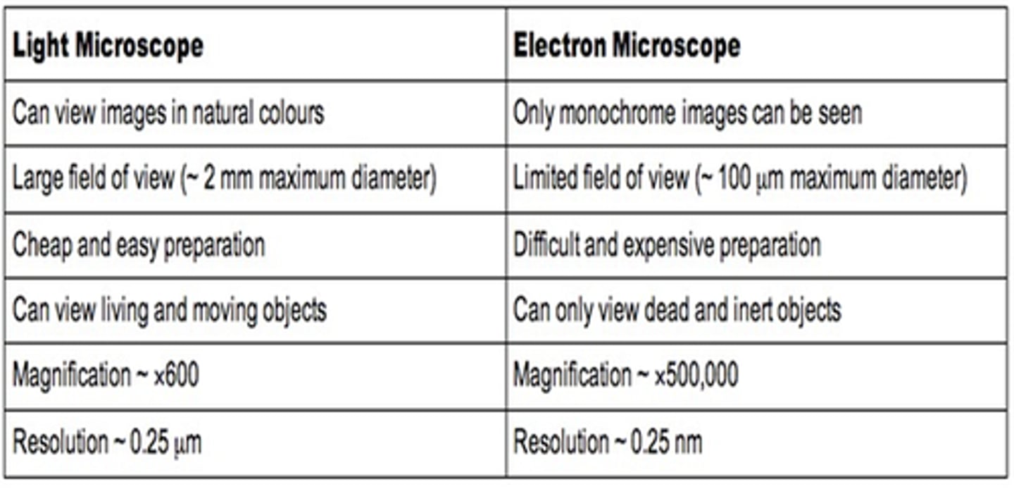
Comparison points for Electron Microscopes
-Expensive to buy and produce electron beam
-Large and requires a special room
-Lengthy and complex sample prep
-Vacuum is required
-All images black and white
-Magnifies over 500000 times
-Specimens are dead
-Electron beam can damage specimens as they must be stained with an electron-dense chemical.

Why might organelles appear to be different sizes down a microscope?
Cut in a transverse or longitudinal planes, natural variation in shape, some may have just divided or be growing, an artefact may have deformed it
Advantages of filming when doing microscopy
Can use time lapse, continuous record, you don't need to be constantly looking, not dependent on drawing or describing ability, easy to see detail
Positively charged dyes
Crystal violet, methylene blue
Negatively charged dyes
Nigrosin, Congo red
Why do we use staining?
To increase contrast because cells don't absorb much light
The crystal of cells and other cell structures are often transparent.
What is methylene blue?
Is an all-purpose stain
What is differential staining?
Some stains bind to specific cell structures staining each structure differently so the structures can be easily identified.
Acetin Orcein binds to....
DNA + stains chromosomes dark red.
Eosin stains.....
Cytoplasm.
Sudan red stains....
Lipids.
Iodine in KI solution stains....
The cellulose in plant cell walls yellow
+
Starch granules blue/black might look violet under MS