TMJ & Mastication
1/36
There's no tags or description
Looks like no tags are added yet.
Name | Mastery | Learn | Test | Matching | Spaced | Call with Kai |
|---|
No analytics yet
Send a link to your students to track their progress
37 Terms
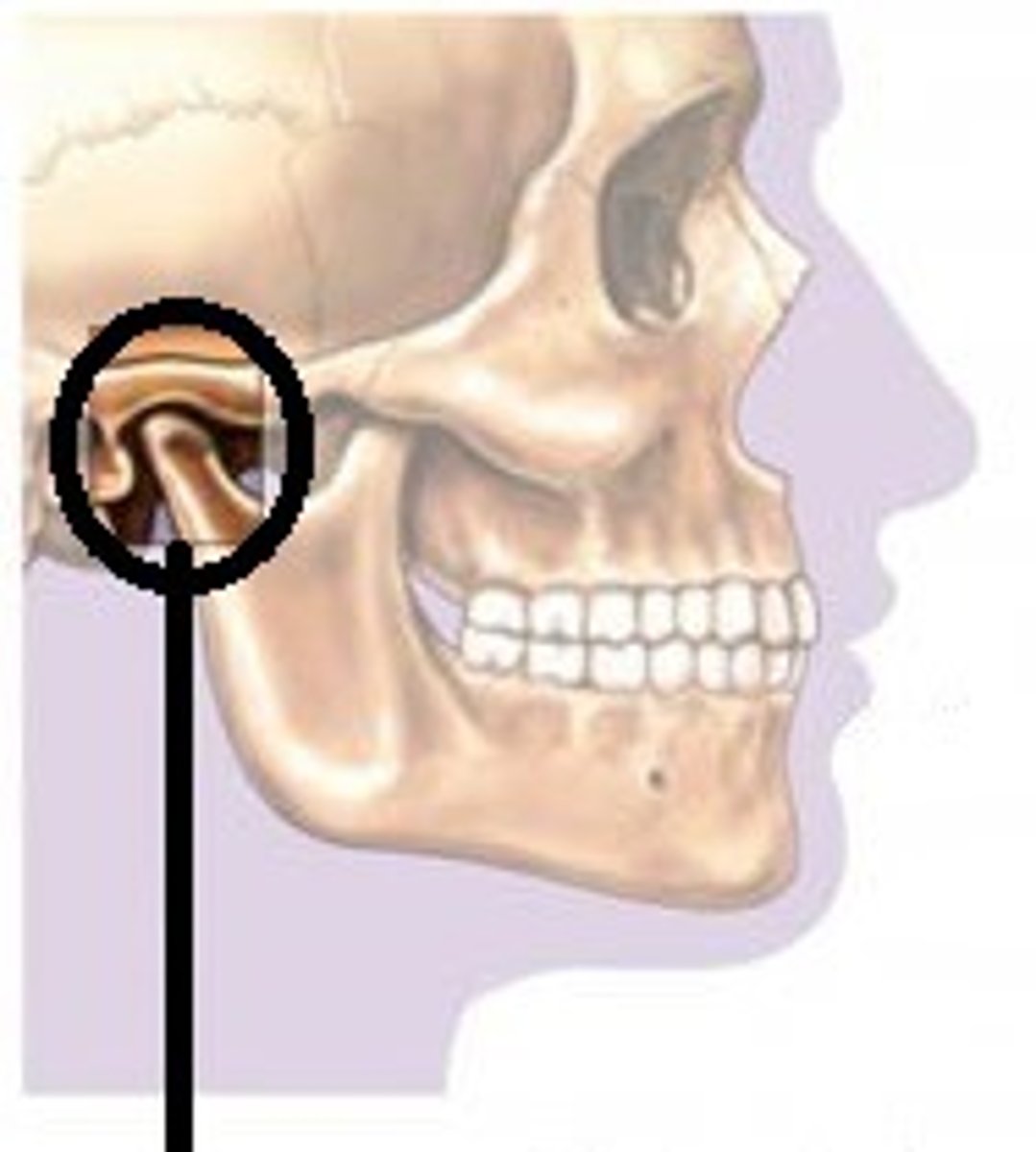
TMJ
temporomandibular joint; articulation between the articular fossa of the temporal bone of the skull with the condyle of the mandible
TMJD
Temporal mandibular joint dysfunction
Frontal Bone
forms the forehead
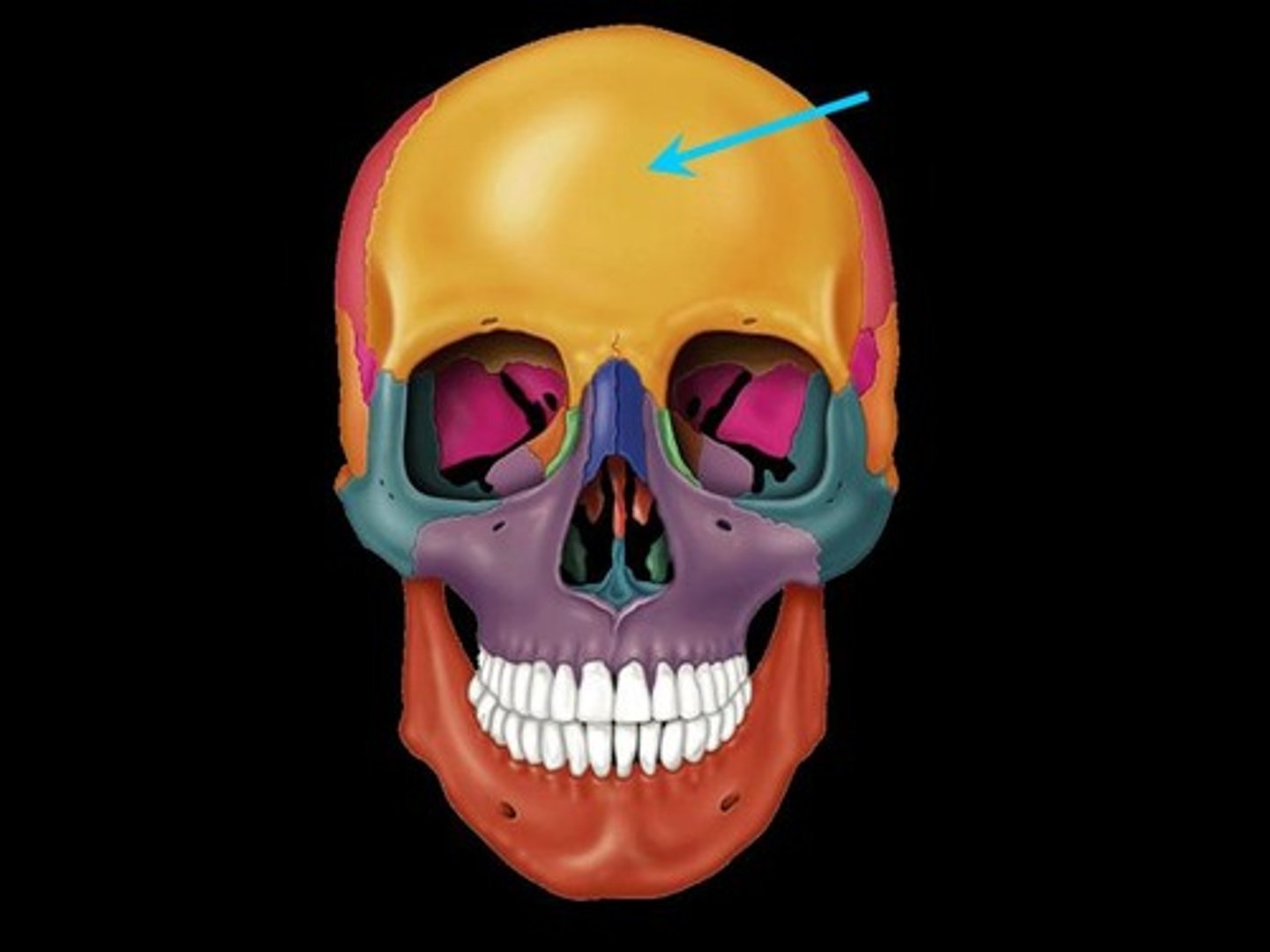
Parietal Bone
either of two skull bones between the frontal and occipital bones and forming the top and sides of the cranium
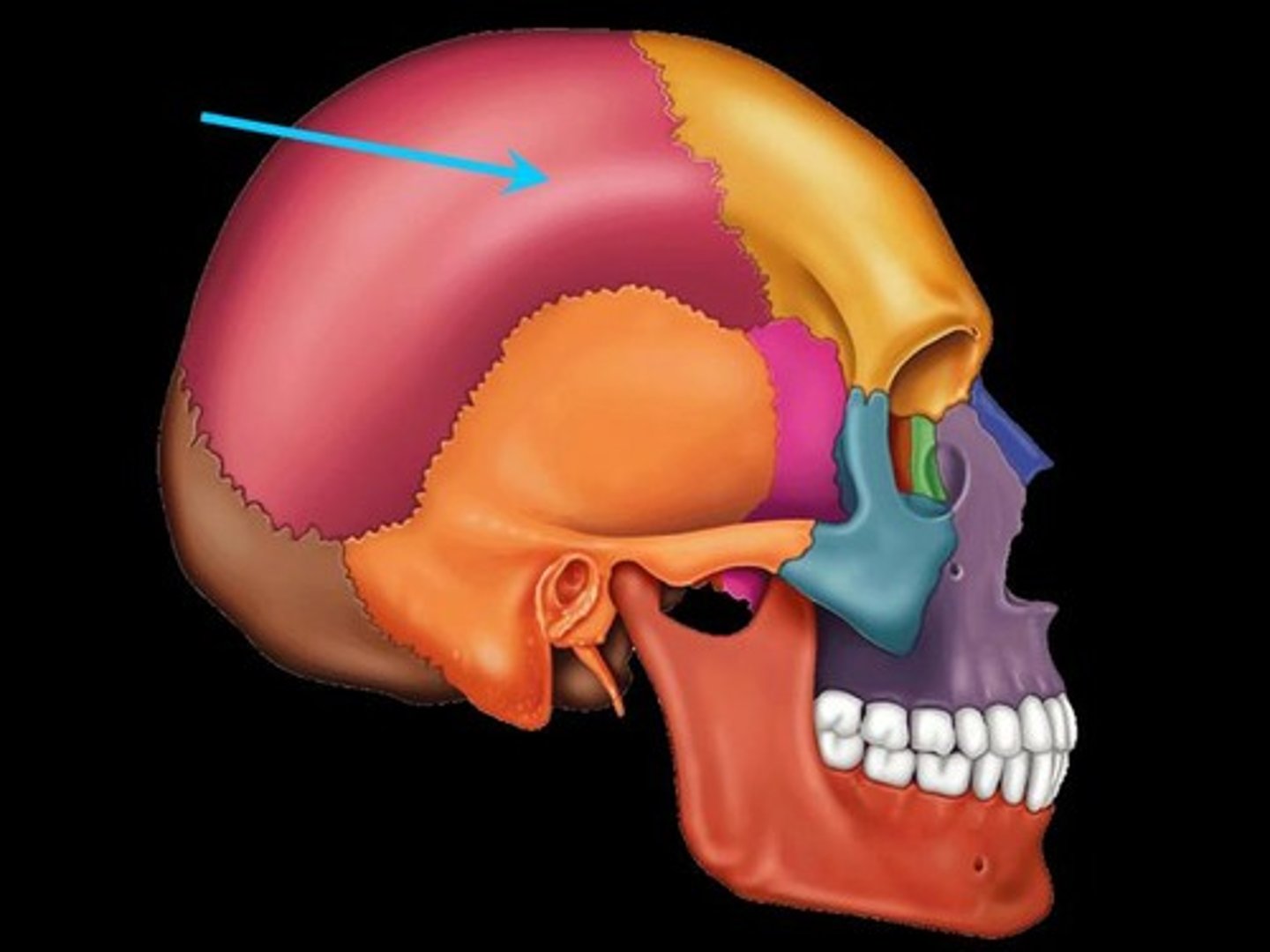
Occipital Bone
The bone that protrudes at the base of the skull is the

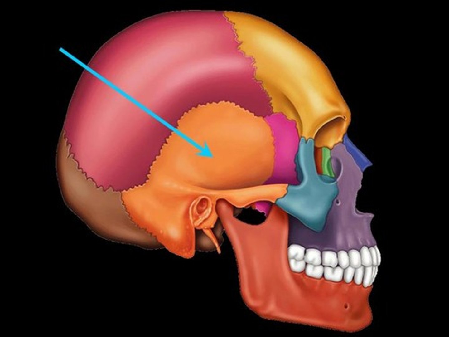
Temporal Bone
bone that forms parts of the side of the skull and floor of the cranial activity. There is a right and left temporal bone.
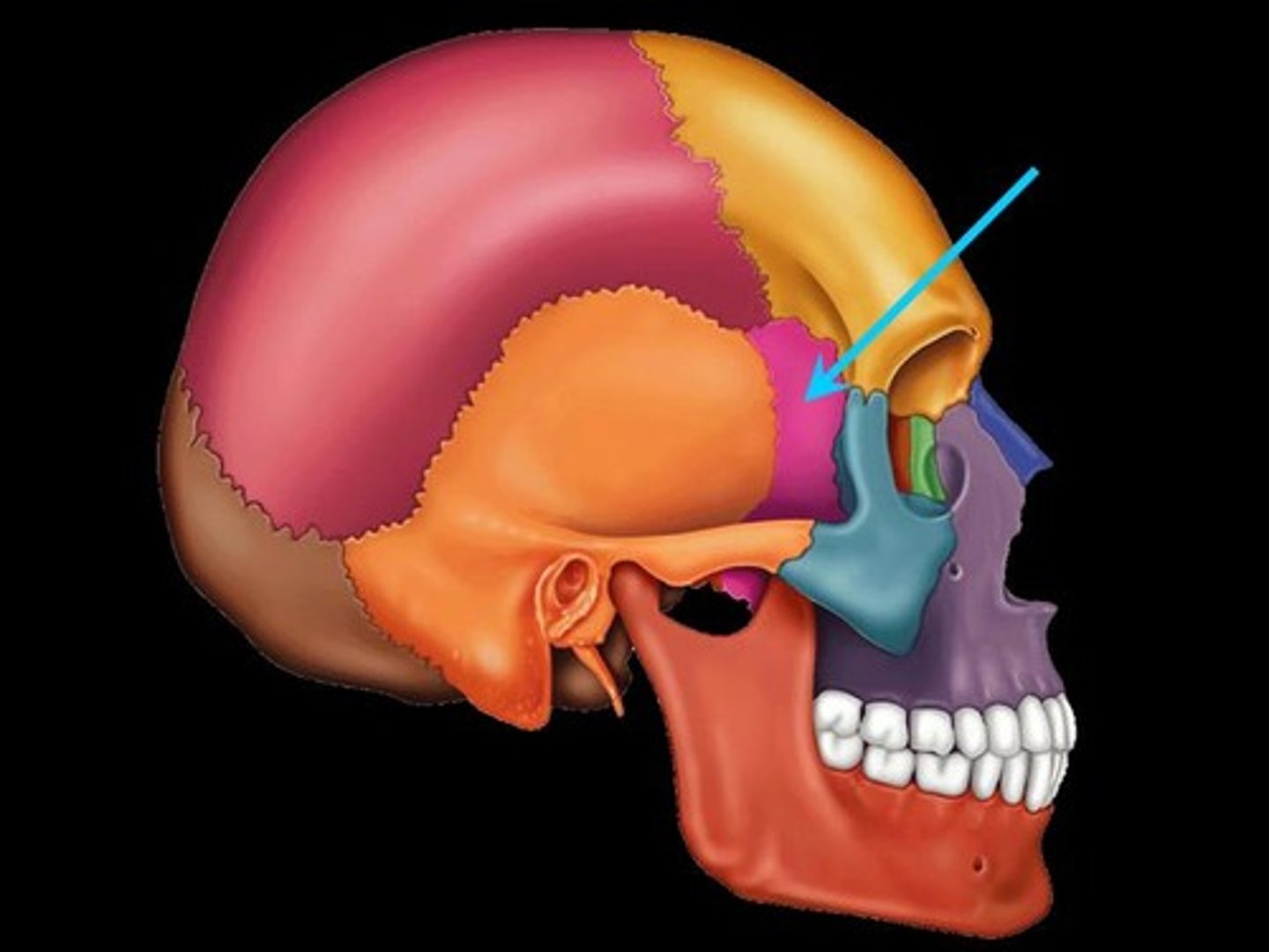
Sphenoid Bone
Located at the lateral base of the skull anterior to the temporal bone.
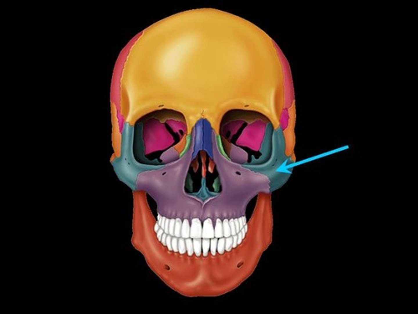
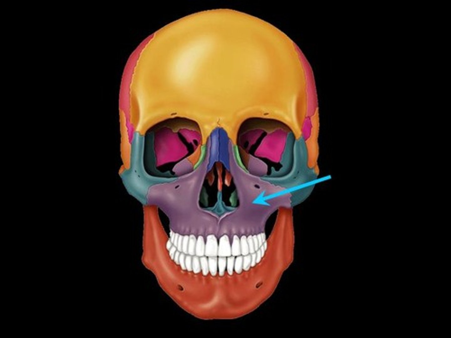
Zygomatic Bone
cheekbone
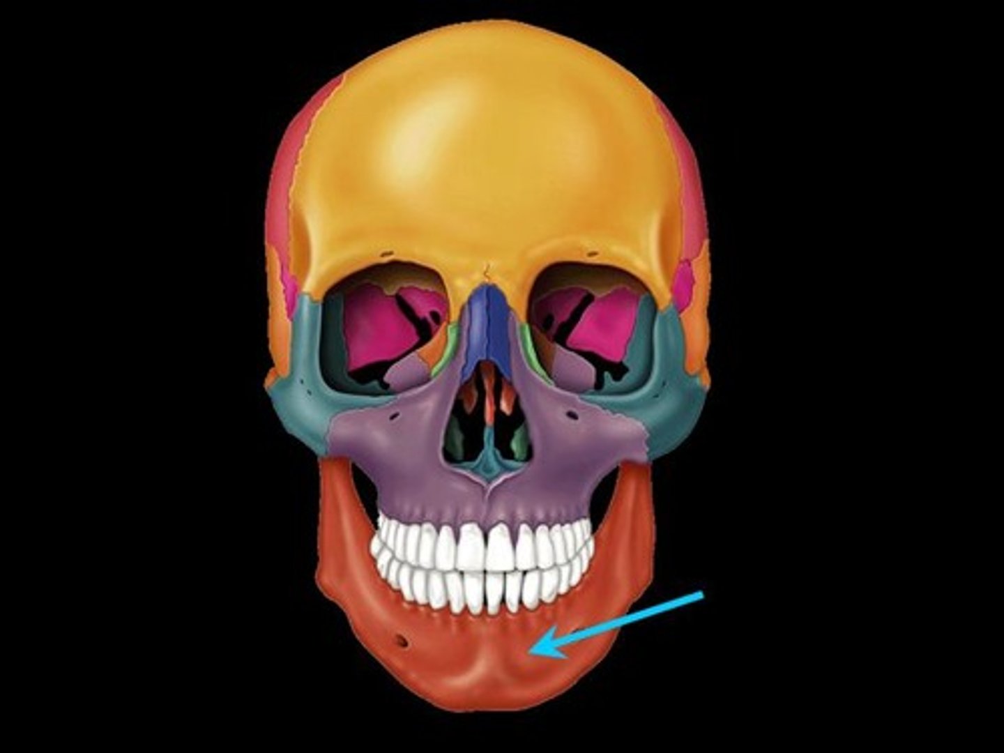
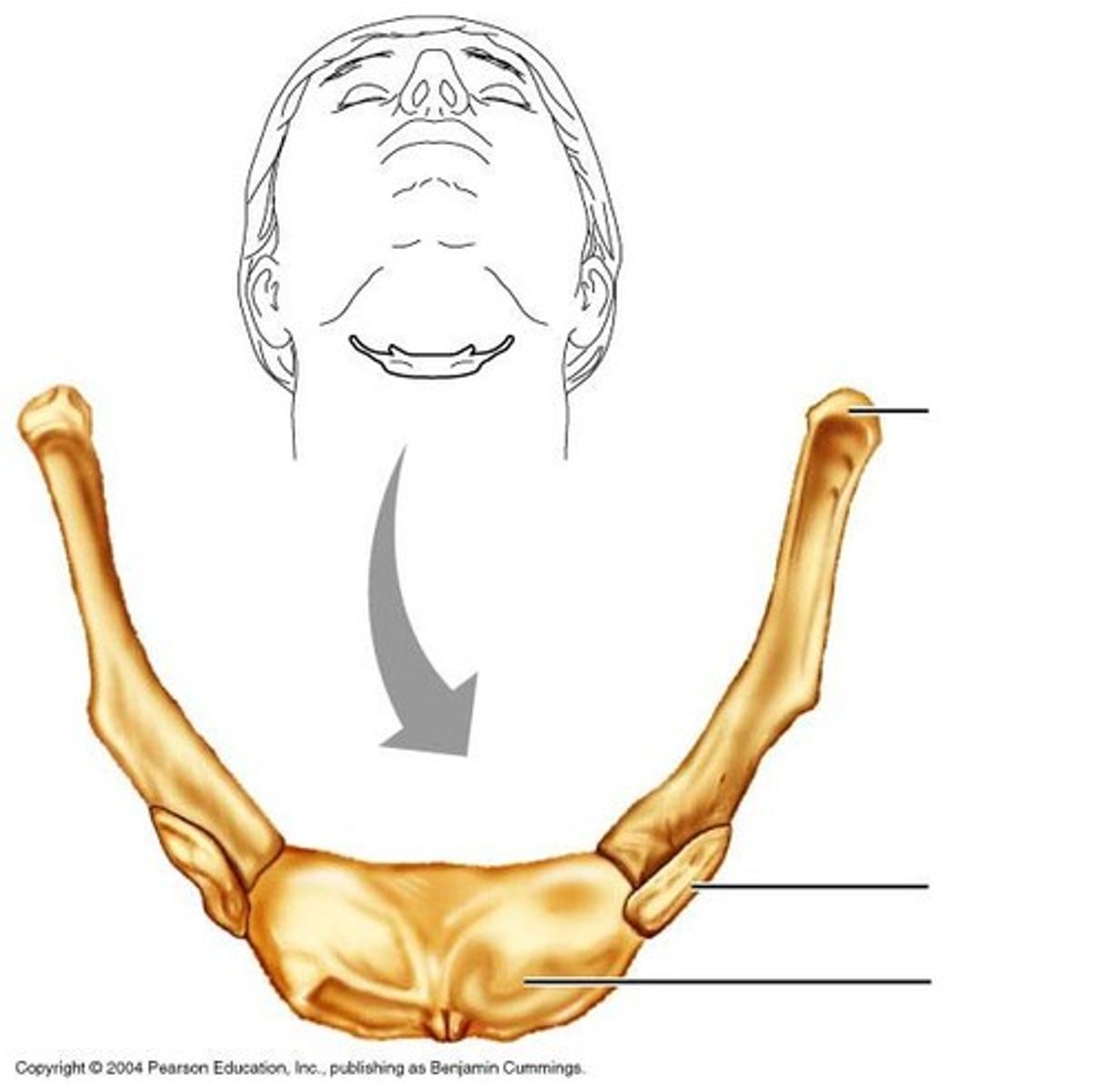
Mandible
Jaw bone
Maxilla
upper jaw bone
Nasal Bone
Bridge of nose

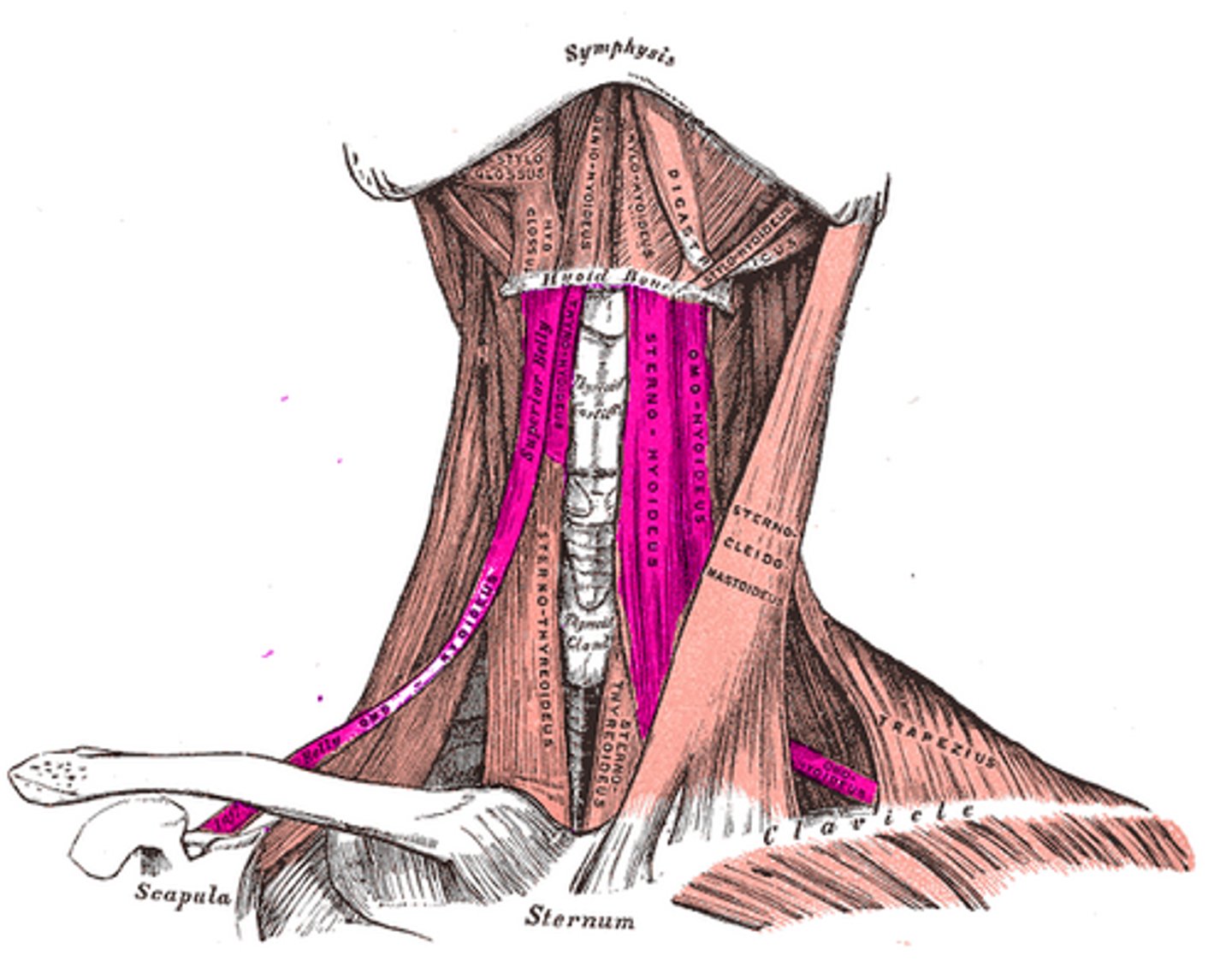
Hyoid Bone
bone that is suspended in the mid neck region and provides a moveable base for the tongue
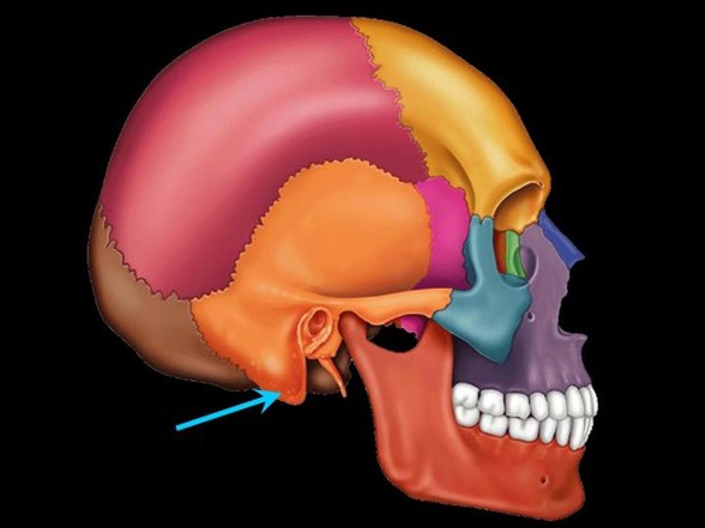
Mastoid Process
round projection on the temporal bone behind the ear
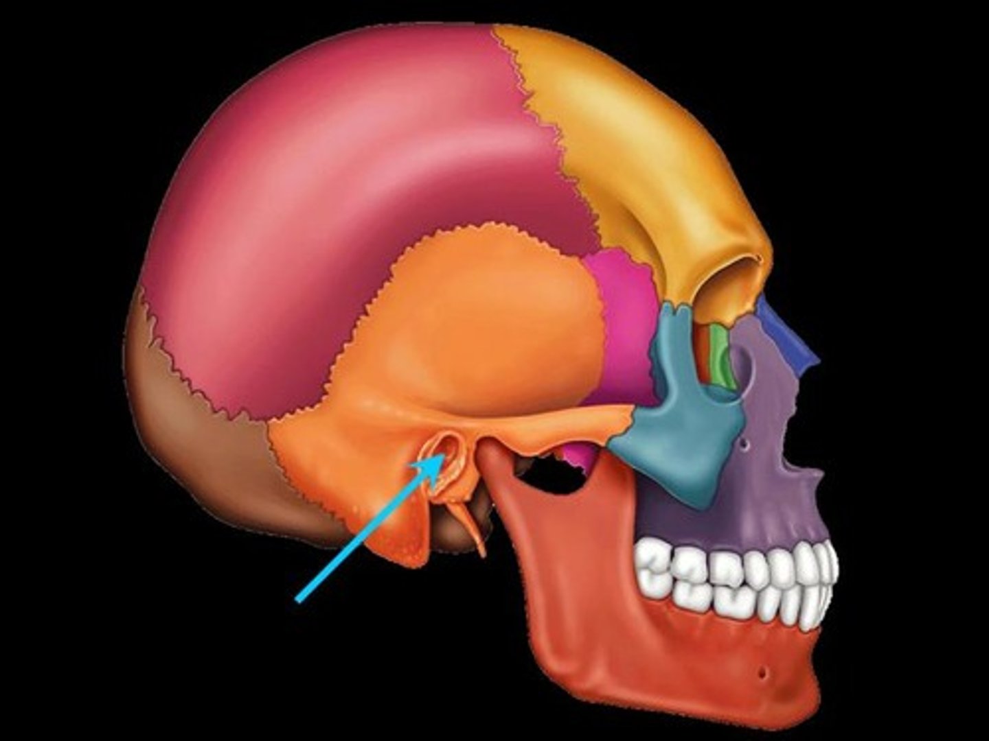
External Auditory Meatus
the opening of the exernal auditory canal of the outer ear
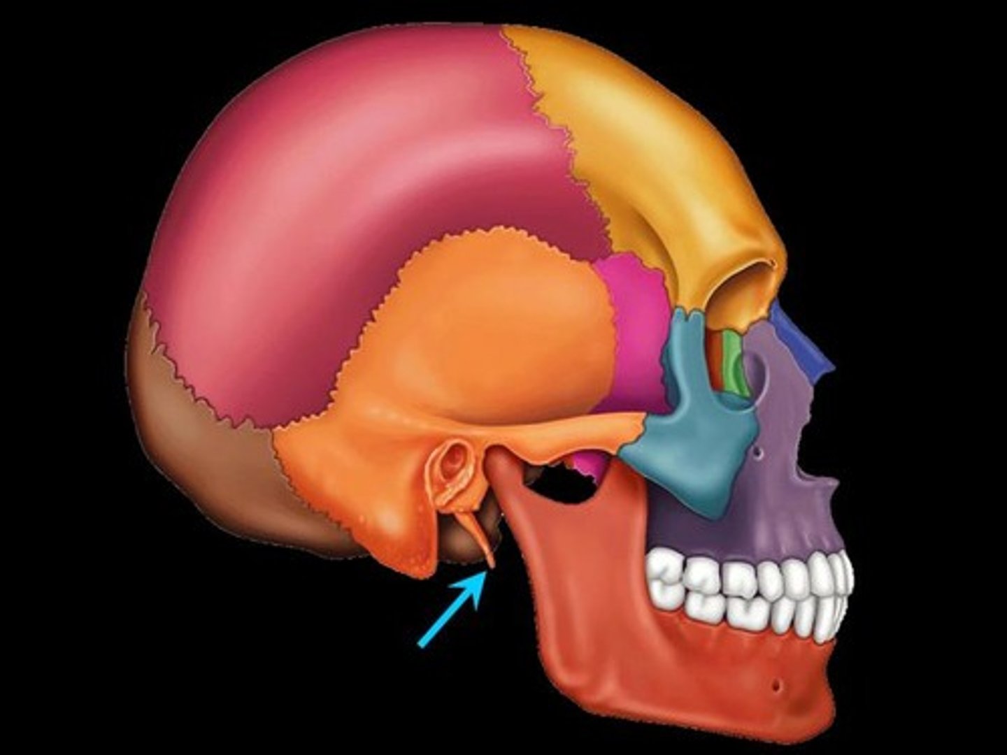
Styloid Process of the Temporal Bone
A slender projection positioned down and forward from the temporal bone on the inferior surface
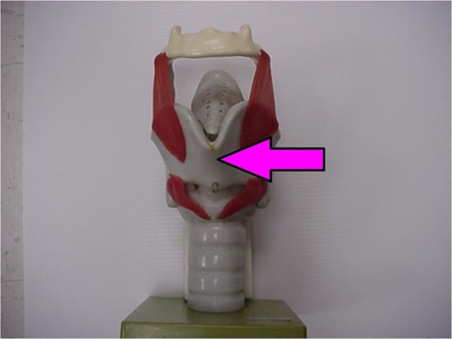
Thyroid Cartilage
The largest of the 9 cartilages of the larynx; AKA Adam's apple; lies just inferior to the hyoid bon at about the level of C3-C4
TMJ Depression
Opening the mouth
TMJ Elevation
Closing the mouth
TMJ Lateral Deviation
Movement of the mandible from right to left and left to right sides
TMJ Protrusion
moving the mandible anteriorly
TMJ Retrustion
moving the mandible posteriorly
Resting Position of the Mandible
lips closed, teeth are several millimeters apart
TMJ prime movers
all innervated by the Trigeminal Nerve (cranial nerve V); temporalis, masseter, medial pterygoid, lateral pterygoid
Temporalis action
Bilateral: elevation of the mandible (closing the mouth); retrusion of the mandible
Unilateral: ipsilateral lateral deviation
Temporalis location
originates on the temporal fossa; inserts on the coronoid process and ramus of the mandible
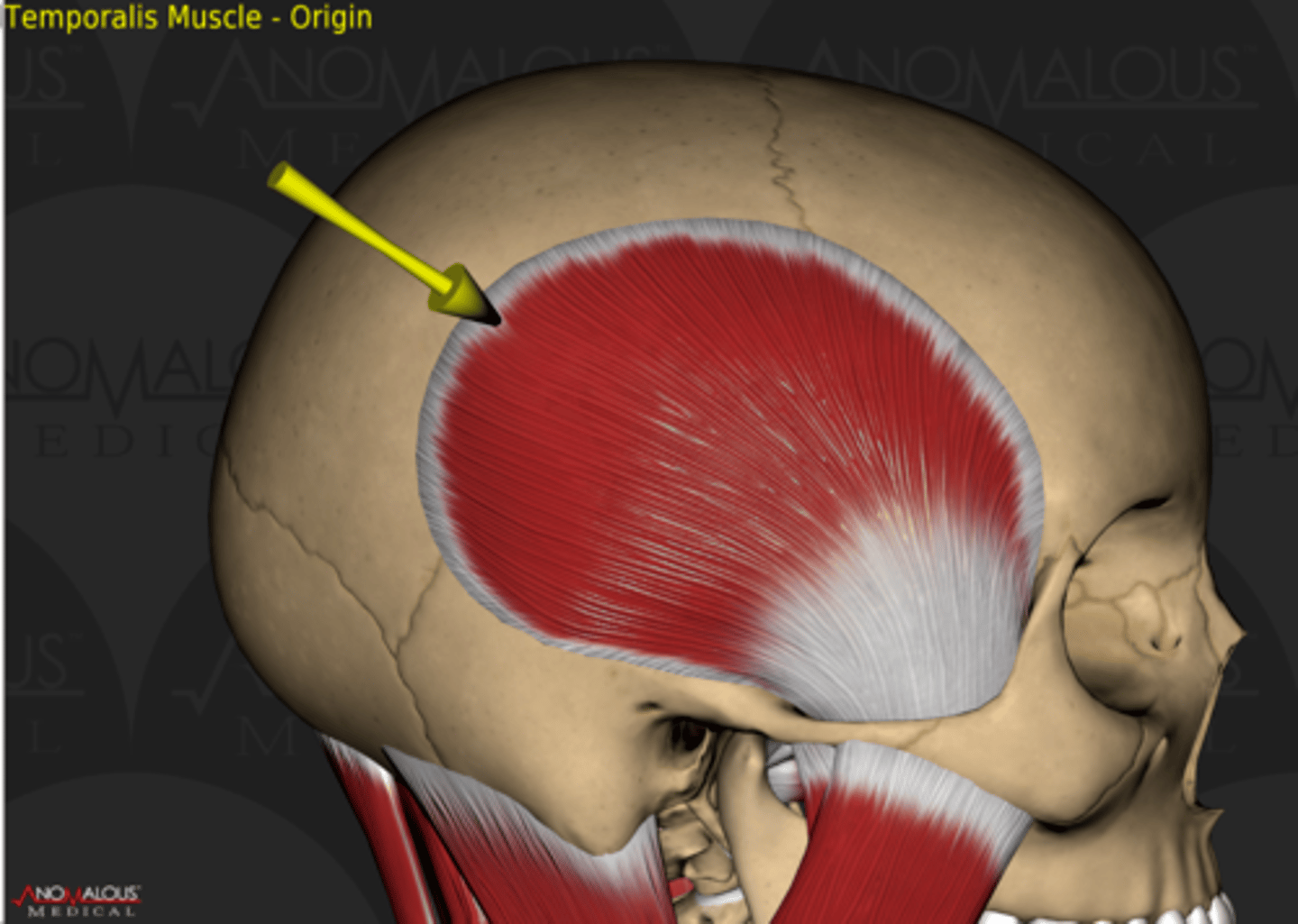
Temporalis innervation
Trigeminal Nerve (cranial nerve V);
Masseter action
Bilateral: elevation of the mandible (closing the mouth);
Unilateral: ipsilateral lateral deviation
Masseter location
Located between the zygomatic arch of the temporal bone and the mandible
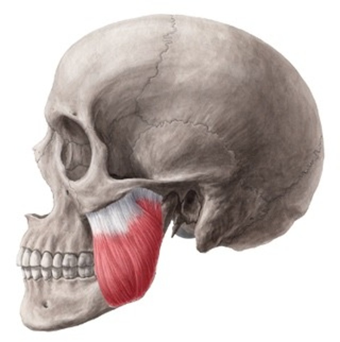
Masseter innervation
Trigeminal Nerve (cranial nerve V);
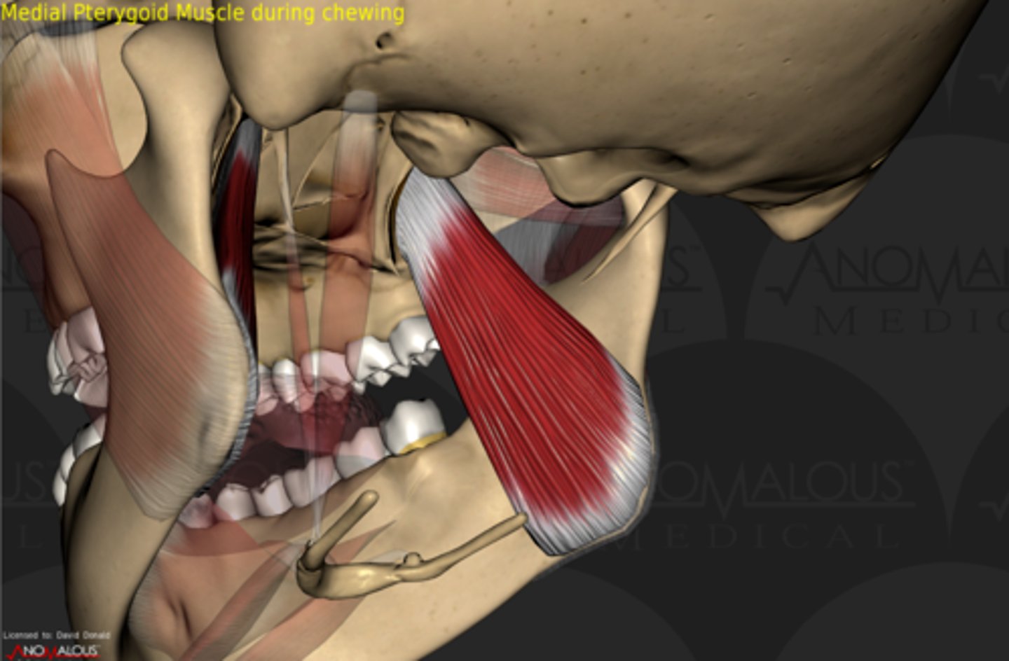
Medial Pterygoid action
Bilateral: elevation of the mandible (closing the mouth); protrusion of the mandible
Unilateral: contralateral lateral deviation
Medial Pterygoid location
internal angle of the ramus of the mandible

Medial Pterygoid innervation
Trigeminal Nerve (cranial nerve V);
Lateral Pterygoid action
Bilateral: depression of the mandible (opening the mouth); protrusion of the mandible
Unilateral: contralateral lateral deviation
Lateral Pterygoid location
inside the mouth near the condyle of the mandible
Lateral Pterygoid innervation
Trigeminal Nerve (cranial nerve V);
Suprahyoid Muscles
Mylohoid, Geniohyoid; Stylohyoid, Digastric; a group of muscles located superior to the hyoid bone; they connect the hyoid bone to the skull; their primary function is to elevate the hyoid and they also assist in mandibular depression (when the infrahyoid muscles stabilize the hyoid bone)
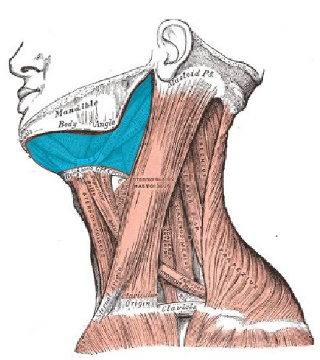
Infrahyoid Muscles
Sternohyoid, Sternothyroid, Thyrohyoid, Omohyoid; a group of muscles located inferior to the hyoid bone and function to depress the hyoid bone. They also stabilize the hyoid bone, thereby allowing the suprahyoid muscles to depress the mandible