unit 4 : Nervous system and brain
1/166
There's no tags or description
Looks like no tags are added yet.
Name | Mastery | Learn | Test | Matching | Spaced | Call with Kai |
|---|
No analytics yet
Send a link to your students to track their progress
167 Terms
3 functions of the nervous system
1) monitors the external and internal environments
2) Processes and integrates information (specifically the CNS )
3) Coordinates a response to the incoming information and sends commands to effectors of the body
Major divisions of the nervous system
1) central nervous system
2) Peripheral nervous system
What is PNS the link to?
Between our CNS and the outside world
Central nervous system
Consists of brain and spinal cord and is housed in dorsal cavity of the body
Peripheral Nervous System
this division consists of all neural tissue outside of the CNS. It is composed of ganglia, nerves and receptors
2 subdivisions of PNS
1. Sensory (afferent) division
2. Motor (efferent) division
Motor (efferent) division
delivers motor commands from CNS to the muscle and glands of the body, which are called effectors
Sensory (afferent) division
consists of specialized structures called receptors that detect sensory information and send it to the CNS
Subdivisions of the Sensory Division of the PNS
1)The somatic sensory division
2)The visceral sensory division
somatic sensory division
detects information from regions of the body that are external to the ventral cavity, which includes the skin, skeletal musculature and bones
visceral sensory division
includes sensory receptors that detect stimuli in the viscera of the body, including the digestive, urinary and reproductive organs
Subdivisions of the Motor Division of the PNS
1) somatic motor division
2) visceral motor division (autonomic nervous system)
Subdivisions of the Autonomic Nervous System (ANS)
1) sympathetic division
2) parasympathetic division
somatic motor division
The effector targets are the skeletal muscles of the body
visceral motor division
has effector targets that include cardiac and smooth muscle as well as glands of the body
sympathetic division
the ‘fight or flight’ system
parasympathetic division
the ‘rest and digest’ system
What are 2 Cell Types in Nervous Tissue
1. Neurons
2. Neuroglia (glial cells)
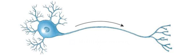
Neurons
1. The basic unit of the nervous system. The neurons carry out the functions of the nervous system. To carry out these functions, neurons communicate with one another and with other types of cells (such as muscle cells or glands).
2. Neurons are not capable of mitotic division; if neurons are damaged they are not replaced. They also have a very high demand for oxygen and glucose
Neuroglia (glial cells):
1. Support the neurons
2. The glial cells actually outnumber the neurons (for everyone 1 neuron there are about 10 glial cells) but they are much smaller than the neurons
3.Also, unlike the neurons, the glial cells can mitotically divide.
5 basic features of neurons
1) dendrites
2) soma (cell body)
3) axon hillock
4) axon
6) axon (synaptic) terminals
Soma (cell body)
Houses most of the organelles
Dendrites
short cytoplasmic extensions. They receive incoming information
Axon hillock
transition region from soma to axon, site of action potential initiation
Axon
a single, long projection that carries
electrical signals away from the soma, towards the
distal ends of the axons
Axon (synaptic) terminals
very distal, enlarged region of the axon which will communicate with the target of the neuron
Synaptic End Bulb
Dilated knob-like end
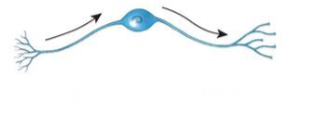
Structural classifications of neurons
1) multipolar neuron
2) unipolar neuron
3) bipolar neuron
Multipolar neurons
2 or more dendrites and 1 axon. These are the most common types of neurons
Unipolar neurons
there is a single extension from the soma, and dendrite and the axon are continuous
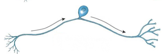
Bipolar neurons
there is one dendrite and one axon that extend from the cell body. These are the most rare
type of neuron
Functional classifications of neurons
1) afferent neuron (sensory neuron)
2) efferent neuron (motor neuron)
3) interneuron
afferent neuron (sensory neuron)
-sends signals toward the CNS
-It generates action potentials from sensory receptors at Its peripheral end
-It has a long axon and is found mainly in the PNS
efferent neuron (motor neuron)
-sends signals away from the CNS to an effector organ
-It has a long peripheral axon in the PNS
Interneuron
is found entirely within the CNS.
It lies between afferent and efferent
neurons
Neuroglia in the CNS
Astrocytes
Oligodendrocytes
Microglia
Ependymal cells
Astrocytes
Has many functions, overall will support the
neuronsHelp to maintain the blood brain barrier (a protective structure of the brain)
Oligodendrocytes
These cells form the myelin in the CNS
Myelin
is a lipid sheath that wraps around the axons of some neurons in the nervous system
Myelin increases the speed of action potential movement down an axon
myelinated axons
Axons that are wrapped in myelin
unmyelinated axons
Axons that are not wrapped in myelin
Microglia
They are phagocytes
They phagocytize pathogens and wastes
in the CNS
Ependymal cells
These cells line the central canal of the spinal cord and chambers in the brain called ventricles, both of which are filled with a fluid called cerebrospinal fluid.
The ependymal cells help to produce and circulate the cerebrospinal fluid
Neuroglia in the PNS
satellite cells
schwann cells
satellite cells
small cells which surround the neuron cell body
in the PNS (their name comes from the resemblance to satellites around a planet)
Schwann cells
create the myelin in the PNS
(another name they are called is neurolemmocytes)
The myelinated axons of PNS
are wrapped in concentric layers of the Schwann cell plasma membrane, which
creates a lipid sheath around the cell
nodes of Ranvier
The gaps between the myelinated segments
Glossy shiny matter in nervous system
Is due to the lipid layers that create the myelin, myelinated axons are a shiny, glossy white
White matter in the nervous system
In the CNS where there are myelinated axons, the nervous tissue has a white appearance
Gray matter in the nervous system
Areas of the CNS where there are unmyelinated axons or cells body and dendrites appear gray
Ion channels
is a transmembrane protein in the plasma membrane that allows specific ions to move into or out of the cell
2 categories of ion channels
1) leaked
2) gated
leak channel
is always opens and always allowing current to pass through
Classes of Gated Ion Channels
Chemically (ligand)-gated
Voltage-gated
Mechanically-gated
Mechanically-gated
opened when a mechanical stimulus is present, such as stretch, pressure
Think “ketchup bottle” you must squeeze the bottle to force the ketchup through the gate
Voltage-gated
opened when there is a depolarization of the membrane potential
Chemically (ligand)-gated
open when a ligand, or extracellular chemical
messenger, binds to the receptor region
of the channel• Channels will only open when a
specific chemical is compatible with
that channel.• Neurotransmitter or chemical does
NOT pass through channel. It only
opens the ‘door’.• Only the ion the channel is permeable
to will pass through the ‘door
Polarized
Neurons, like all other cells in the body, are polarized
This means the cell has an electric charge that is created by a difference in distribution of positive and negative charges across the plasma membrane.
Membrane potential
separation of the positive and negative charges creates a potential difference
(This is analogous to the potential of a battery)
Voltage
is also a term that is used for potential difference
The unit for voltage is Volts (V)
In the cells, the voltage is small and so is measured in mV.
potential difference in a resting cell
is known as resting membrane potential (RMP)
Typically, the RMP of a neuron is -70mV
Average concentration in ICF
150mM K+
15mM of Na+
Average concentration of ECF
5mM K+
150mM Na+
Major factors of RMP
1) unequal distribution of ions (mostly Na+ and K+,but other ions as well) across the plasma membrane. The membrane is selectively permeable to these ions. Meaning more K+ is allowed to leave than Na+ to come in due to leaky channel differences
2) the large proteins are negatively charged anions that cannot leave the cell
3) Na+/K+ pump (3Na+ pumped out and 2K+ pumped in
The central nervous system is composed of what?
The brain and spinal cord
The brain
Is an extremely complex organ responsible for an individuals behavior, personality, and intellect.
It carries out these and many other functions via the interactions of neurons
Rostral
Towards the nose / forehead
(Synonym with anterior )
Caudal
Toward the tail (cord)
(synonymous with posterior)
Cortex
The outer gray matter located at the surface of the brain
Nuclei
Groups of neuronal cell bodies
Corpus callous
Nerve fibers that connect the left and right hemispheres
Internal capsule
Nerve fibers that connect the brain stem and the cerebral cortex
Septum pellucid
Membrane connecting the corpus callous and fornix
4 regions of the brain
1) the brain stem 2) cerebellum 3) diencephalon 4) cerebrum
Cerebrum
• The most rostral part of brain
Covers the diencephalon and the brain stem
Divided into 2 cerebral hemispheres which make up 80% of total brain mass
Wrinkled surface
Fissures
Deep groves that are associated with the cerebrum. They separate different regions of the brain
Transverse cerebral fissure
Separates cerebral hemisphere from the cerebellum
Longitudinal fissure
Separates the right and left hemispheres
Gyri
Elevated ridges on the cerebrum
Sulci
Shallow grooves adjacent to the gyri
Purpose of gyri and sulci
To increase the surface area of the brain
3 main regions of the cerebral hemispheres
1) outer gray matter (cerebral cortex)
2) white matter internal to the cerebral cortex
3) gray matter within the white matter
What is the ratio for k+ channels to Na+ channels
There are 25 k+ channels for every 1 Na + channel
Depolarization
Changes in membrane potential that cause the potential to become more positive ( or less negative)
Probability of producing impulses increases
Membrane potential moves towards 0mV
Hyper polarization
Changes that cause the membrane potential to become more negative
Probability of producing impulse decreases
Away from zero
2 types of electrical signals created by ionic movement
Graded potential
Action potentials (AP’s)
Graded potential
Mediated by ligand-gated ion channels or mechanically gated ion channels
Initiated by dendrites (or soma) in response to synaptic input from another neuron
Local changes in membrane potential that decay rapidly as the current (carried by the ions) moves from the initial site of entry into the cell
Are called graded potentials because their magnitude varies directly with the strength of the stimulus (the stranger the stimulus the larger the voltage change and the further the current goes )
Action potentials (AP’s)
Mediated by voltage gated or leaky ion channels
All or none (either happen completely if threshold is reached or not at all)
Do not vary in amplitude or strength (always the same magnitude)
Very rapid ,very large change in membrane potential
Polarity of membrane reverses during action potential
Used for long-distance communication
Do not decay
Do not decrease in strength with distance they are same magnitude at the synaptic terminals as they are when they are initiated at the axon hillock
What are 2 voltage gated channels responsible for the action potential?
Voltage gated Na+ and voltage gated K+ channel
3 different states voltage - gated Na+ can be in
1) Closed state: closed but capable of opening
2) Open (Activated ) state: open with Na+ ions passing through
3) Inactivated state: closed,not capable of opening
States in which voltage gated k+ channel can be in
1) closed state
2) open (activated) state
Voltage gated Na+ channels kinematic
Fast to open and close (Like a sports car)
Voltage gated K+ channel kinematic
Slow to open and close ( like a semitruck)
Tetrodotoxin (TTX)
blocks vgNa+ channels only
Found naturally produced in some species like pufferfish
1-2mg is lethal. LD of 50. 5.0-8.0 microgram/kg. Less than the tip of a pinhead can unalive a human. No antidote exists
TEA (Tetraethylammonoium)
man made for laboratory research to block vgK+ channels only
No antidote. No reported cases of unalivement
What is needed for action potential to be initiated by the opening of the voltage-gated
channel?
the membrane potential has to depolarize to the threshold potential
What is the threshold potential in mV?
-55 mV
What causes membrane potential to depolarizes to the threshold from RMP?
graded potentials in the neuron caused by synaptic activity
What happens during depolarization?
Once threshold (-55mV) is reached, voltage-gated Na+ channels open and Na+ moves
into the cell (Na+ influx) causing a rapid depolarization to +30mV