HBS: Nervous System
1/48
There's no tags or description
Looks like no tags are added yet.
Name | Mastery | Learn | Test | Matching | Spaced | Call with Kai |
|---|
No analytics yet
Send a link to your students to track their progress
49 Terms
Functions of the nervous system
-detect incoming stimuli (sensory input)
-interpret stimuli (brain)
-respond to stimuli (motor output)
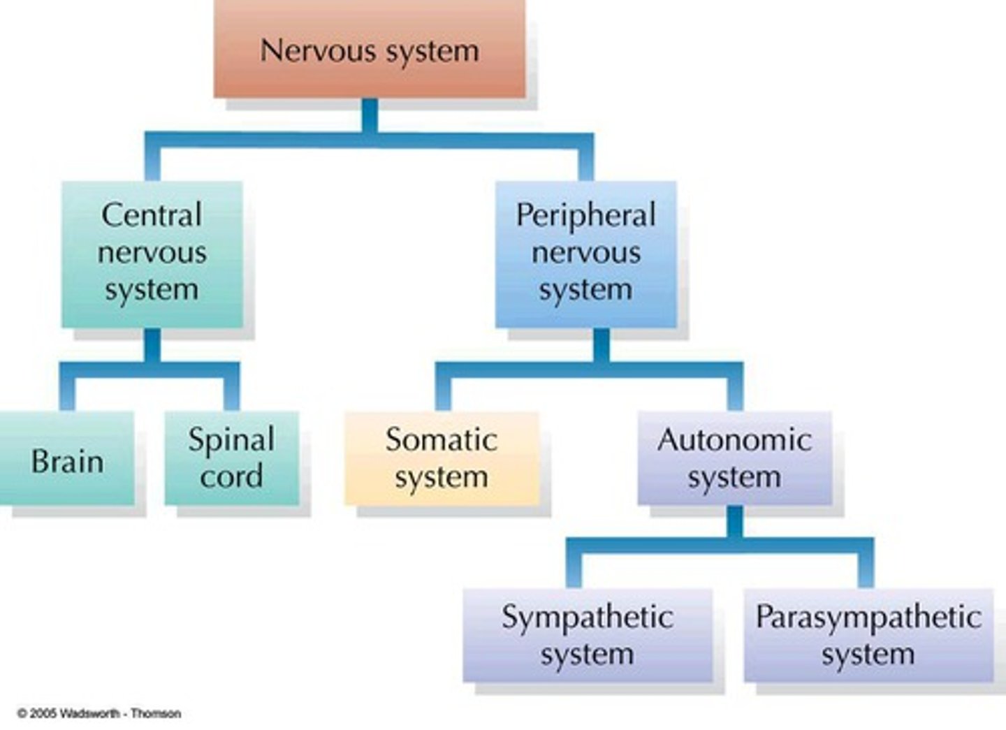
Nervous system flowchart
(see image)
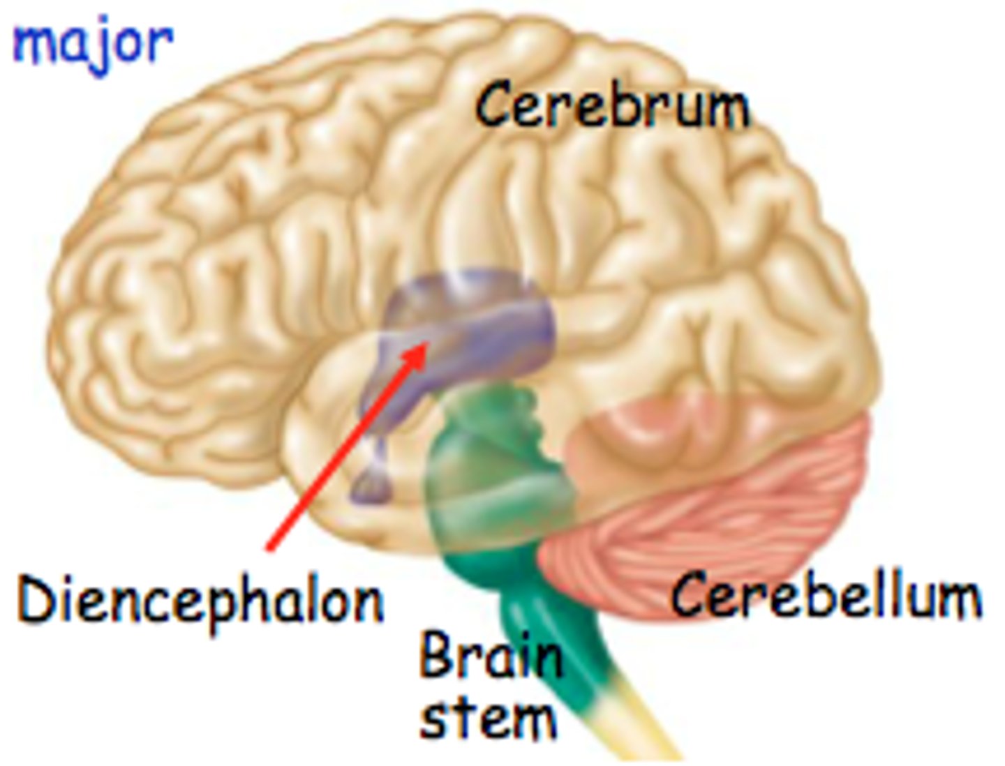
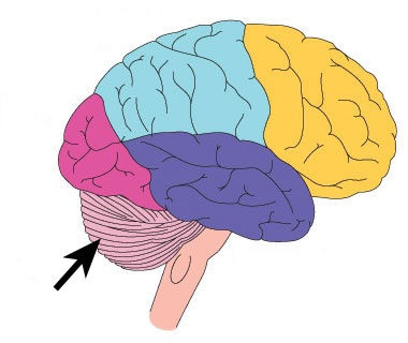
Regions of the brain
cerebrum, diencephalon, brain stem, cerebellum
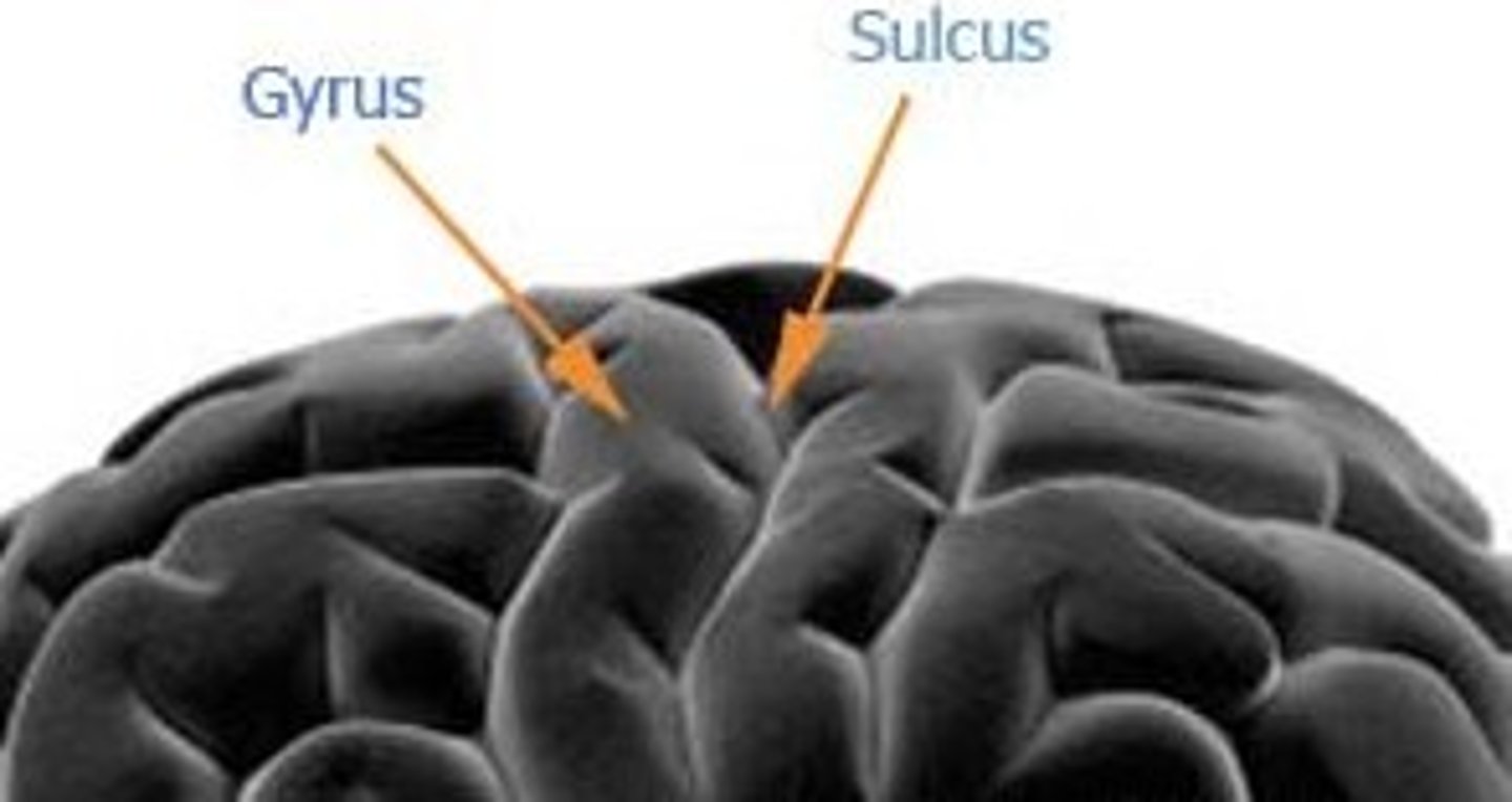
Gyri
ridges of the brain

Sulci
grooves of the brain
Where is gray matter in relation to white matter?
Gray matter is 1/4 inch before the white matter
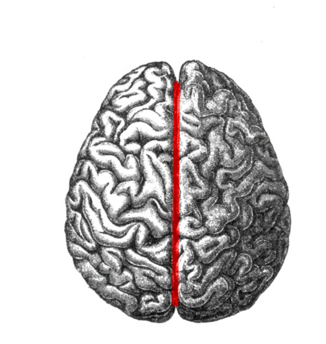
longitudinal fissure
separates left and right hemispheres
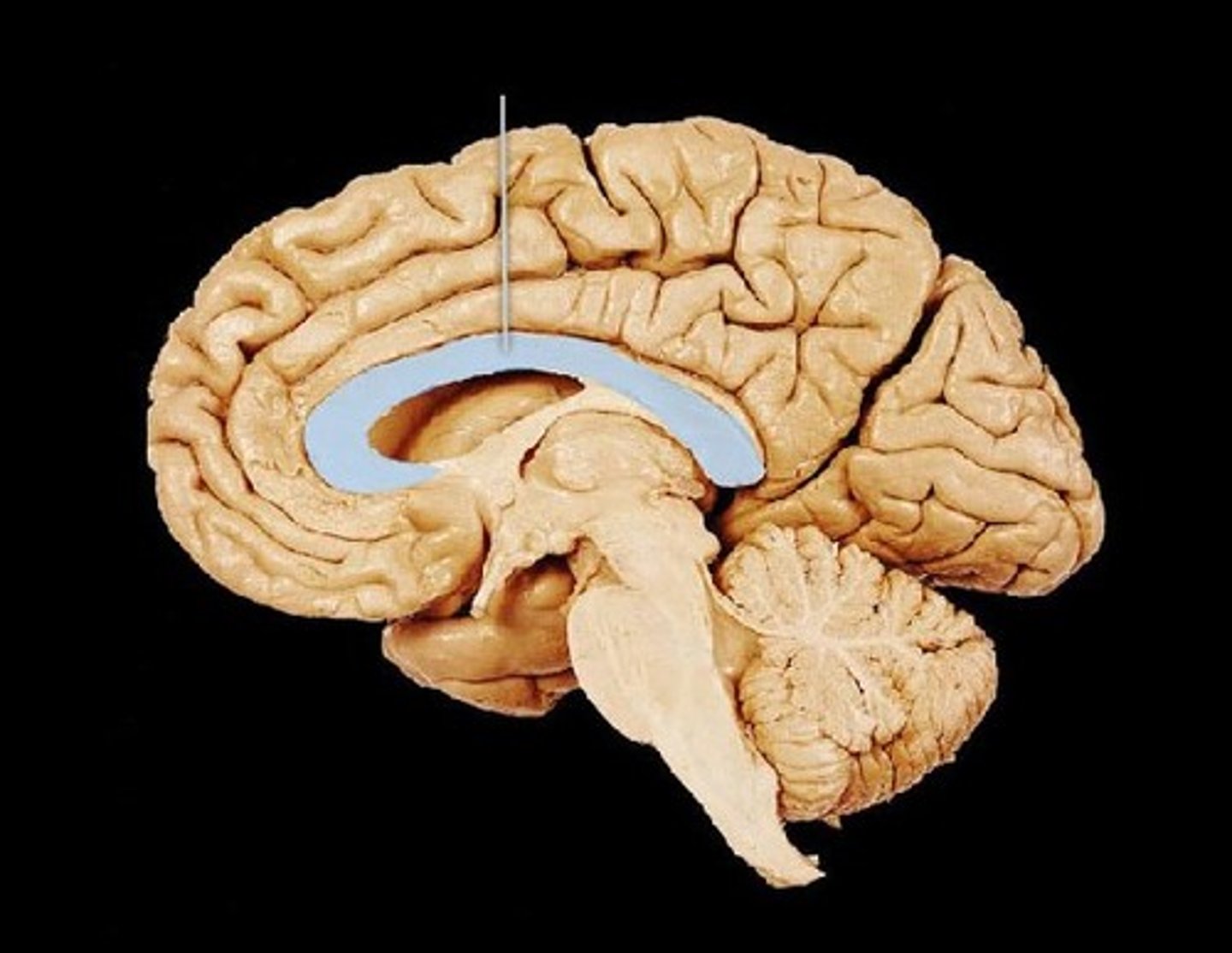
corpus callosum
connects the two hemispheres
gray matter (in cerebrum)
unmyelinated neurons that control somatic sensory receptors/motor receptors
white matter (in cerebrum)
myelinated neurons that send impulses throughout the brain from one hemisphere to the other
Lobes of cerebrum
frontal, parietal, temporal, occipital
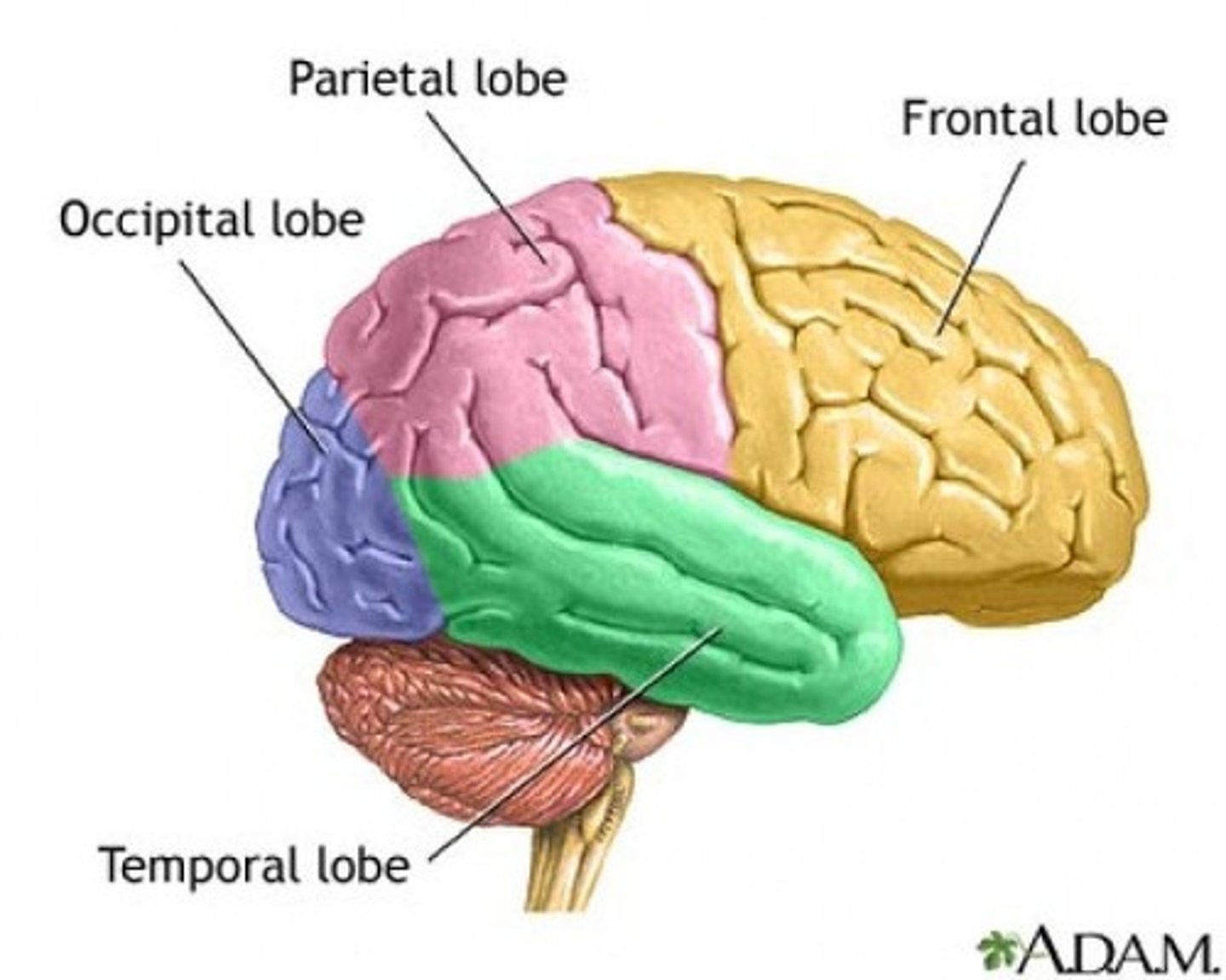
Frontal lobe function
higher intellectual reasoning, socially acceptable behavior, voluntary movement control
Parietal lobe function
interprets somatosensory sensations (skin... perception of touch, pressure, pain, temperature, position, movement, and vibration)
temporal lobe function
interprets hearing/smell
Occipital lobe function
visual cortex, interprets sight
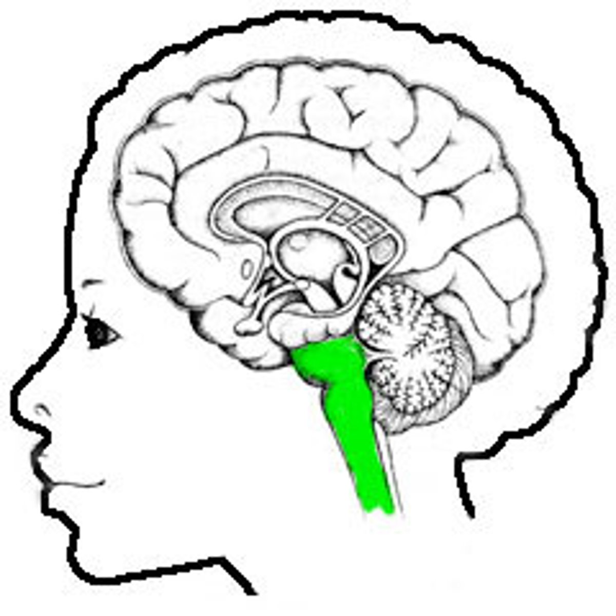
brain stem function
attaches brain to spinal cord
medulla oblongata function
Control center for heart rate, blood pressure, breathing, swallowing, vomiting (vital functions)
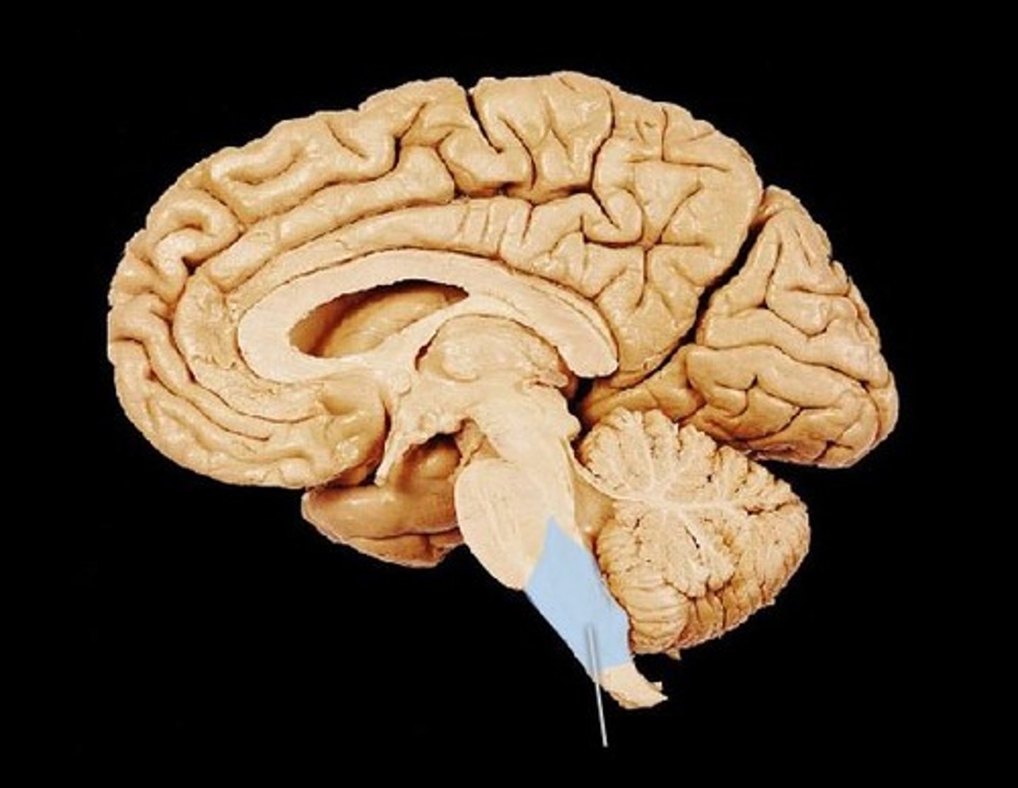
pons function
composed of fiber tracts, controls breathing
midbrain function
composed of tracts/nerve fibers that control reflex centers for vision/hearing
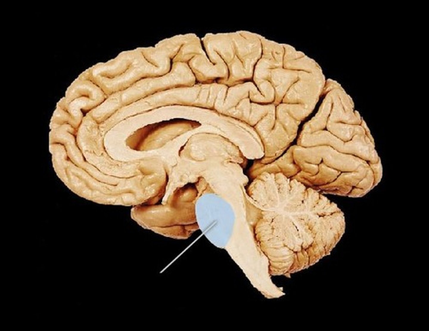
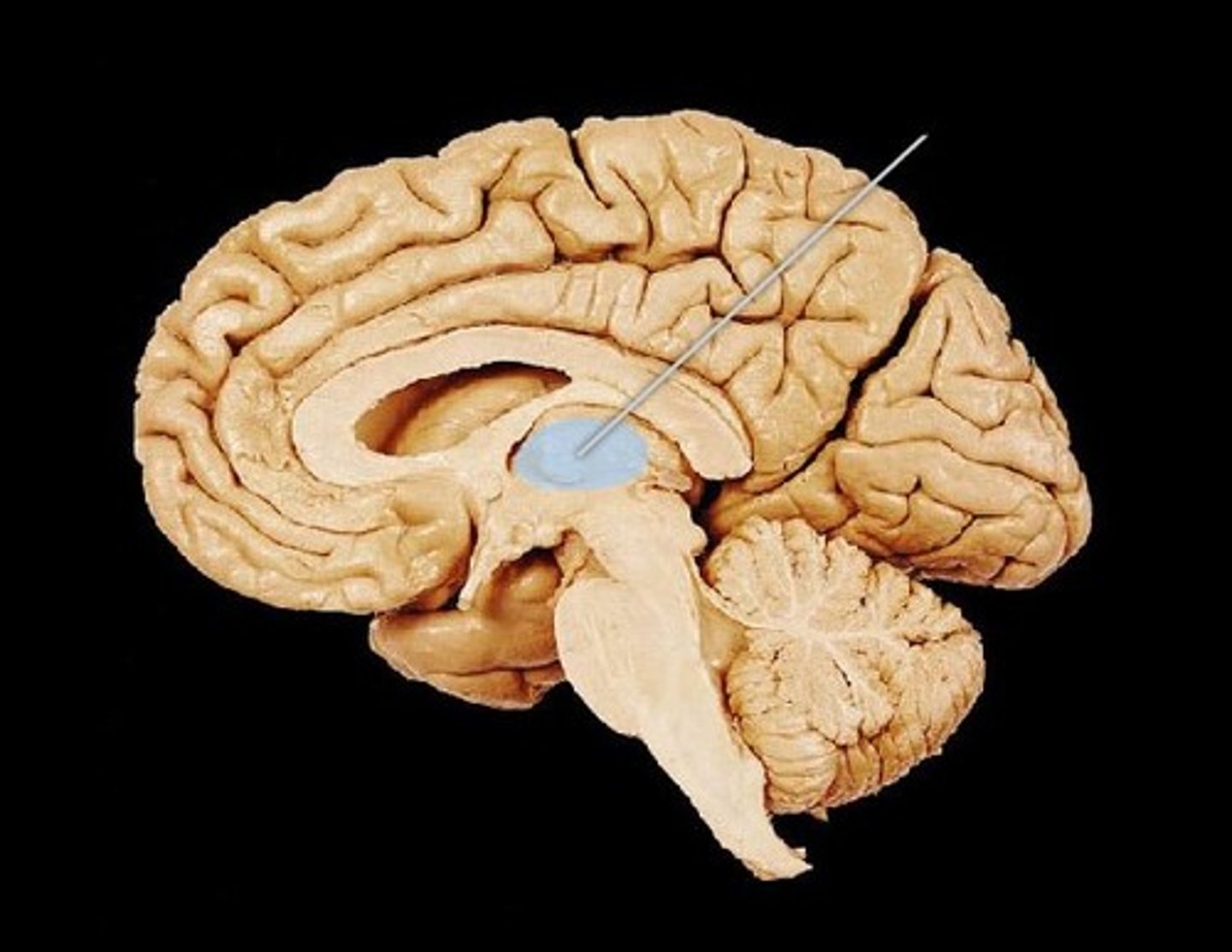
thalamus function
relay station for sensory impulses, pain... transfers impulses to correct cortex for interpretation
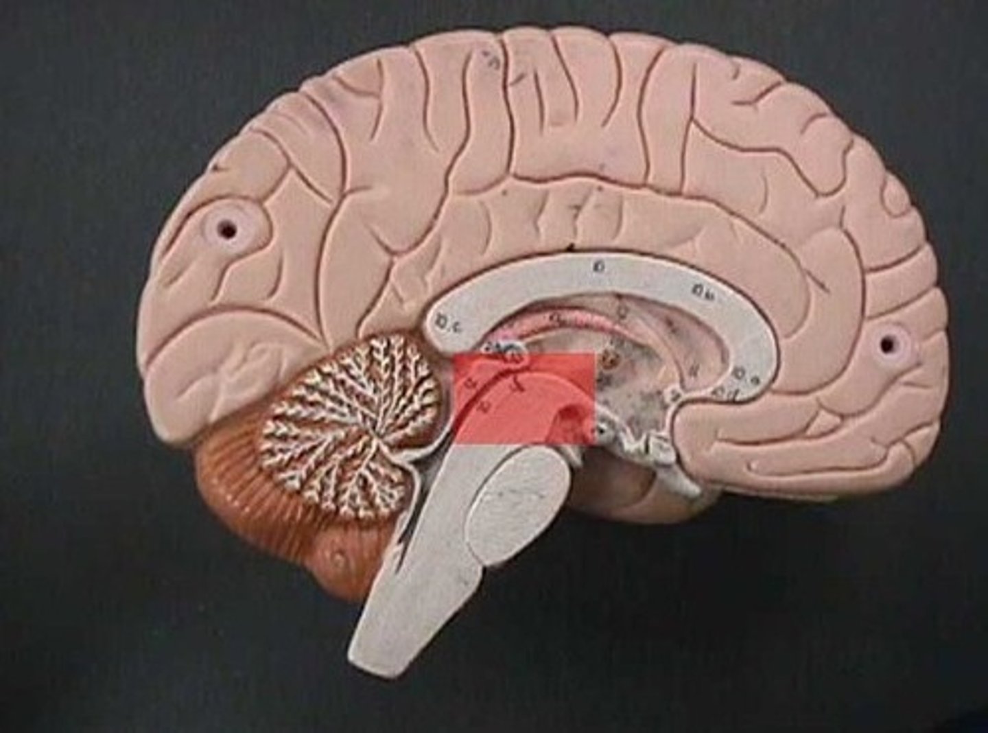
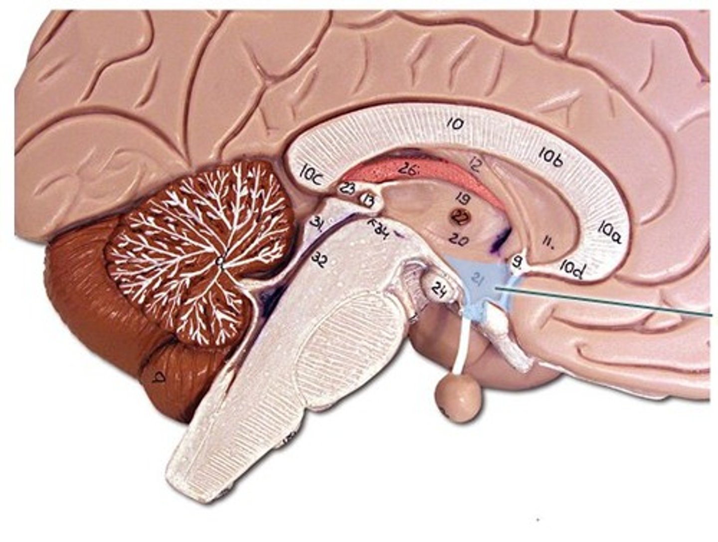
hypothalamus function
Regulates body temperature, metabolism, controls water balance, important in emotions
Cerebellum function
Controls muscle balance and coordination
iris
Muscle that controls size of pupil, pigmentation gives color to the eye, regulates amount of light entering the eye
Cornea
transparent tissue covering the front of the eye
pupil
hole in the center of the eye where light passes through
Fovea
focal point of the eye
optic nerve
sends image to the brain
aqueous humor
clear watery fluid found in anterior side of eye
vitreous humor
clear, jelly-like fluid that keeps the shape of the eye
lens
transparent tissue that bends light passing through the eye
retina
layer of tissue on back side of eye that contains cells responsive to light
choroid
Middle layer that contains blood vessels in eye
Sclera
outer covering that helps maintain the shape of the eye (whites of the eyes)
Optic disk
Blind spot... A hole in the retina where the optic nerve connects to the retina
cilliary body
Contains a ring of muscles that surround the lens and control its shape
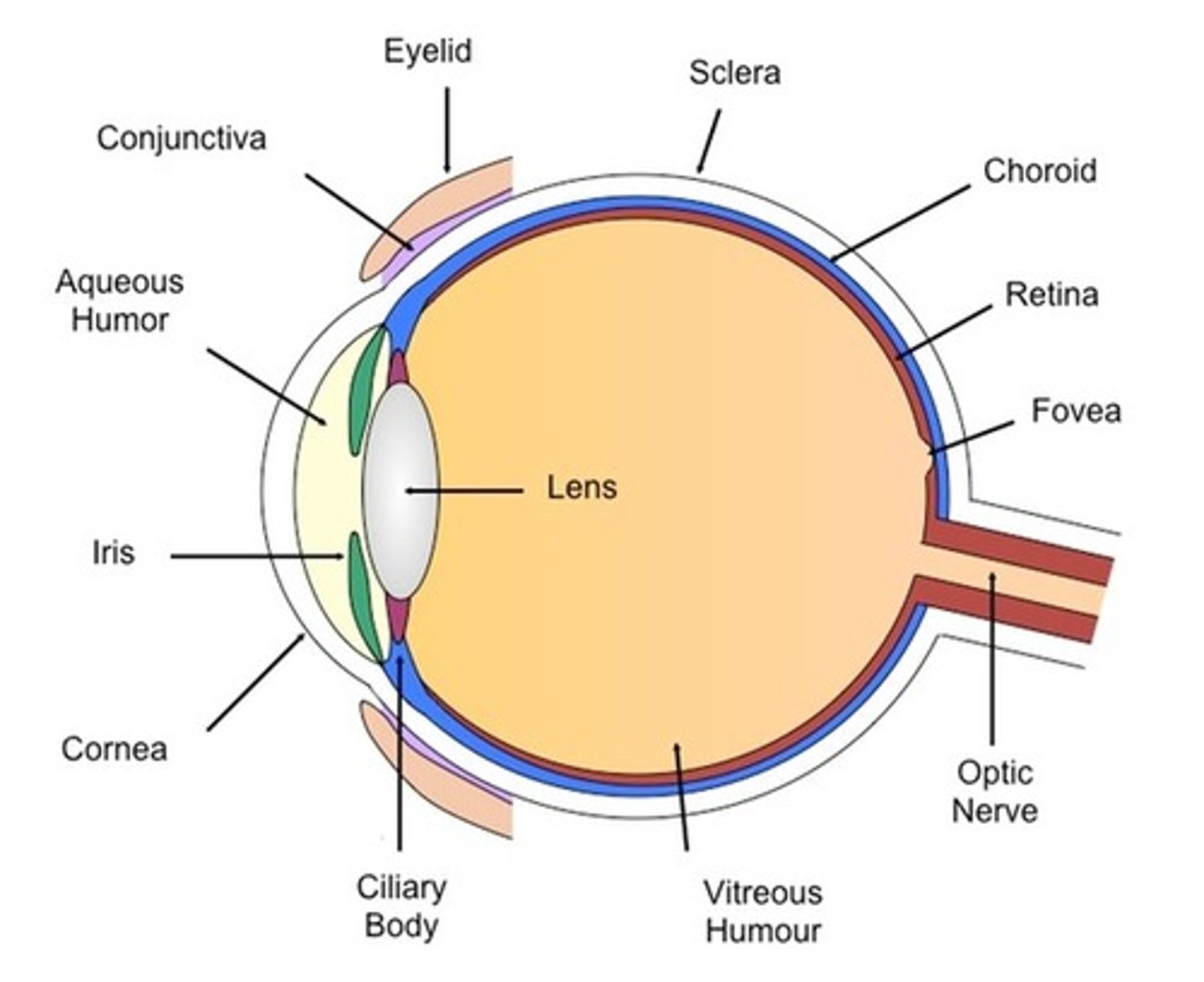
Eye diagram
(see image. the image doesn't have all of the parts we need to know... make sure you look at your colored diagrams!)
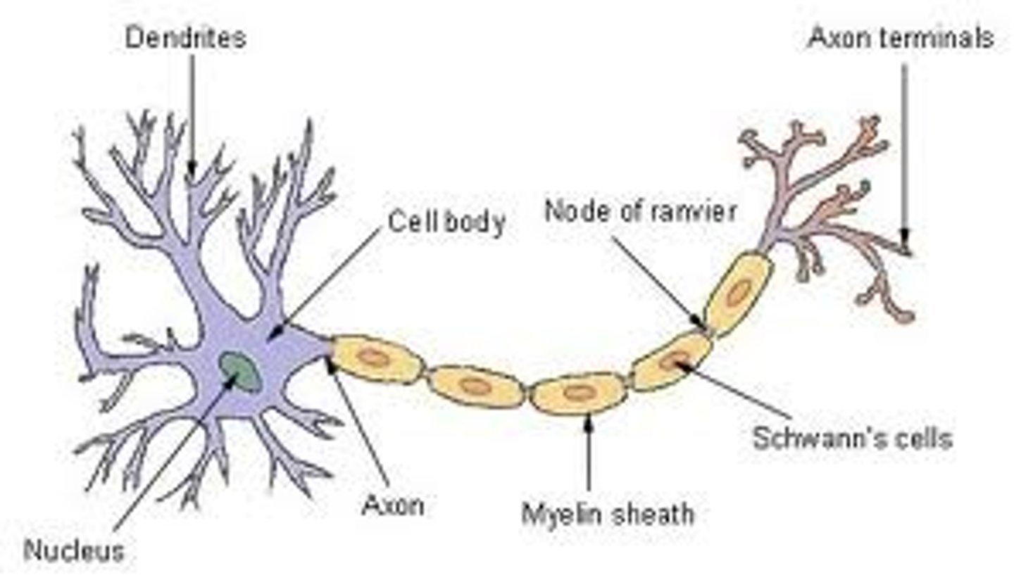
Dendrites
detect stimuli
Axon hillock
Acts like a funnel for info to travel down the axon
Axon
directs stimulus down the neuron
myelin sheath
protective layer to axon
Nodes of Ranvier
helps move the stimulus down the axon
Cell body of neuron
contains nucleus and organelles
Nucleus of neuron
Contains genetic info
Axon terminal
end of axon, sends stimulus to the next neuron
Neurillema
protects Schwann cells
Schwann cells
produce myelin sheath
neuron diagram
(see image + your own diagram)
Synapse
Gap between neurons
Neurotransmitters
what is released from the axon terminal... neurotransmitters cross the gaps between neurons (synapses)