Central Nervous System
1/740
Earn XP
Description and Tags
Year 1 - Semester 2
Name | Mastery | Learn | Test | Matching | Spaced | Call with Kai |
|---|
No analytics yet
Send a link to your students to track their progress
741 Terms
Which 2 structures make up the CNS?
brain and spinal cord
What protects the CNS?
bony structures and meninges
What are the 3 main regions of the brain?
forebrain, midbrain, hindbrain
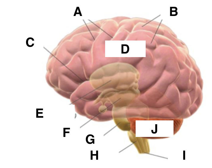
A
sulci


B
gyri


C
fissure


D
cerebrum


E
diencephalon (thalamus and hypothalamus)


F
midbrain


G
Pons


H
medulla oblongata


I
spinal cord


J
cerebellum

How are the 3 regions of the brain defined?
embryological origin
Where does the CNS receive afferent information from?
peripheral nervous system
processes involved in brain development
neurulation, neuronal proliferation, neural migration, apoptosis, synaptogenesis, myelination
neurulation
process where the flat neural plate folds and fuses to form the neural tube
neural migration
process during which neurones move around the CNS and out to their specific locations in the PNS
synaptogenesis
the formation of synapses between neurones
myelination
the formation of myelin sheaths around the axons of neurones
What is the cerebrum divided into?
left and right cerebral hemispheres
Where do the cerebral hemispheres arise from?
the embryonic prosencephalon
Which structures make up the diencephalon?
thalamus and hypothalamus
Which structure in the brain do most of the cranial nerves arise from?
brain stem
Function of cerebellum
to organise and refine motor activity, to coordinate gait, to maintain posture, to control muscle tone and voluntary muscle activity, to compare intended movements with the outcome
notochord
a cylinder of mesodermal cells which are responsible for inducing formation of the neural plate via secretion of chemical signals (including sonic hedgehog protein)
neurulation process
notocord secretes sonic hedgehog protein which causes the neural plate to bend medially to form the neural groove
neural groove deepens and its ectodermal walls thicken at the dorsal lips to form the neural folds
superficial ectoderm either side of the dorsal surface of the neural tube proliferate to form the neural crest
neural folds fuse to close the neural tube, beginning in the cervical region and proceeding rostrally and caudally
Which embryonic germ layer does the neural plate form from?
(dorsal) ectoderm
What causes spina bifida
incomplete caudal fusion of the neural folds in the neural tube
What structures does the neural crest give rise to?
cells in PNS, meninges, some structures of the head
Where does the sonic hedgehog protein work predominantly?
ventral region
neuropores
the holes at the rostral and caudal ends of the neural tube which close last
In which breeds is it common for the neural tube to not fully fuse?
tailless breeds
Which section of the neural tube do brain vesicles develop from?
rostral pole
What are the 3 primary brain vesicles which develop first?
prosencephalon, mesencephalon, rhombencephalon
Which region of the brain develops from the prosencephalon?
forebrain
Which region of the brain develops from the mesencephalon?
midbrain
Which region of the brain develops from the rhombencephalon?
hindbrain
What are the 5 secondary brain vesicles?
telencephalon, diencephalon, mesencephalon, metencephalon, myelencephalon
At what stage does the embryo develop the primary brain vesicles?
3-4 weeks
At what stage does the embryo develop the secondary brain vesicles?
5 weeks
Which structure in the brain develops from the telencephalon?
cerebrum
Which structures in the brain develop from the diencephalon?
eye cup, thalamus, hypothalamus, epithalamus
Which structure in the brain develops from the mesencephalon?
midbrain
Which structure in the brain develops from the metencephalon?
pons and cerebellum
Which structure in the brain develops from the myelencephalon?
medulla oblongata

A
diencephalon


B
cephalic flexure


C
rhombic lip


D
developing cerebellum


E
pontine flexure


F
cervical flexure

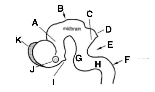
G
pons


H
medulla


I
infundibular stalk


J
optic vesicle

functions of cerebrospinal fluid
to provide nutrition to CNS tissues, to act as a cushion and physically support the CNS, to act as a volume buffer to accomodate small amounts of CNS swelling, to maintain a stable environment for neurones, to help the movement of neurotransmitters around the CNS
Which structures form brain hemispheres?
telencephalic vesicles
holoprosencephaly
a single-lobed brain and a single central eye caused by failure of the prosencephalon to divide
How does CSF access inner CNS cells in the brain?
an interconnected series of ventricles in the brain which leads into a central canal in the spinal cord

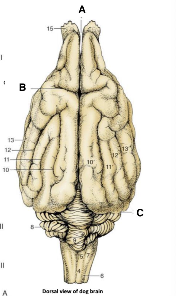
Which number corresponds to the lateral ventricle of the brain?
2
Which number corresponds to the third ventricle of the brain?
3

Which number corresponds to the fourth ventricle in the brain?
7
Which structures make up grey matter?
nerve cell bodies, dendrites and synapses
Which structures make up white matter?
axons of neurones
nuclei
clusters of nerve cell bodies within the CNS
ganglia
clusters of nerve cell bodies within the PNS
What gives white matter its white appearance?
the presence of fat in the myelin around the neurone axons
glial cells
non-neuronal supporting cells in the CNS and PNS
tracts
bundles of axons in the CNS
nerves
bundles of axons in the PNS
corpus callosum
a major tract of white matter which connects the 2 cerebral hemispheres in the brain together
corpus striatum
basal nuclei in deep parts of the brain hemispheres which are interwoven with white matter tracts
function of corpus callosum
to allow exchange of information between the 2 brain hemispheres
What type of epithelium initially lines the neural tube?
pseudostratified columnar epithelium
Which 3 layers does epithelium in the neural tube differentiate into?
germinal, mantle, marginal
What is found in the germinal layer of the neural tube?
neuronal and glial progenitors
What is found in the mantle layer of the neural tube?
mitotic cells in interphase or differentiating into neurones or glia
What is found in the marginal layer of the neural tube?
axons of neurones/ white matter
What structure separates the 2 cerebral hemispheres of the brain?
longitudinal fissure
Which structure separates the cerebrum from the cerebellum?
transverse fissure

A
longitudinal fissure


B
cruciate sulcus


C
transverse fissure
What is the main sulcus in the brain?
cruciate sulcus
What is the main gyrus in the brain?
sylvan gyrus

X
cruciate sulcus

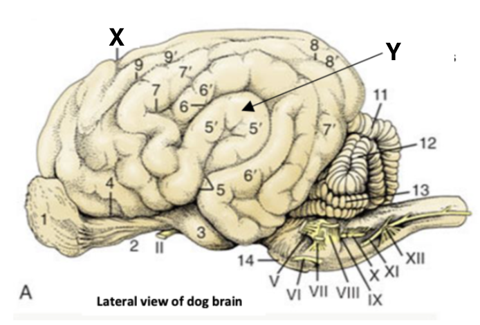
Y
sylvan gyrus

What are the bulges in the cerebrum known as?
gyri
What are the furrows in the cerebrum known as?
sulci
What are the folds in the cerebellum known as?
folia
What are the lobes of the cerebrum?
frontal, parietal, temporal, occipital
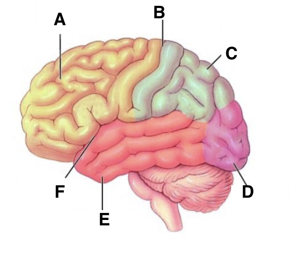
A
frontal lobe


B
cruciate sulcus


C
parietal lobe


D
occipital lobe


E
temporal lobe


F
lateral sulcus

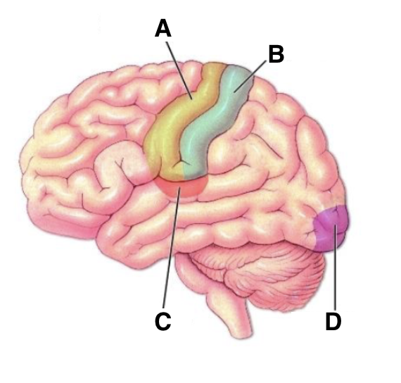
A
motor cortex


B
somato-sensory cortex
