Chapter 8 - THE MUSCULOSKELETAL SYSTEM ALLOWS MOVEMENT
1/68
There's no tags or description
Looks like no tags are added yet.
Name | Mastery | Learn | Test | Matching | Spaced | Call with Kai |
|---|
No analytics yet
Send a link to your students to track their progress
69 Terms
How are muscle cells held together
Muscle cells are held together in bundles. A sheath of connective tissue called the perimysium surrounds each bundle so that it can function as an individual unit. The connective tissue allows adjacent bundles to slide easily over one another as they contract
epimysium
Sheaths of connective tissue called epimysium also hold the bundles together, and towards the end of the muscle they taper and blend to form the tendon.
muscle properties
excitability, contractibility, extensibility and elasticity.
allowing it to…
• be stimulated by a nerve impulse
• shorten in length
• be stretched
• return to their original length.
Muscles can only contract
This means that they can pull bones together, but they cannot push them apart. If muscles contract, pulling a bone in one direction, another set of muscles must contract to pull the bone in the opposite direction. Thus, the muscles that move the parts of the skeleton are always grouped in pairs.
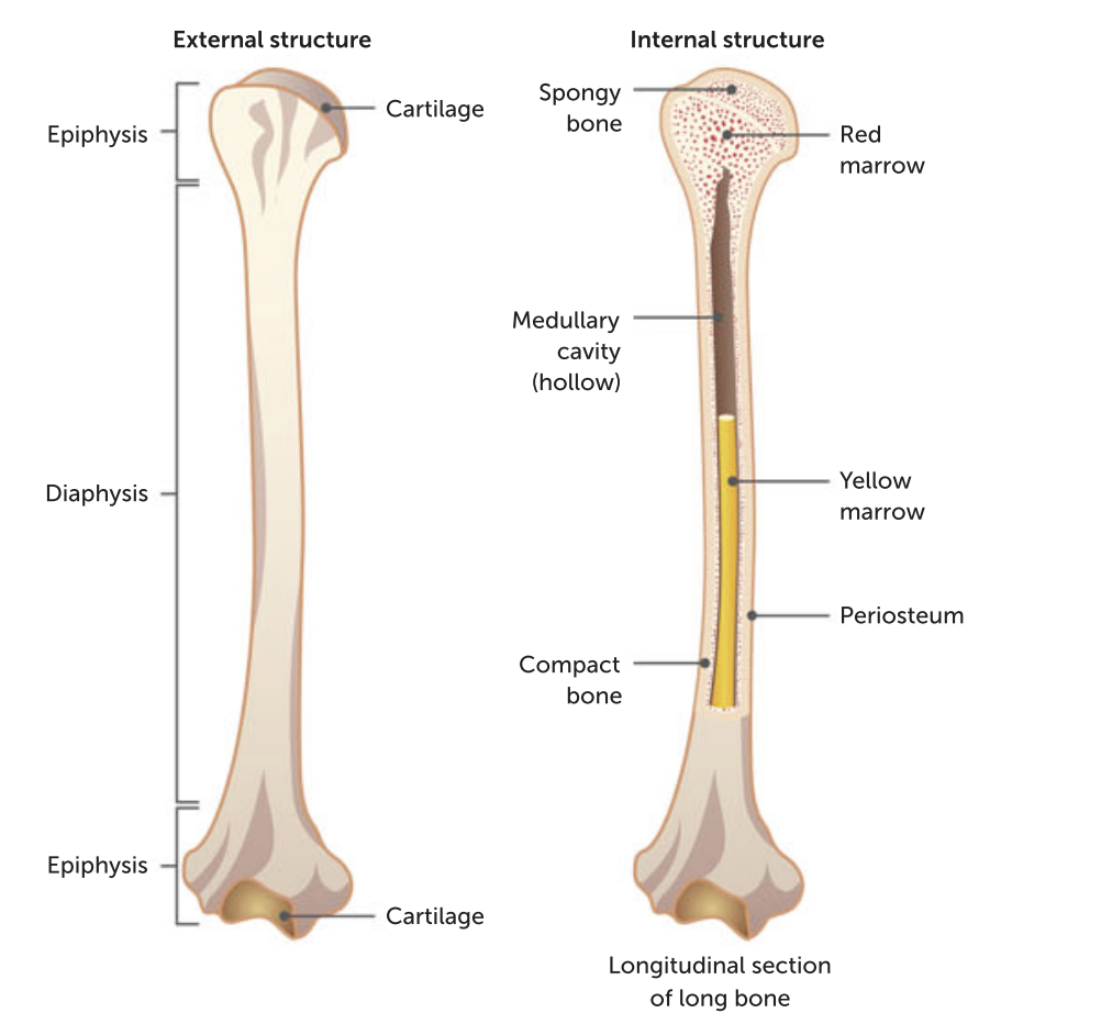
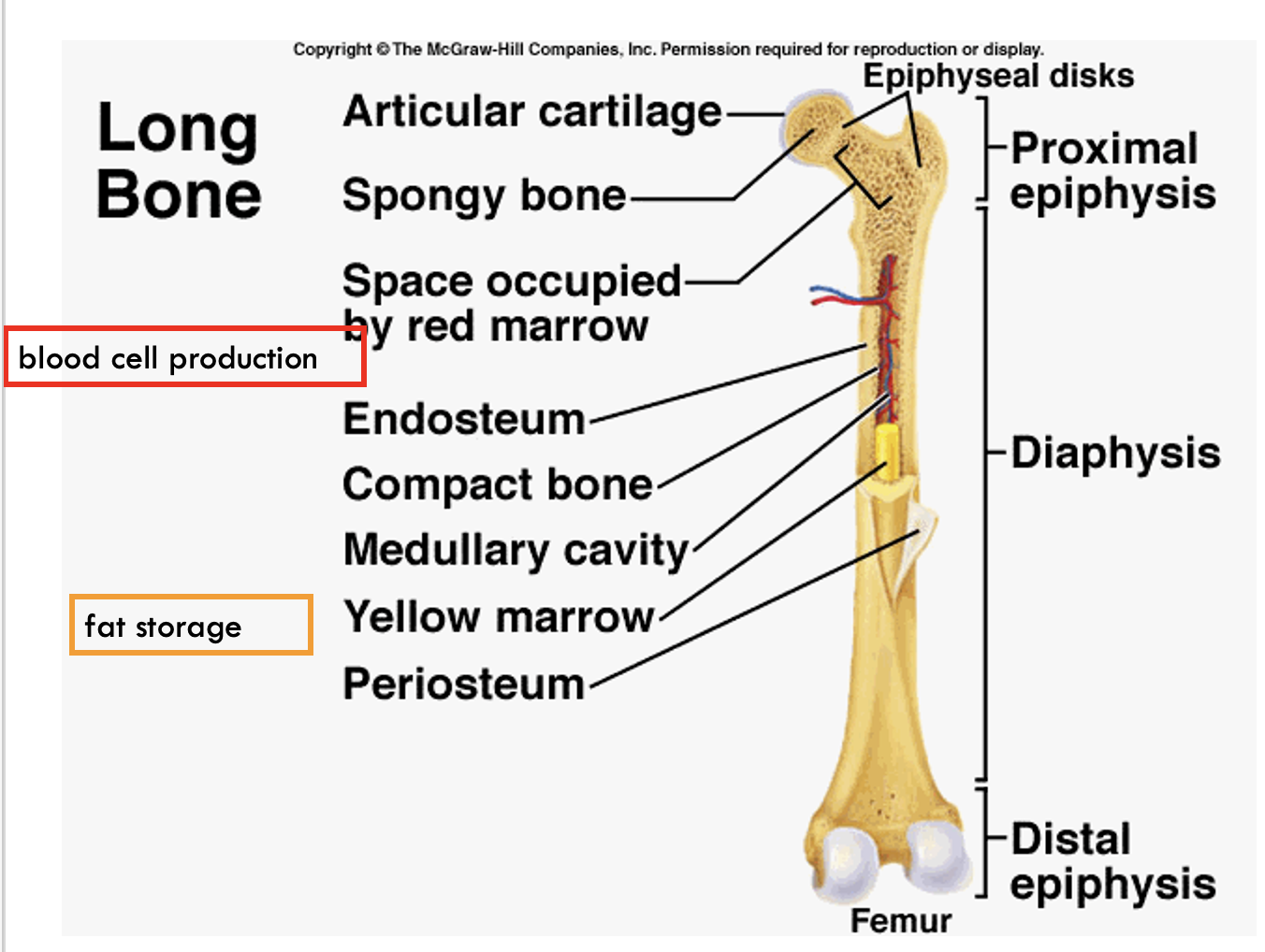
Diaphysis
the shaft making up the main portion of the bone. When the bone is cut lengthwise, the diaphysis is seen to be a hollow cylinder of compact bone surrounding a medullary cavity. This cavity is used as a fat storage site and is often called the yellow bone marrow cavity.
Epiphyses
Epiphyses (singular: epiphysis) – the enlarged ends of the bone, covered with a thin layer of cartilage (articular cartilage). The epiphyses have compact bone on the outside, but their central regions contain spongy or cancellous bone. Cancellous bone is more porous than compact bone, and contains many large spaces filled with marrow. In certain bones, this may be red bone marrow, where blood cell production takes place.
Periosteum
the dense, white, fibrous outer covering of the bone. There is no periosteum at the joints, where the bone is covered with an articular cartilage.
long bone
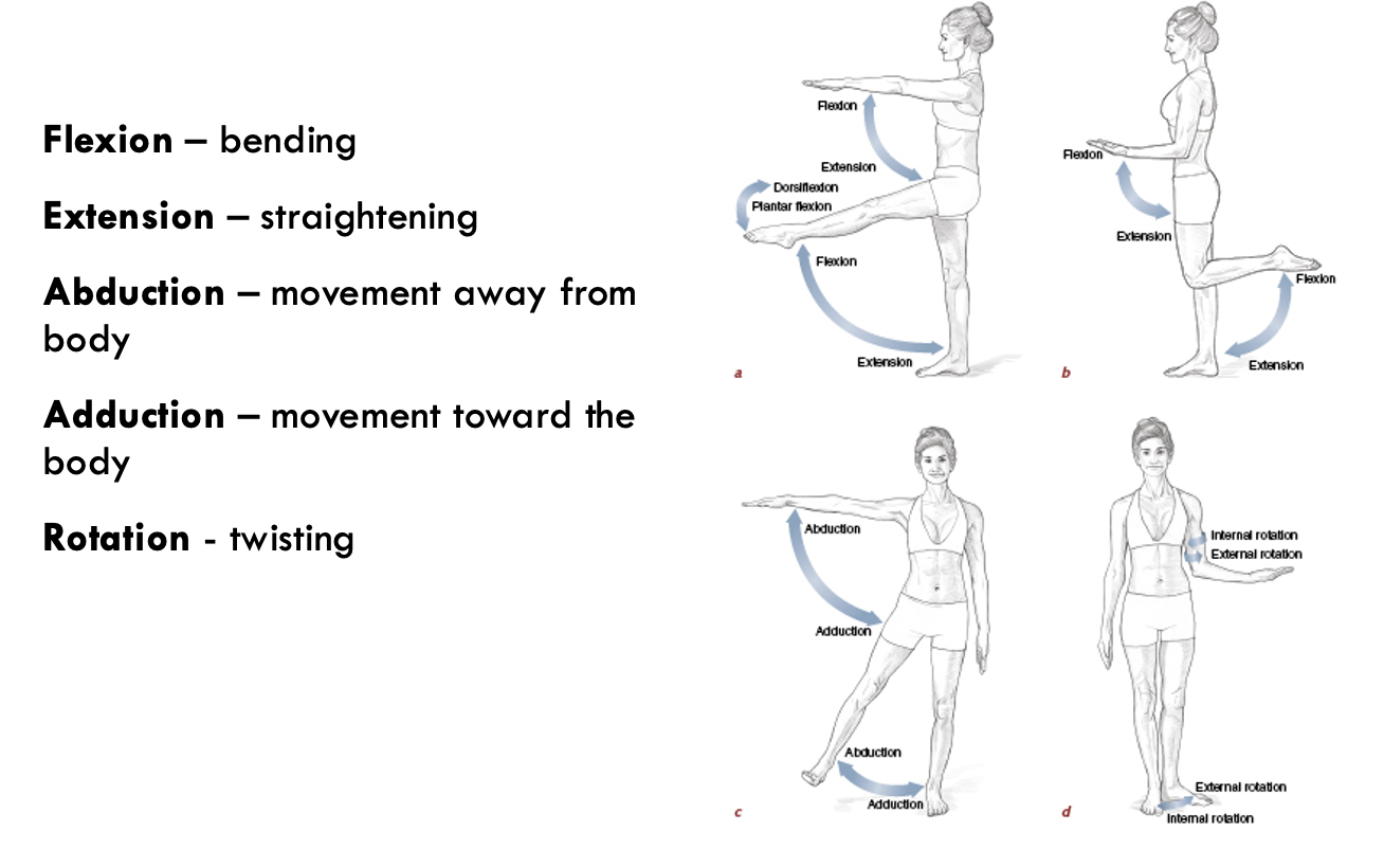
Flexion,
Flexion, or bending, decreases the angle between the articulating bones. This means that the bones come closer together.
Extension
Extension, or straightening, increases the angle between the articulating bones, moving the bones further apart.
Rotation
Rotation is the movement of a bone around its long axis – for example, turning the head from left to right occurs due to rotation at the joint between the first two vertebrae.
The Skeleton
The skeletal framework of the body consist of bone and cartilage which function to provide body support, protection and movement and is facilitated by the structure and function at cell and tissue levels.
Function and location of major bones that make up the axial and appendicular skeleton
The skeletal system consists of
bones, joints, ligaments and cartilages in the body.
Functions of the Skeleton
Support: providing a strong framework that supports the soft tissues
Storage: the storage and release of minerals from bone tissue.
Bone stores calcium and Magneisium
Protection: protection of the delicate organs, e.g. the brain and lungs
Production: the formation of red blood cells in the bone marrow
Movement: allowing movement due to muscle attachment and joints
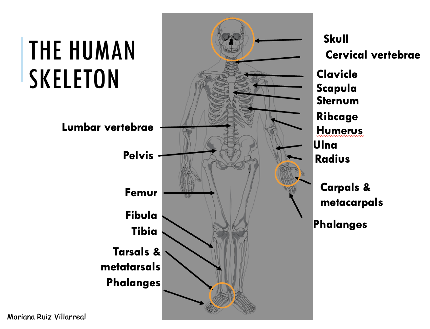
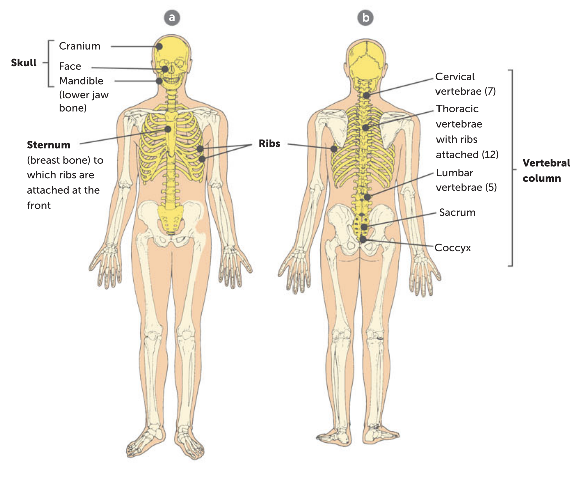
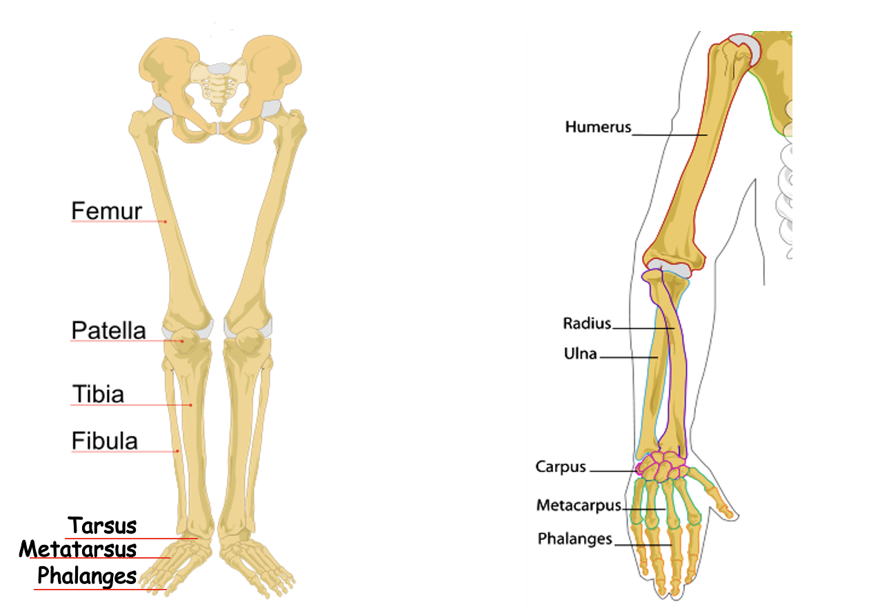
Axial skeleton
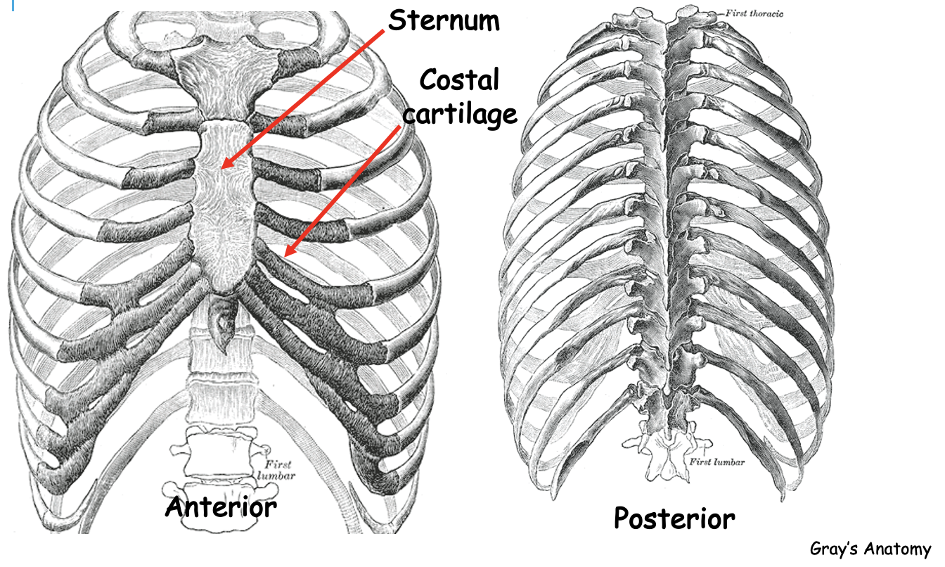
includes the bones that make up the central axis of the body – the skull, vertebral column, ribs and sternum.
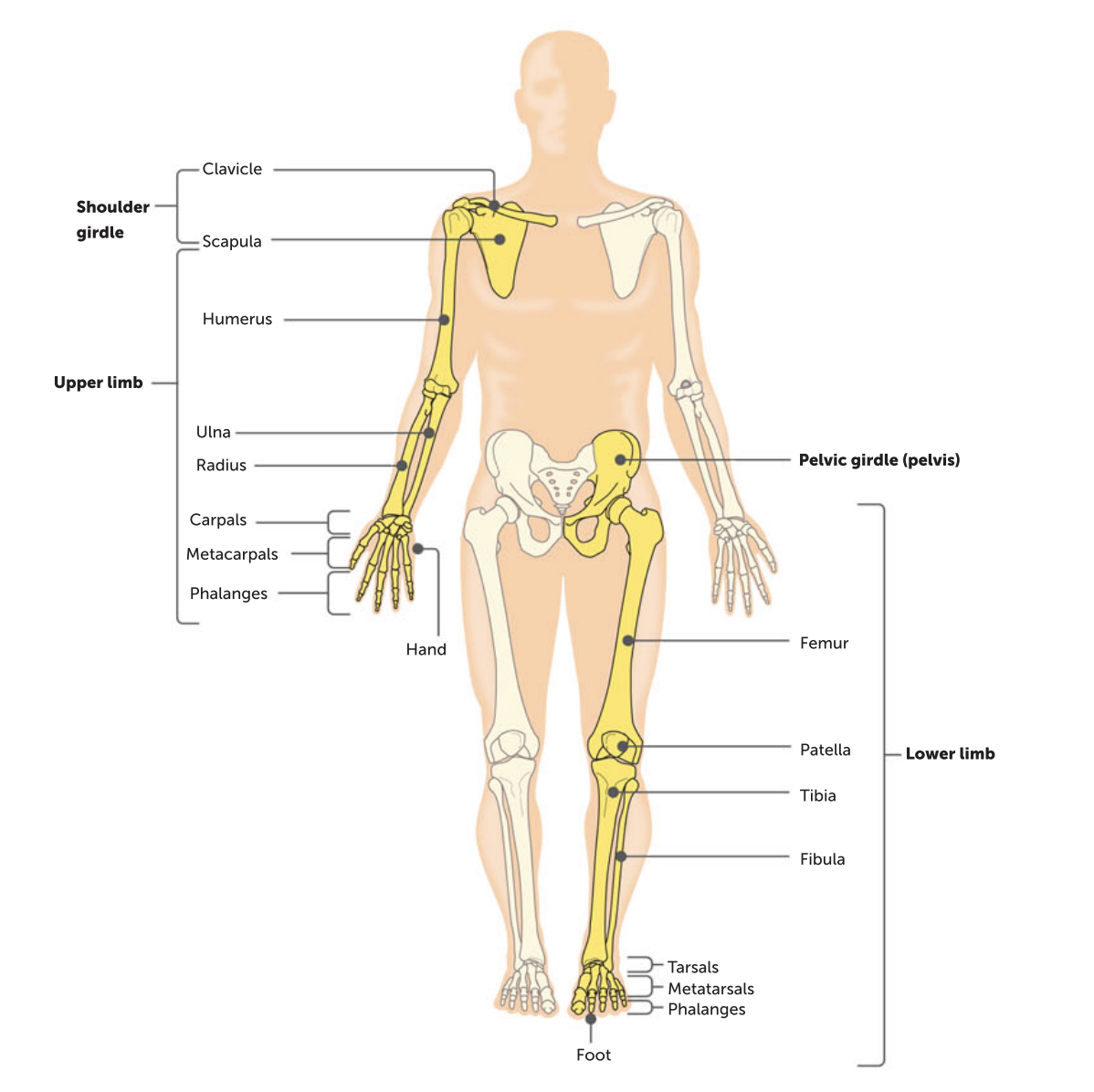
Appendicular skeleton
includes the bones that make up the upper and lower limbs and the pectoral and pelvic girdles.
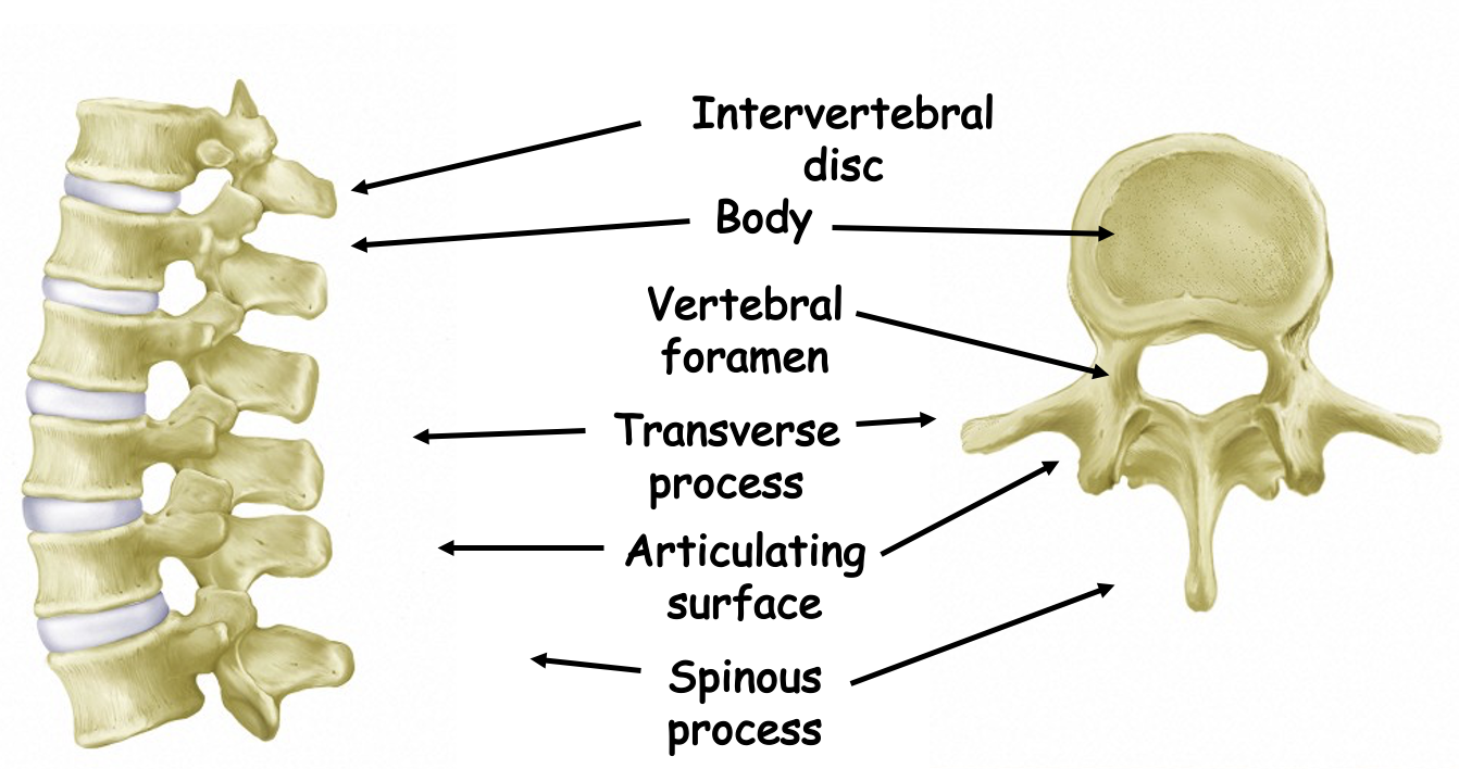
Structure of a vertebra
Ribcage
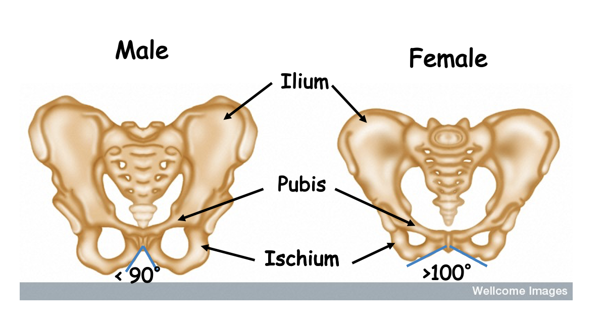
Pelvic girdle
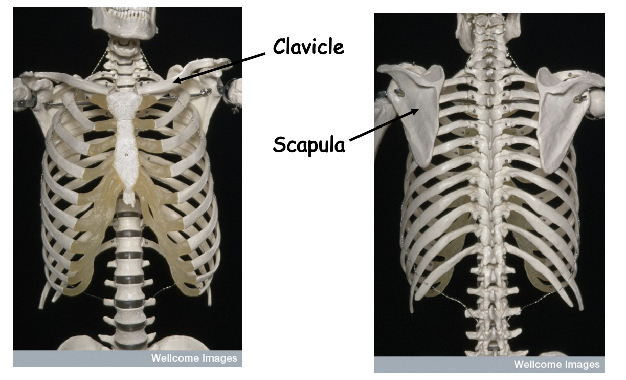
Pectoral girdle
The upper and lower limbs
Cartilage
A type of connective tissue containing fibres of collagen
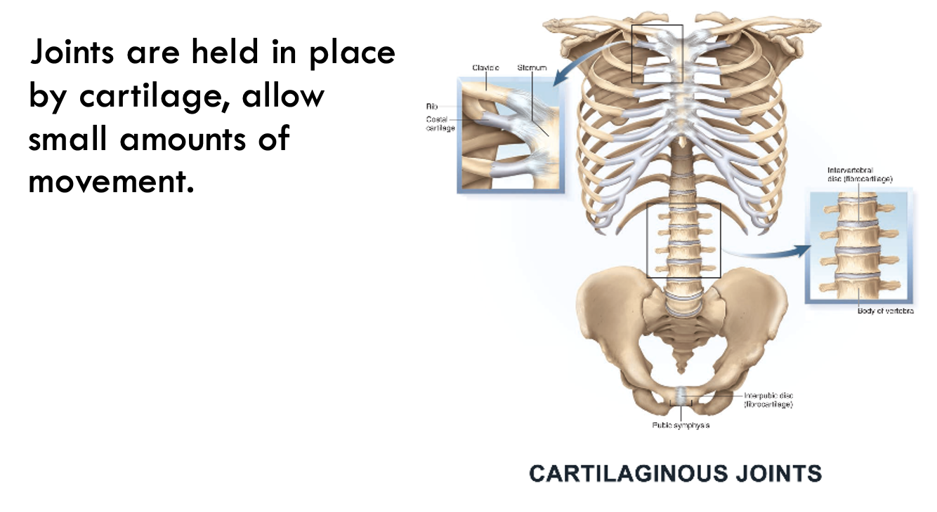
Cartilaginous joint
A joint at which only limited movement occurs between the bones; the bones are held in place by cartilage as between the ribs and
Chondrin
A matrix of protein and carbohydrate in cartilage
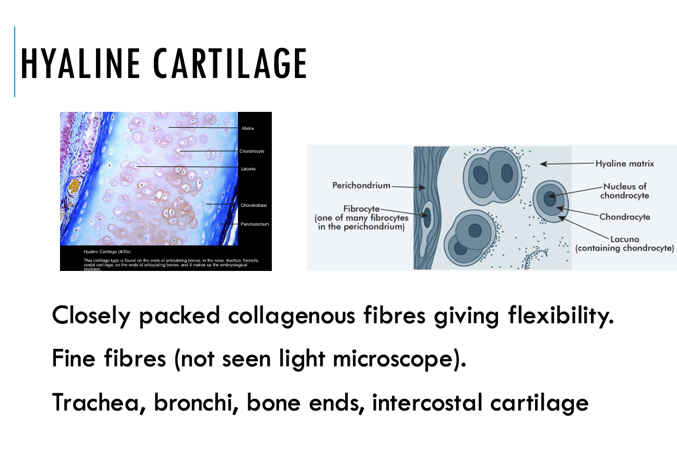
Hyaline cartilage
Flexible supporting cartilage
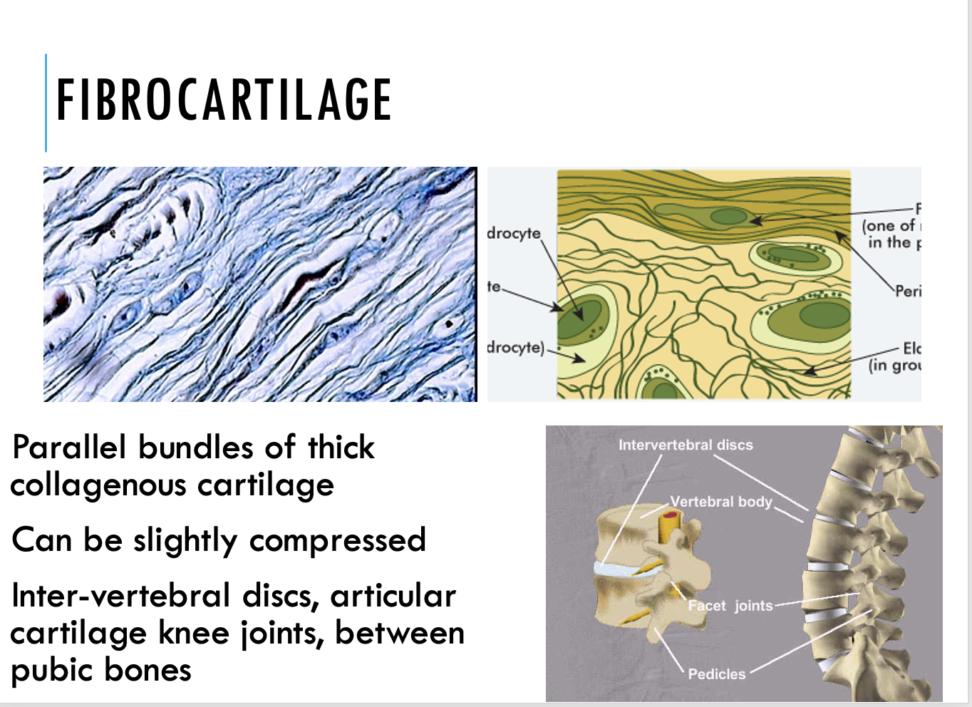
Fibrocartilage
A type of cartilage that contains bundles of fibres
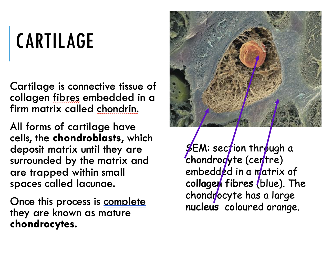
Bone
Bone is made up of cells separated from each other by a non-cellular material called matrix. Bone is maintained by a rich blood supply.
Living bone includes
blood vessels
nerves
Collagen
Three special types of cells :
osteoblasts (bone forming cells)
osteoclasts (bone cells that break down old bone)
osteocytes (mature osteoblasts that can no longer form bone).
The non-living part of bone, which provides strength, is made up of
inorganic salts calcium and phosphate
matrix of collagen fibres.
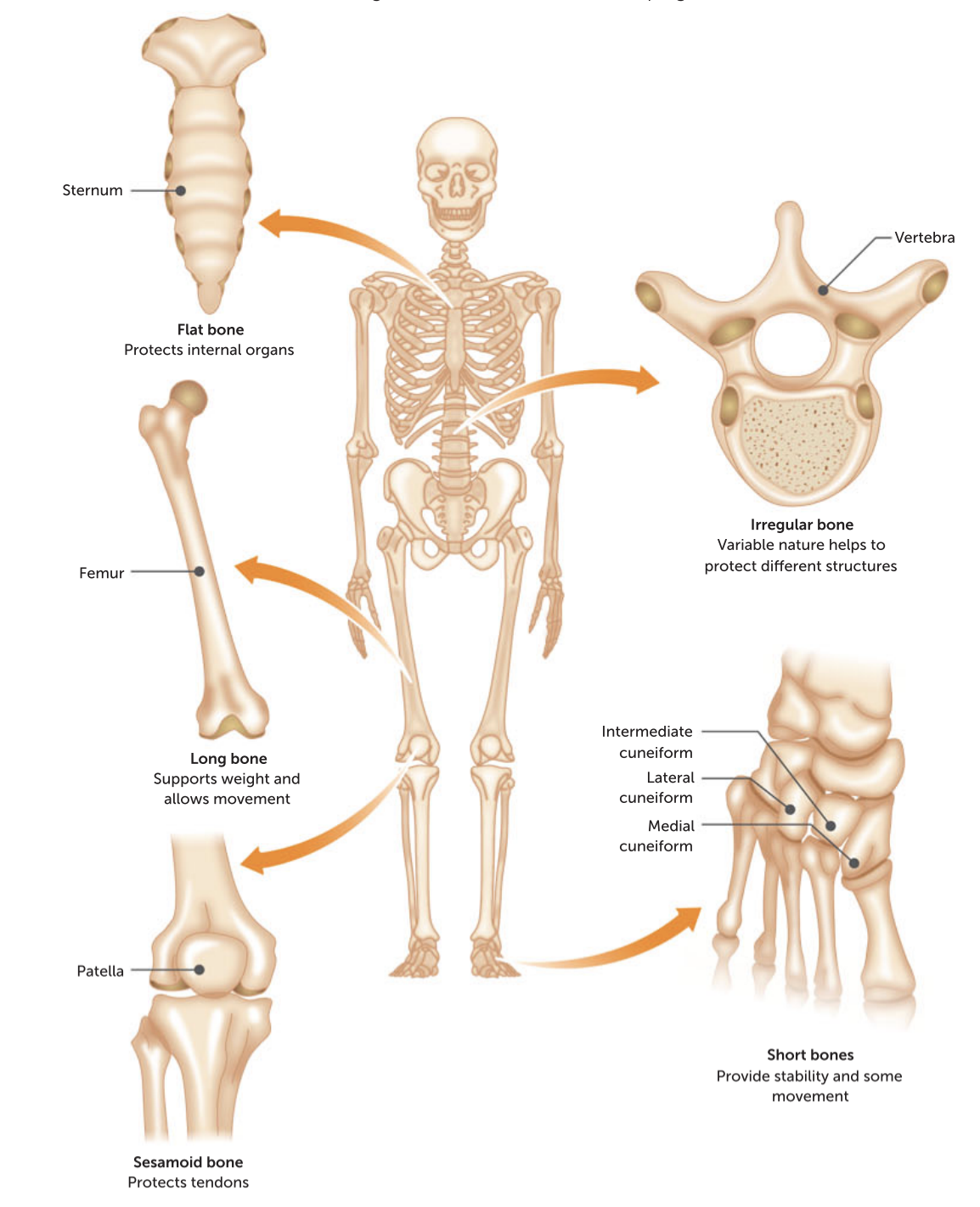
Bones can be classified according to their shape and their structure.
Classification by shape
Long bone – e.g. Thigh bone (femur)
Short bone – e.g. wrist bones (carpals)
Flat bone – e.g. shoulder blade (scapula)
Irregular bone – e.g. vertebrae
Sesamoid bones develop in tendons – e.g. kneecap (patella) – enhances mechanical advantage (lever action).
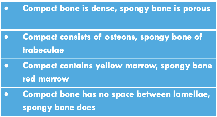
There are two types of bone
compact bone (very hard and dense)
spongy bone - porous, consisting of a network of small bony plates.
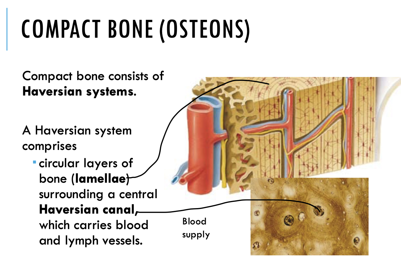
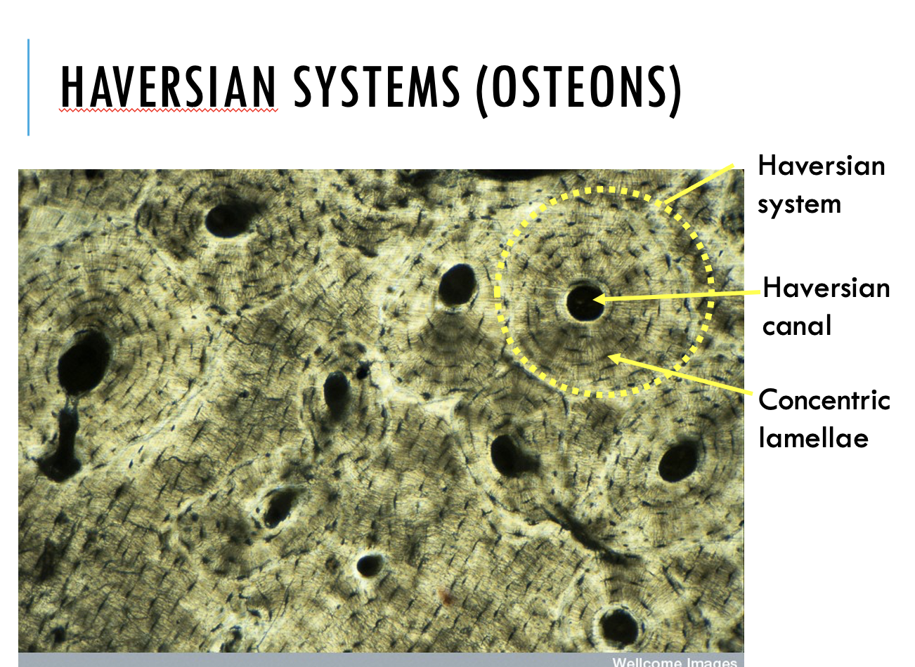
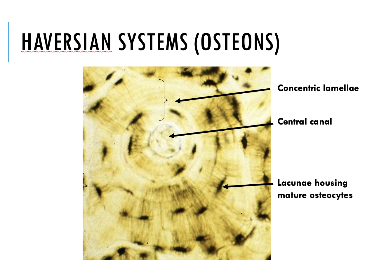
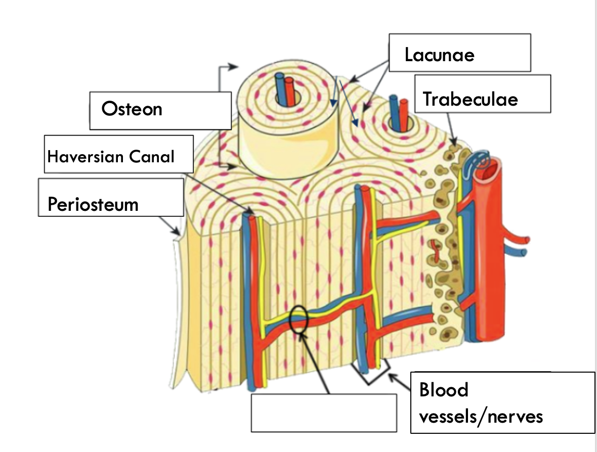
A Haversian system comprises
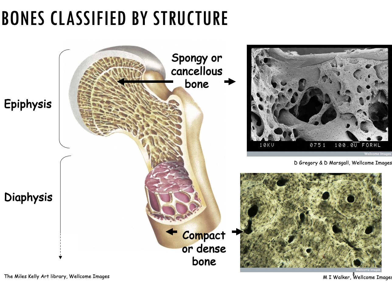
Spongy (cancellous) bone
The microscopic structure of spongy bone is quite different from compact bone.
Instead of regular osteons, spongy bone has an irregular arrangement of thin bony plates called trabeculae.
Nerves and blood vessels pass through the spaces between the bones.
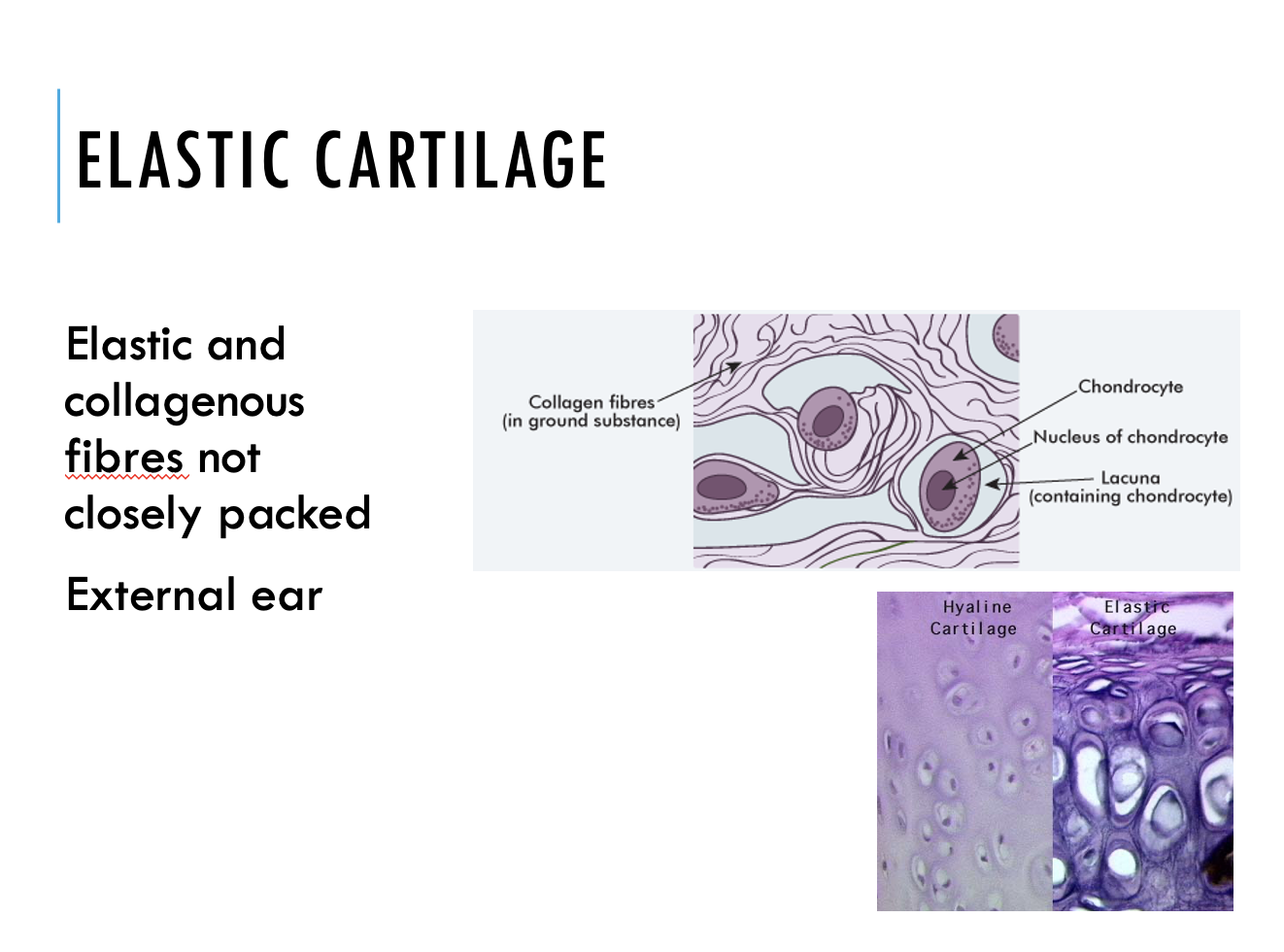
Cartilage
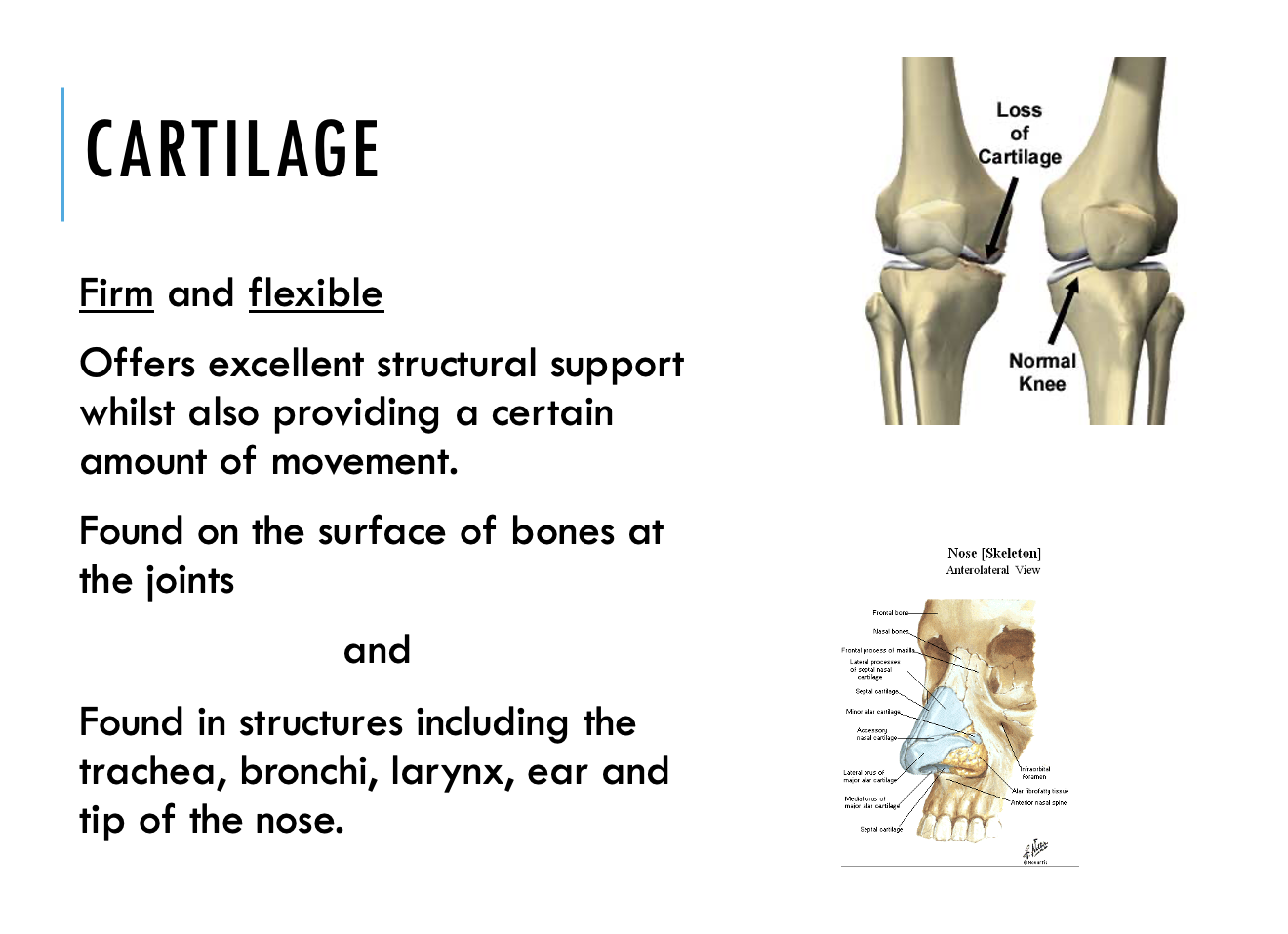
cartilage
Hyaline cartilage
Elastic cartilage
Fibrocartilage
Why the Matrix is Crucial
Tissue Diversity: Variations in the matrix determine the physical properties of your organs. A bone matrix is calcified and rigid, whereas the matrix in the eye is transparent and fluid.
Cell Communication: It acts as a reservoir for growth factors, dictating cell growth, survival, and movement.
Wound Healing: During injuries, the matrix releases signals to coordinate cell repair and scar formation.
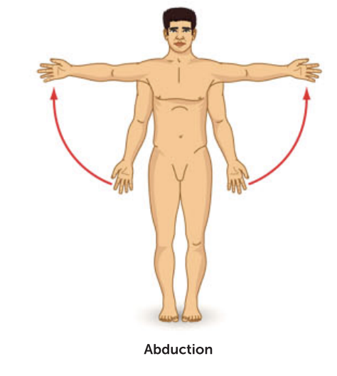
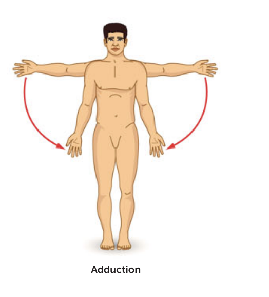
describe certain muscle movements
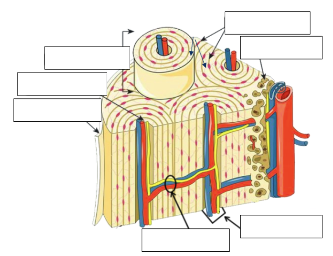
Label the structures found in compact bone
Comparing compact and spongy bone
How are the cells maintained?
·Haversian canals contain blood vessels - these exchange materials with the osteocytes
·canaliculi link the intercellular fluids in the Haversian canal with the lacunae containing the osteocytes (mature bone cells)
·materials diffuse along the canaliculi between the blood vessels and the cells according to the diffusion gradient
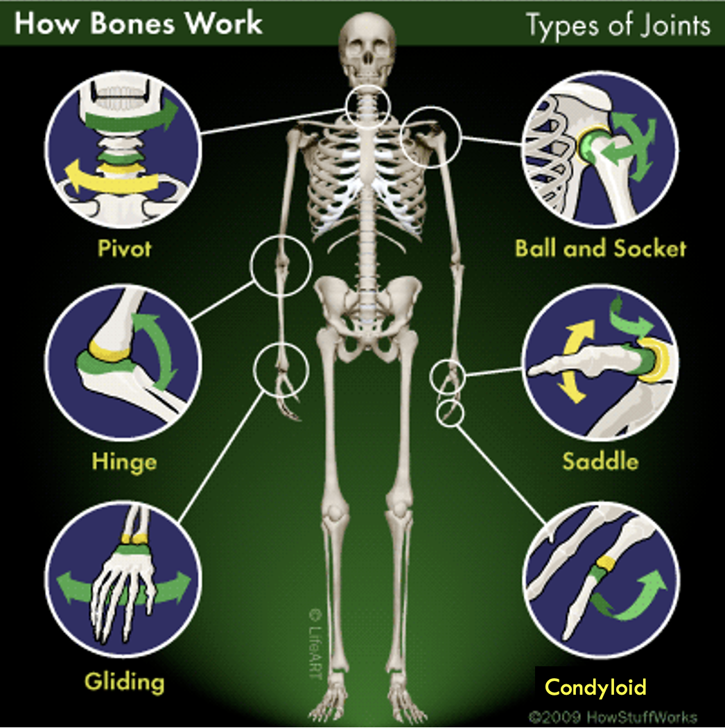
different types of joints
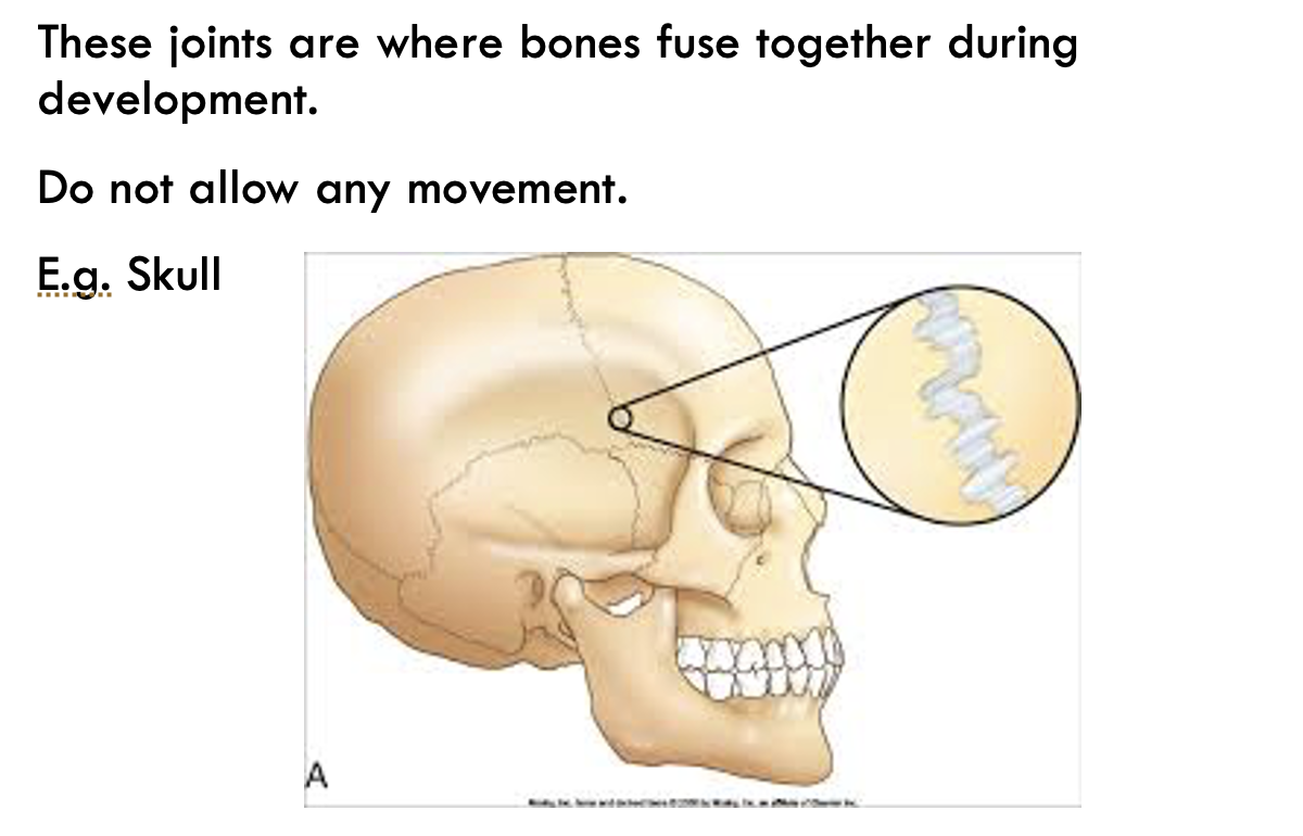
Immovable Joints
Ball and Socket Joints
Hinge Joints
Pivot Joints
Saddle Joint
Fibrous (Immovable) Joint
Cartilaginous Joints
Synovial (Freely Movable) Joints
Most joints are freely movable; however the ultimate range of motion is determined by attached ligaments, tendons and adjoining bones
Ball and Socket Joint
Joints that allow a large amount of movement in all directions.
The ball of one bone fits into the socket of another.
E.g. Shoulder and Hip
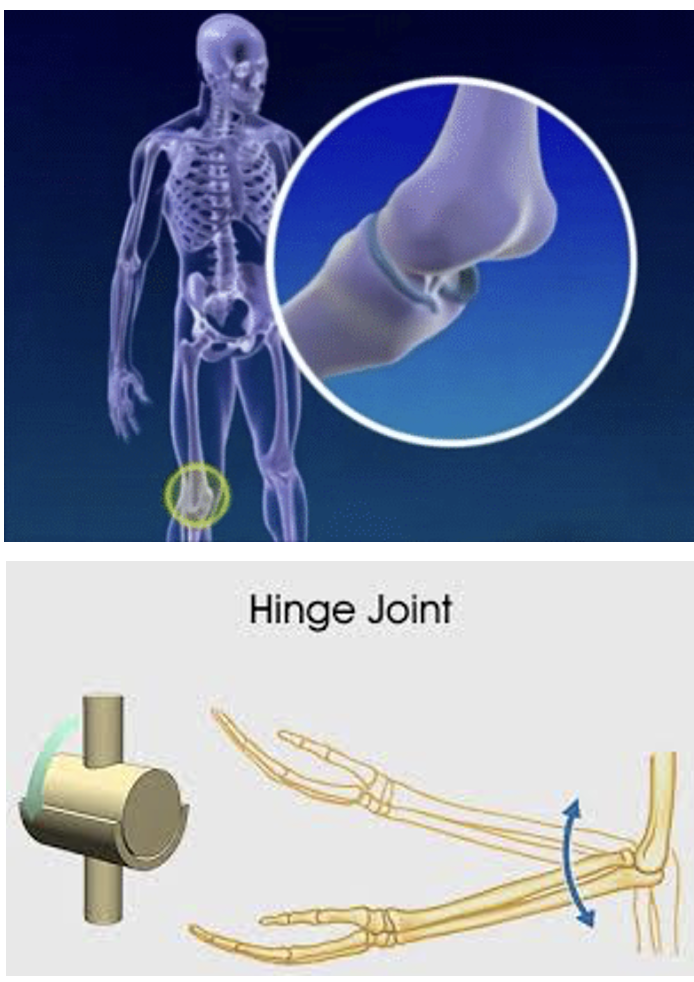
Hinge Joint
Hinge joints allow movement in one direction only – just like a door hinge.
Convex surface of one bone fits into the concave surface of the other
E.g. elbow and knee.
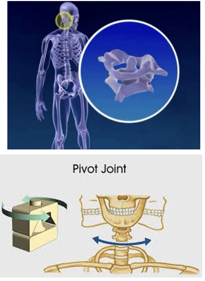
Pivot Joint
Cylindrical boney structure rotating within a circle of bone and ligament.
Rotation around an axis. Allows twisting movement, from side to side.
E.g. 1ST & 2ND neck vertebrae, forearm, between radius and ulna.
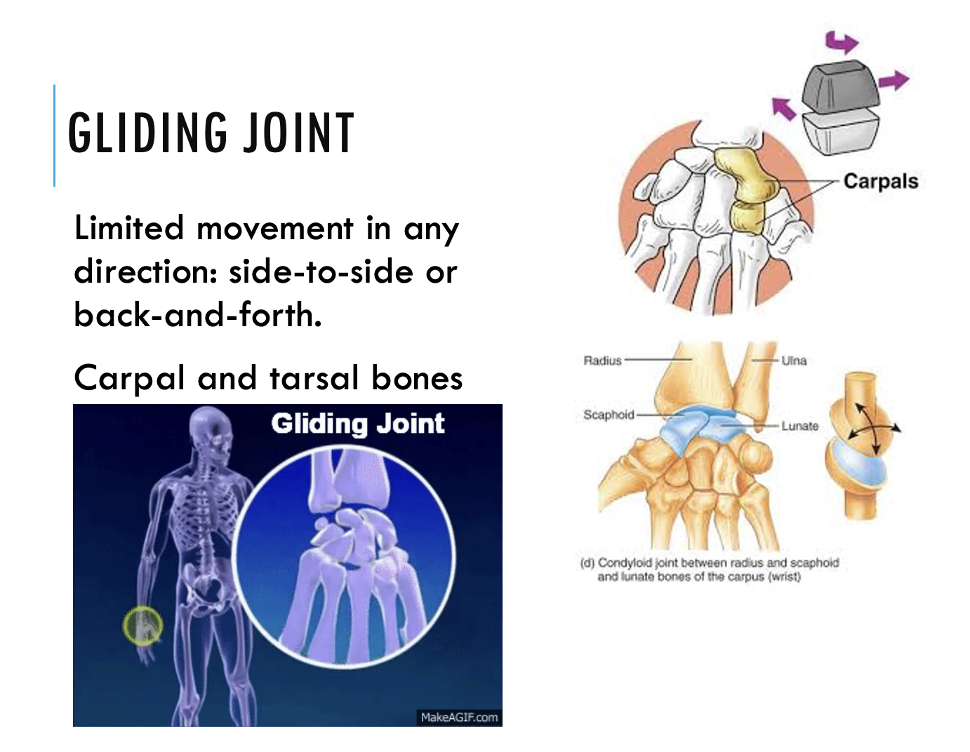
Gliding Joint
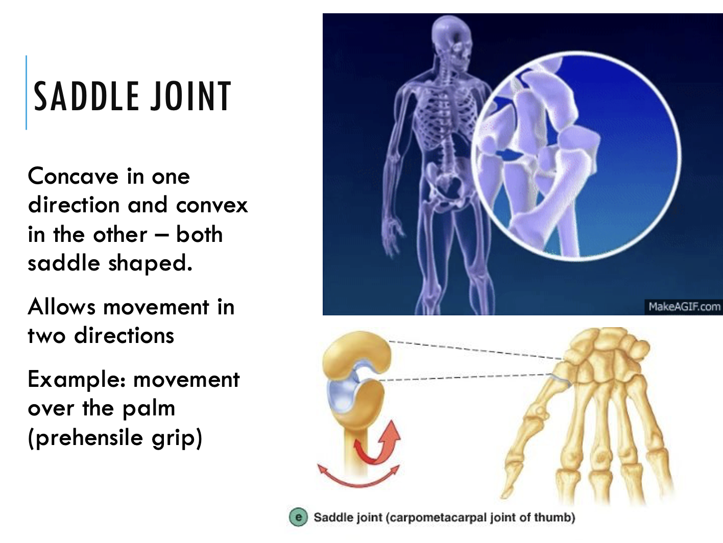
Saddle Joint
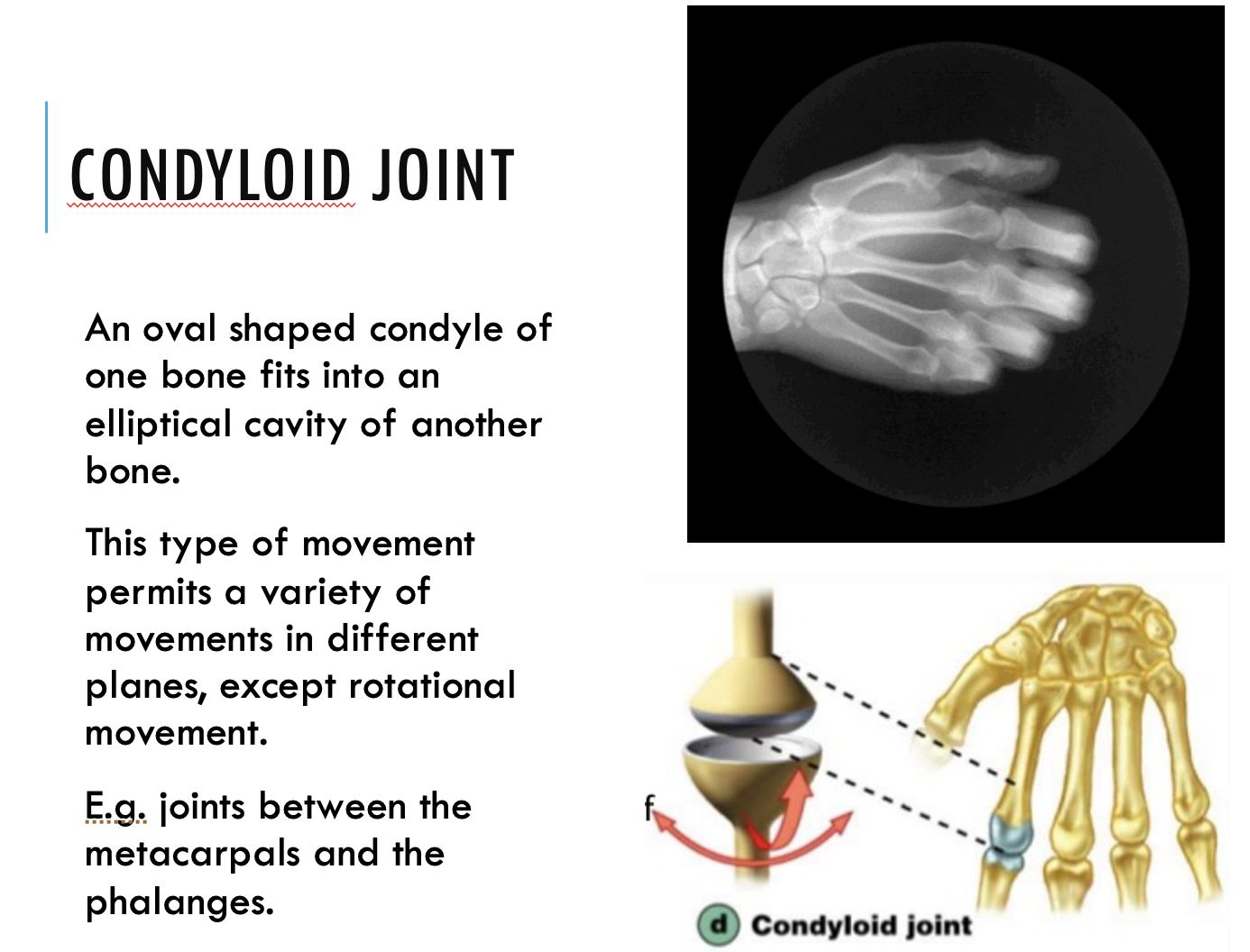
Condyloid Joint
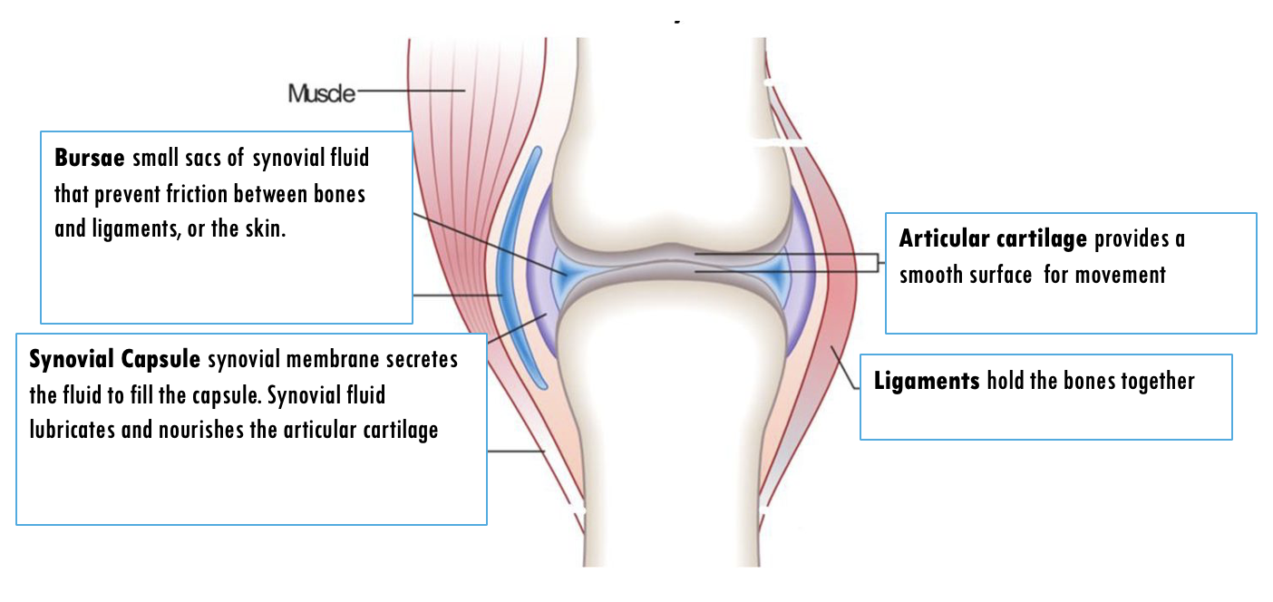
Synovial Joints
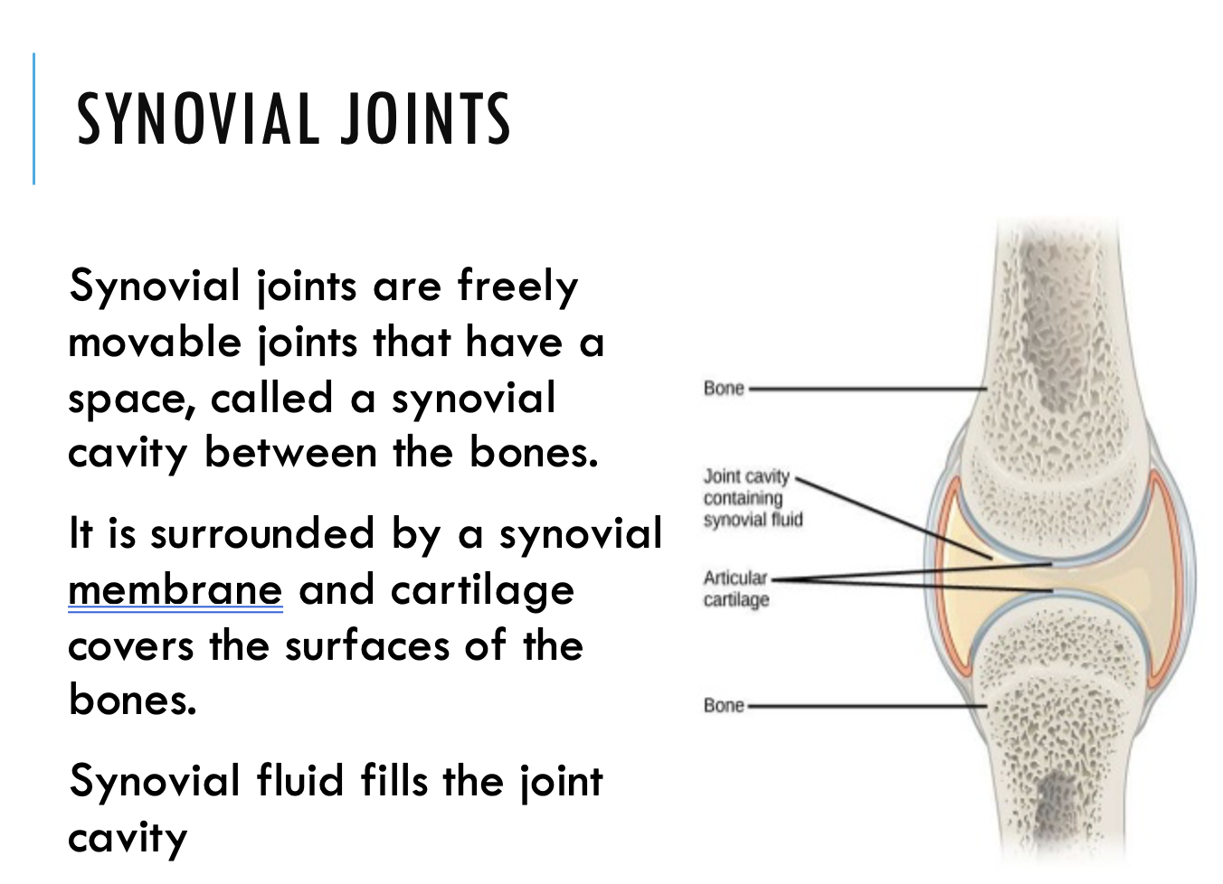
Synovial Joints
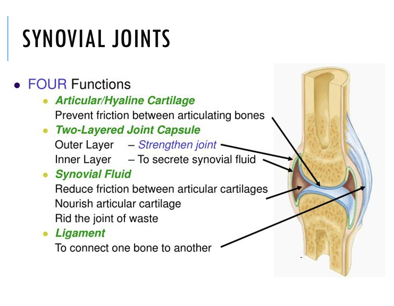
Main Features of a Synovial Joint
Movement at a Joint