Radiology: normal spine
1/106
There's no tags or description
Looks like no tags are added yet.
Name | Mastery | Learn | Test | Matching | Spaced | Call with Kai |
|---|
No analytics yet
Send a link to your students to track their progress
107 Terms
- 7 vertebral segments
- lordosis
- neck
spine, cervical
- 12 vertebral segments
- ribs
- kyphosis
spine, thoracic
- 5 segments
--> can have transition vertebral body
- lordosis
-low back
spine, lumbar
- sacrum (5), fused
- coccyx (3-5), fused
spine, sacrum and coccyx
contains spinal cord, nerve roots, epidural fat, vessels, CSF contained by dura in thecal sac
spinal canal
termination of spinal cord, usually around T12 or L1/L2
conus medullaris
do in conus medullaris, starting at L2/L3 or below
spinal tap
nerve roots in thecal sac surround by CSF
cauda equina
vertebral body
what is the blue arrow pointing to?
pedicle
what is the pink arrow pointing to?

laminae
what is the red arrow pointing to?

vertebral arch
what is the black arrow pointing to?

transverse processes
what is the yellow arrow pointing to?

they allow space for vertebral arteries, injury occurs when spaces are not align or smaller
why are transverse foramina of c-spine important?
spinous process
what is the blue arrow pointing to?
R articular processes
what are the red arrows pointing to?

facet joint
what is the pink arrow pointing to?

spine abnormalities occur to degeneration of joints, synovial joint
- come from articular processes of bodies
facet joints
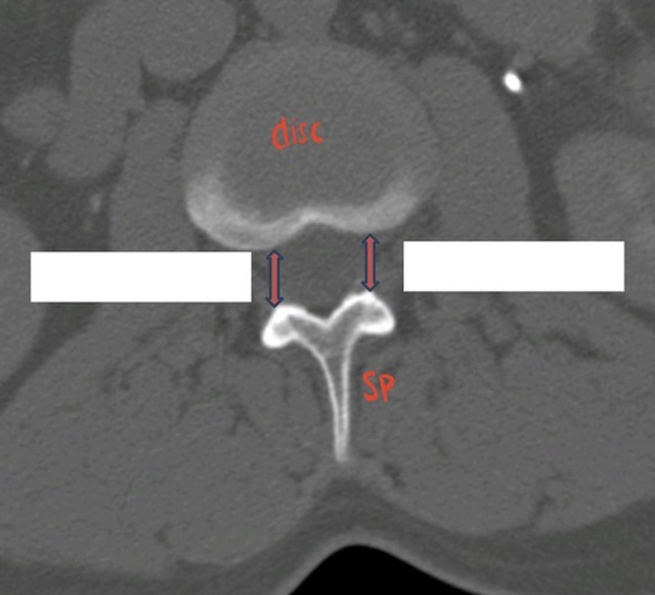
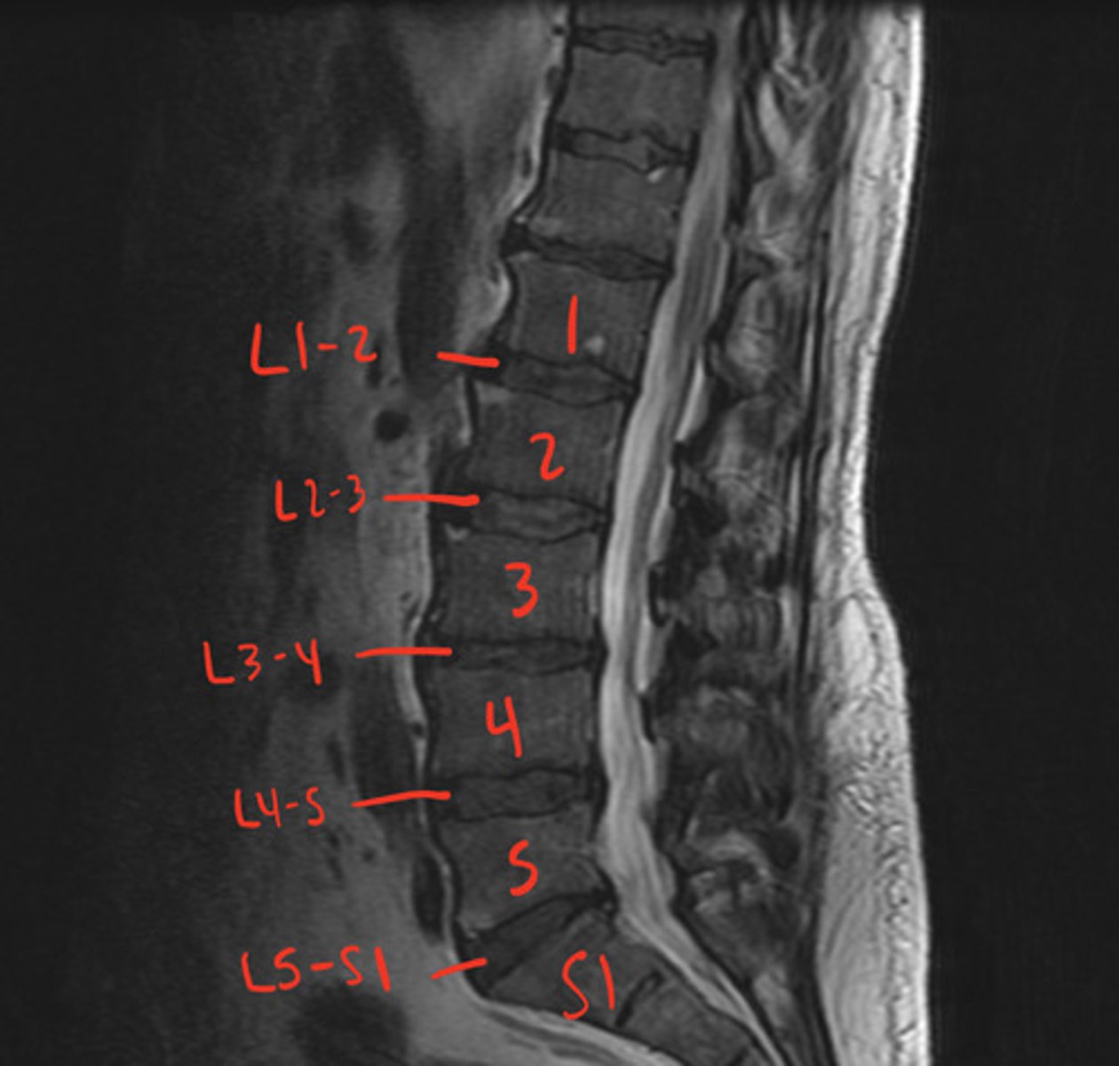
L C5-6 disc space
what disc space is being indicated?
name with vertebra above and one below, needs to include "disc"
how to name a intervertebral disc
intervertebral foramina
where do nerves and vessels of the spinal cord exit through?
neural foramina
--> large and good spinal for spinal nerves
what are the pink arrows, and what is noticeable about these?
connects anterior vertebral bodies
anterior longitudinal ligament
connected posterior vertebral bodies
posterior longitudinal ligament
connected laminae
- canal stenosis when enlarged
ligamentum flavum
1. TP to TP
2. SP to SP
3. tips of SP
1. intertransverse
2. interspinous
3. supraspinous ligaments
- no body
- 2 lateral masses for occipital condyles
- no SP, tubercle instead
- transverse processes have foramina for vertebral a.
cervical spine, C1
- dens, held in place posteriorly by transverse ligament
--> without functional ligament you don't breathe, very important
- allows head to rotate
cervical spine, C2
C7
largest spinous process of C vertebrae
ribs
thoracic spine
lumbar bodies large for weight baring
lumbar spine
- no vertebral bodies
- fused anteriorly and posteriorly
--> anterior and posterior neural formaina
sacrum
screening, first line, don't see disc herniation
imaging modalities for spine, x-ray
good for bony fracture, bony degenerative changes, some bone tumors, not good for discs
imaging modalities for spine, CT scan
- disc and soft tissue
- spinal cord and bone marrow abnormalities
- metastases
imaging modalities for spine, MRI scan
- if no MRI or bad MRI
- just outline, cannot see intrinsic abnormalities
- inject contrast into thecal sac through lumbar puncture
myelography
- bone scans, phosphate accumulates where issue is (non specific)
- prostate and breast cancer
- sacral stress fractures
- spondylolysis
imaging modalities, nuclear medicine
- AP
- Lateral
- 2 obliques
- Open mouth odontoid (OMO)
cervical spine, x-ray
axial, sagittal, coronal places
cervical spine, CT
sagittal and axial, sometimes coronal, multisequence
cervical spine, MRI
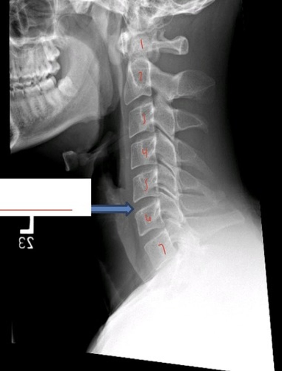
lateral c-spine
what is this imagine?

- less than 5 mm soft tissue at C2
- less than 22 mm soft tissue at C5
5 at 2; 22 at 5
radioluscent
disc spaces are what?
facet joint at C3-4
what is the red arrow pointing at?

spinous process of C3
what is the yellow arrow pointing at?

C6
what is the pink arrow pointing at?

C6-7 disc space
what is the blue arrow pointing at?

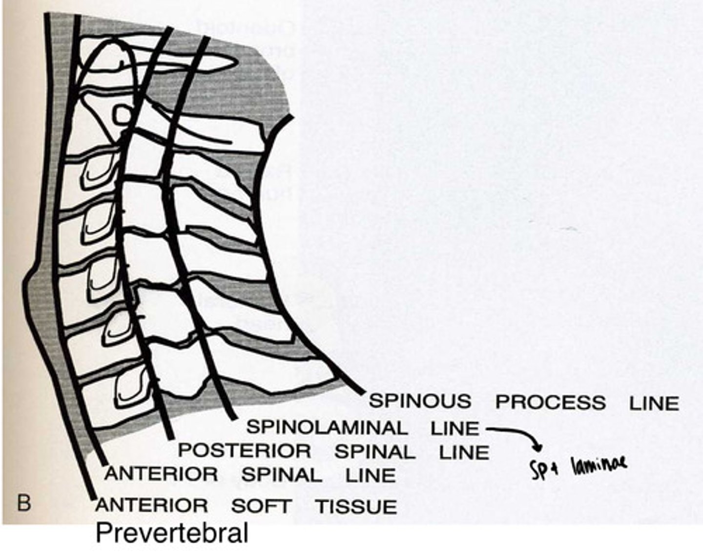
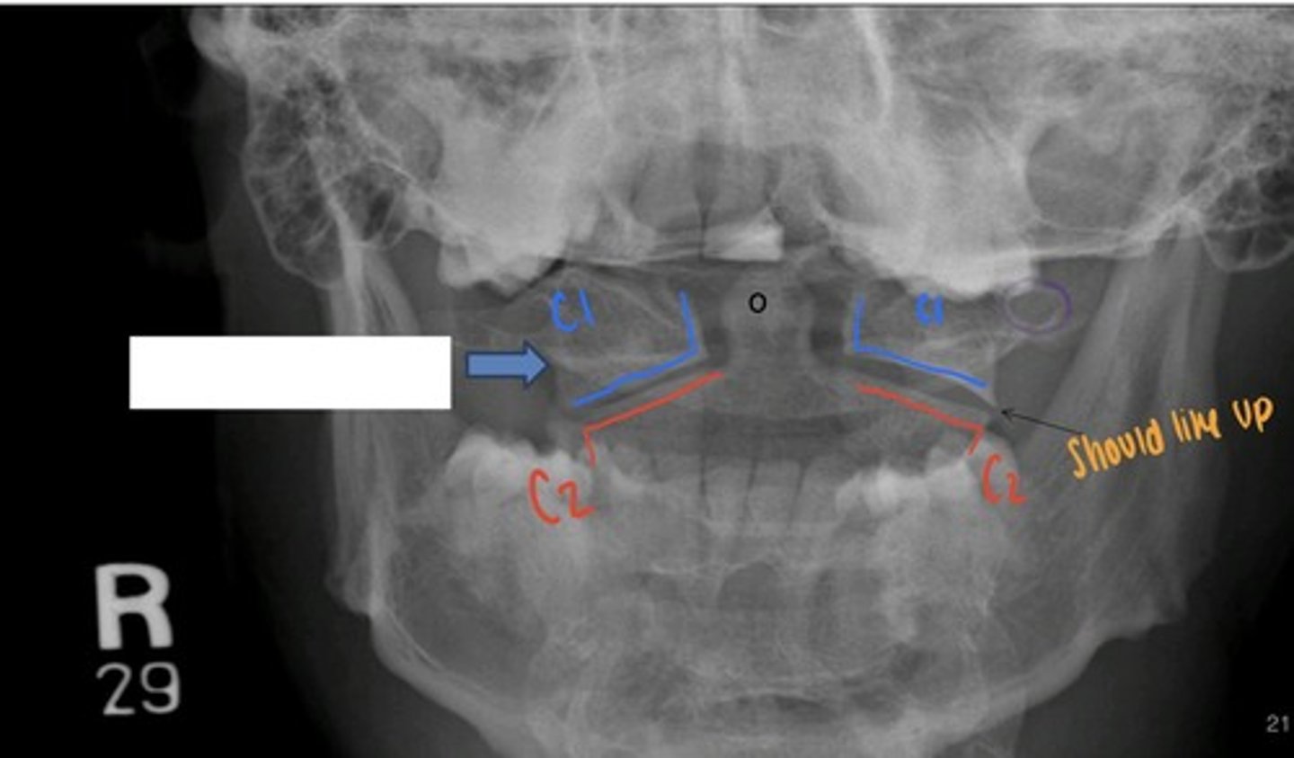
should all line up nicely
cervical spine lines
oblique c-spine, R
what is this image?
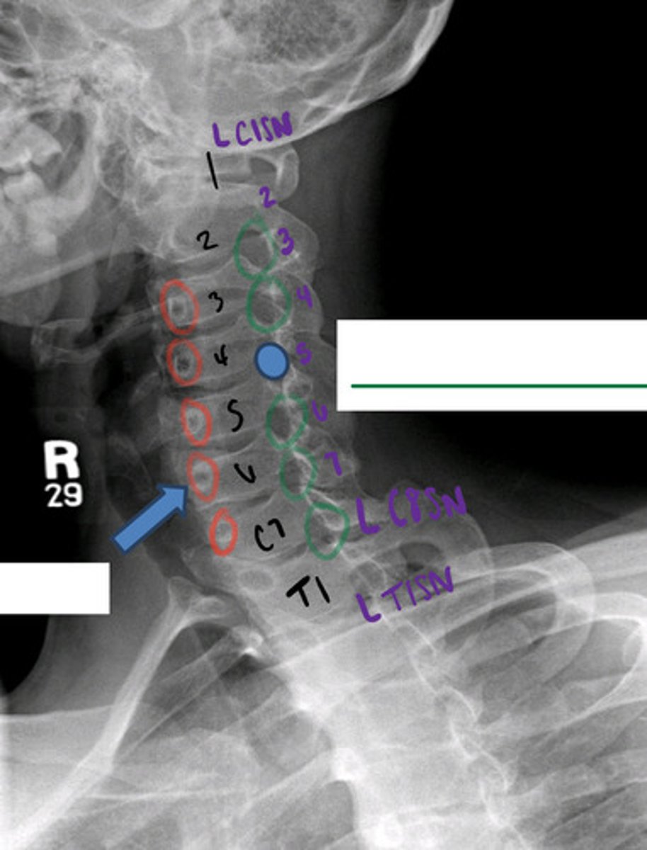
to look at neural foramina
why do we do oblique views
R pedicle C6
what is the blue arrow pointing to?

L neural foramina C4-5
what is the blue circle indicating?

L C5 nerve root
what exits through the L neural foramina C4-5?

oblique c-spine, L
what is this image?
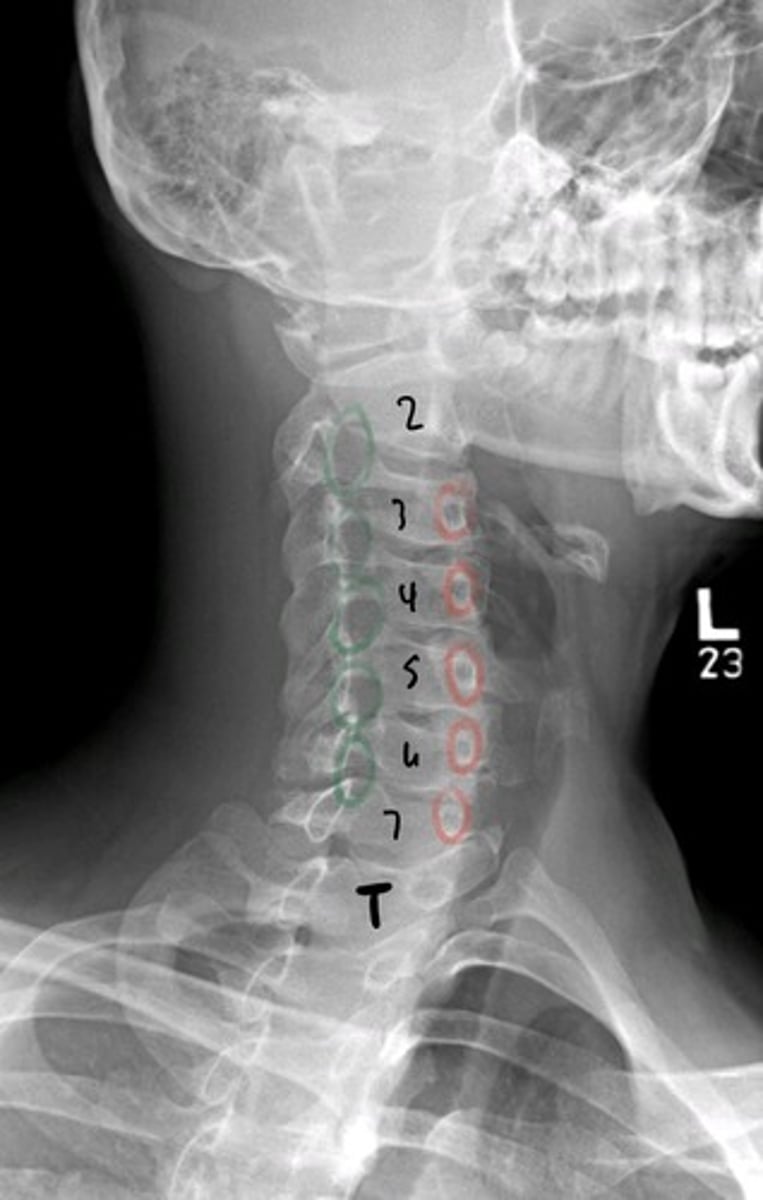
red= L pedicle
green= R neural foramina
what are the red circles indicating?
what are the green circles indicating?

OMO
what is this image?
R lateral mass C1
what is the blue arrow pointing to?

dens of C2
what is the black circle indicating?

transverse foramen of C vertebrae for vertebral a.
what is the purple circle indicating?

AP c-spine
what is this image?
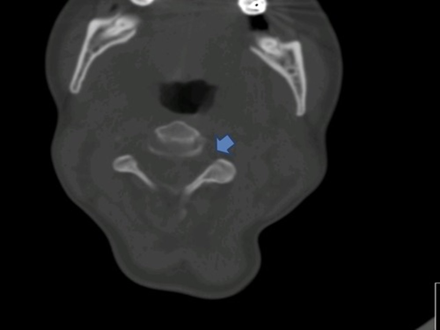
L C4-5 uncovertebral joint
what is the light blue arrow pointing to?

uncinate processes for degenerative disease
lung apexes to pick up cancer
two things to look at on AP c-spine
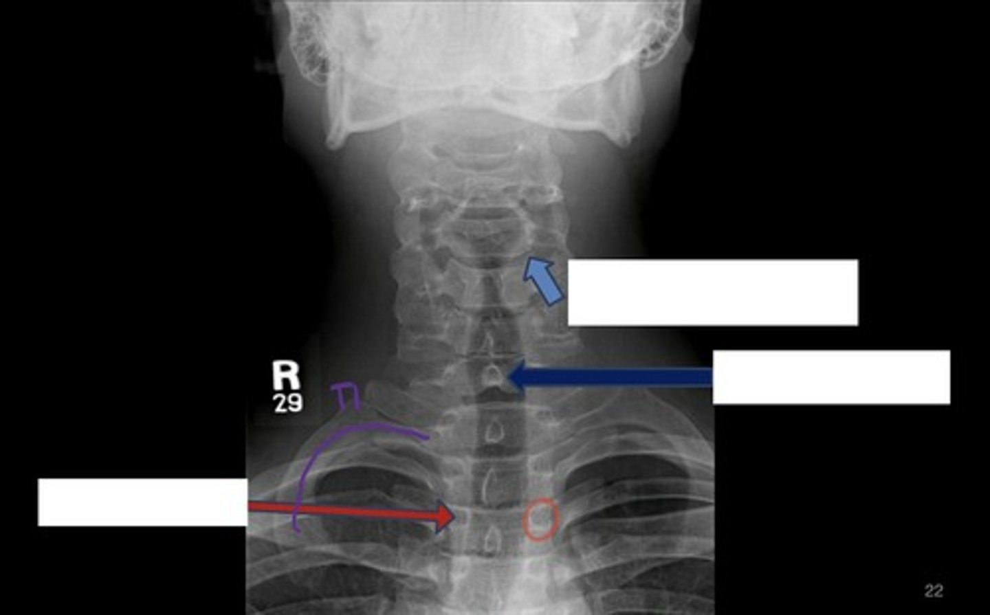
C7 spinous process
what is the dark blue arrow pointing to?

R pedicle of T3
what is the red arrowing pointing to?

the ribs
how to distinguish between C and T vertebra
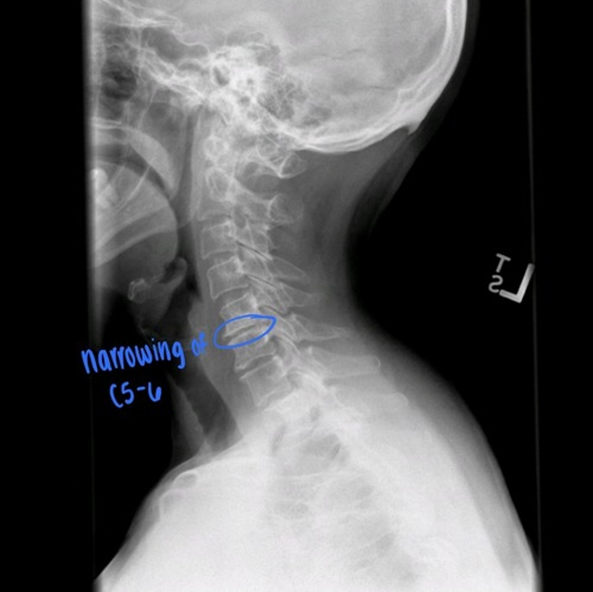
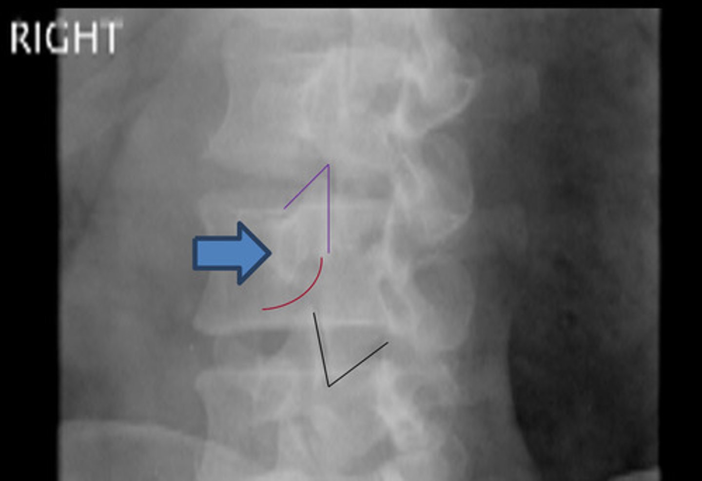
degenerative disc disease of L C5-6
--> smaller than rest of discs
what is the blue circle indicating?
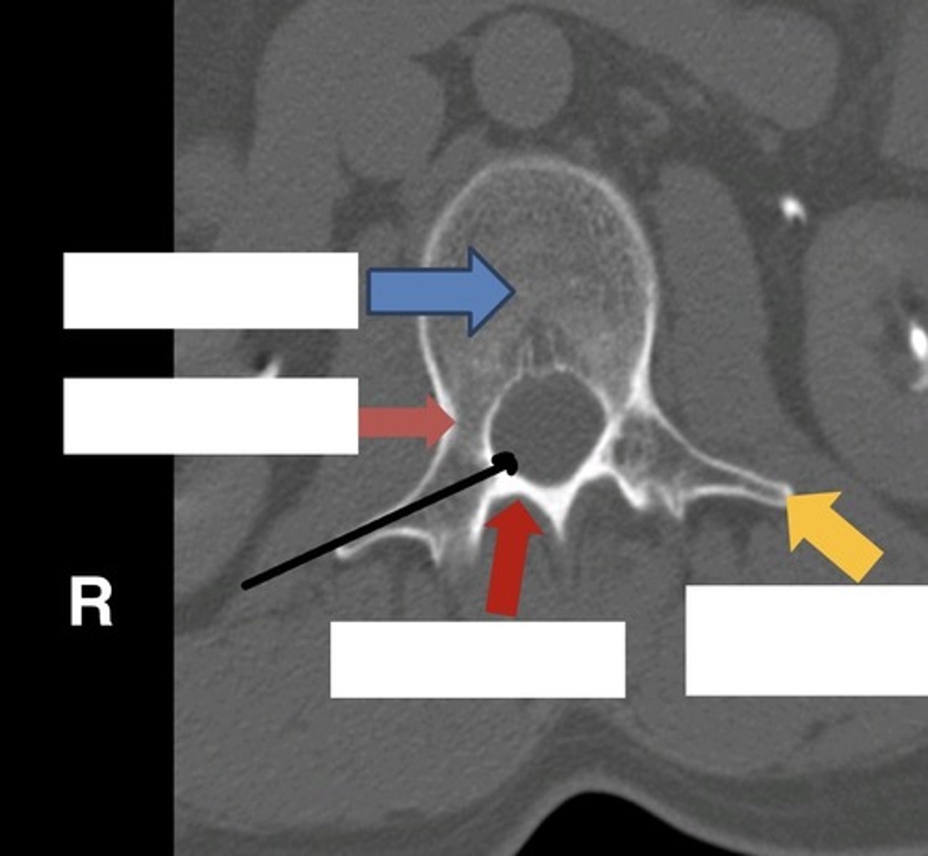
CT C1 axial
what is this image?

dens of C2
what is the light blue arrow indicating?
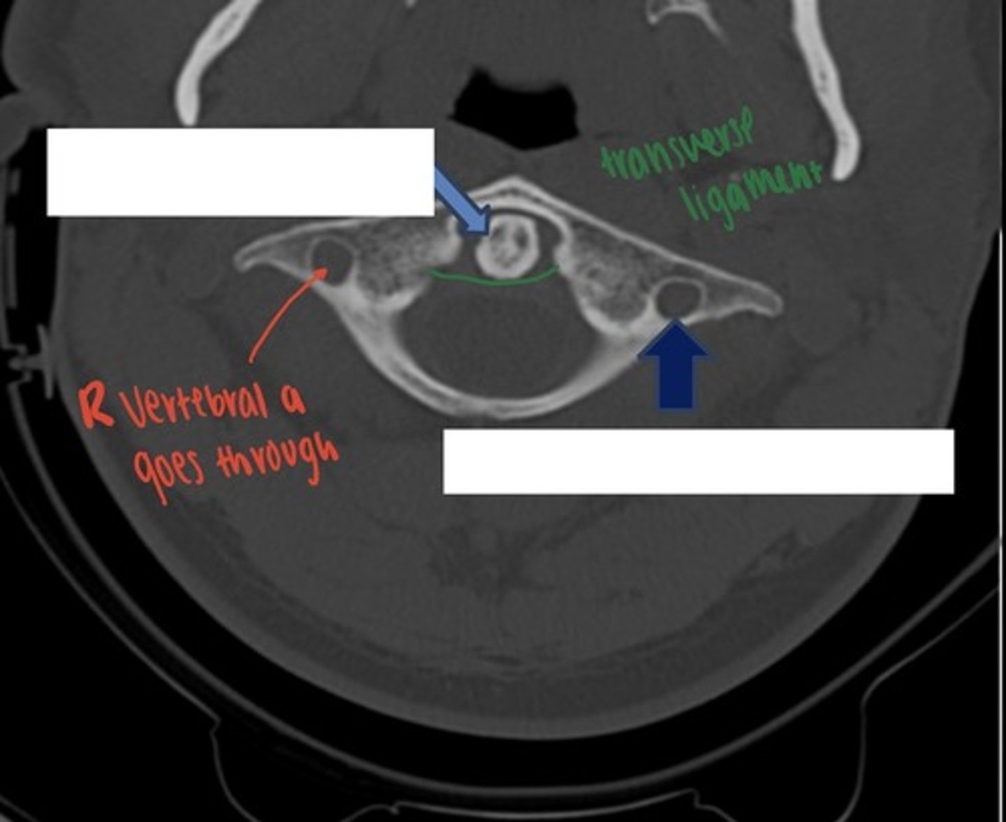
L C1 transverse foramina
what is the dark blue arrow indicating?

neural: nerve roots, fat
transverse: vertebral a.
what exits through the neural foramina and transverse foramina?
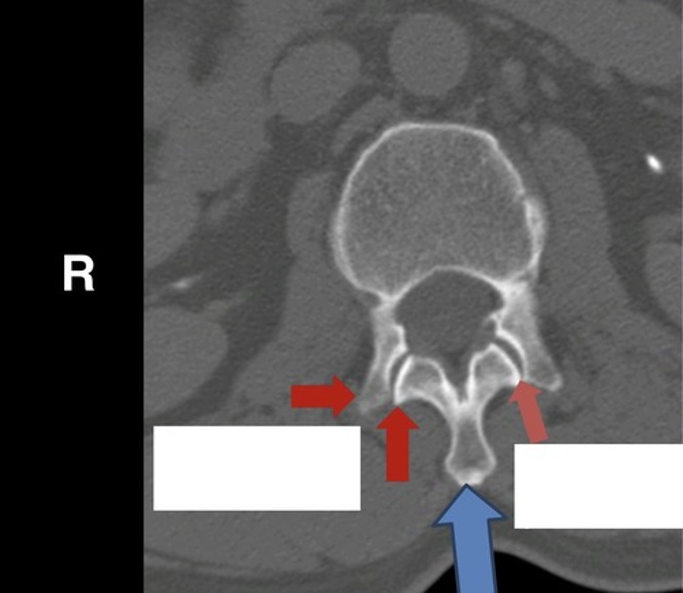
axial c-spine CT neural foramina
--> blue arrow showing neural foramina
what is this image?
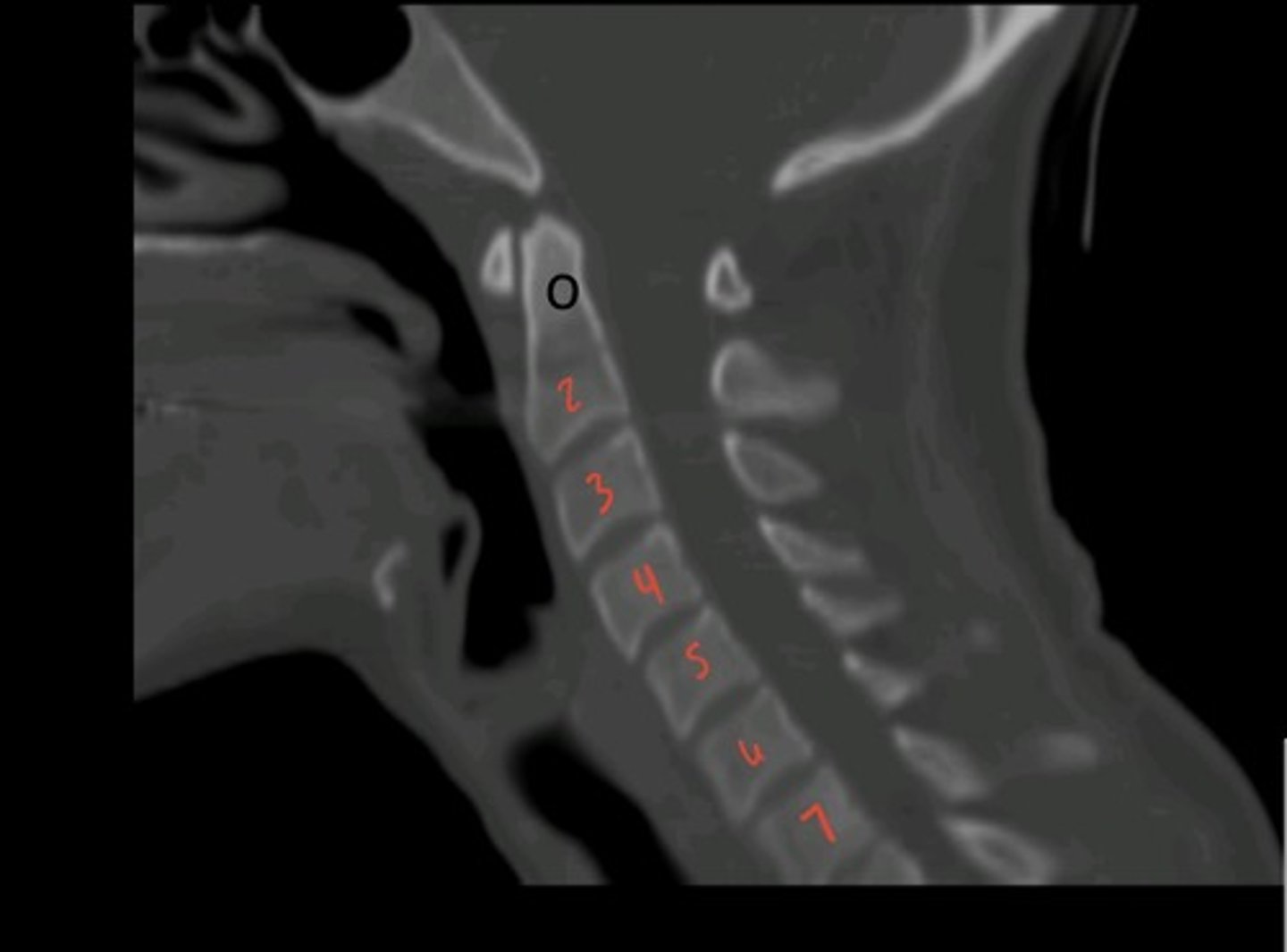
sagittal CT c-spine midline
what is this image?
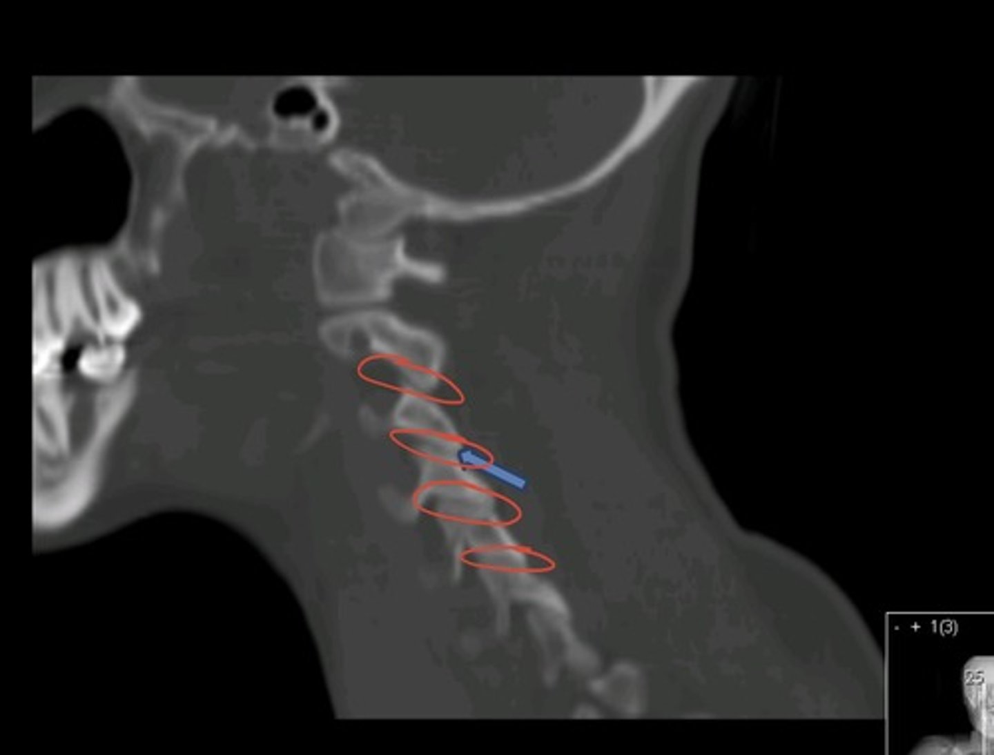
sagittal CT c-spine off midline
--> blue arrow showing facet joint
what is this image?
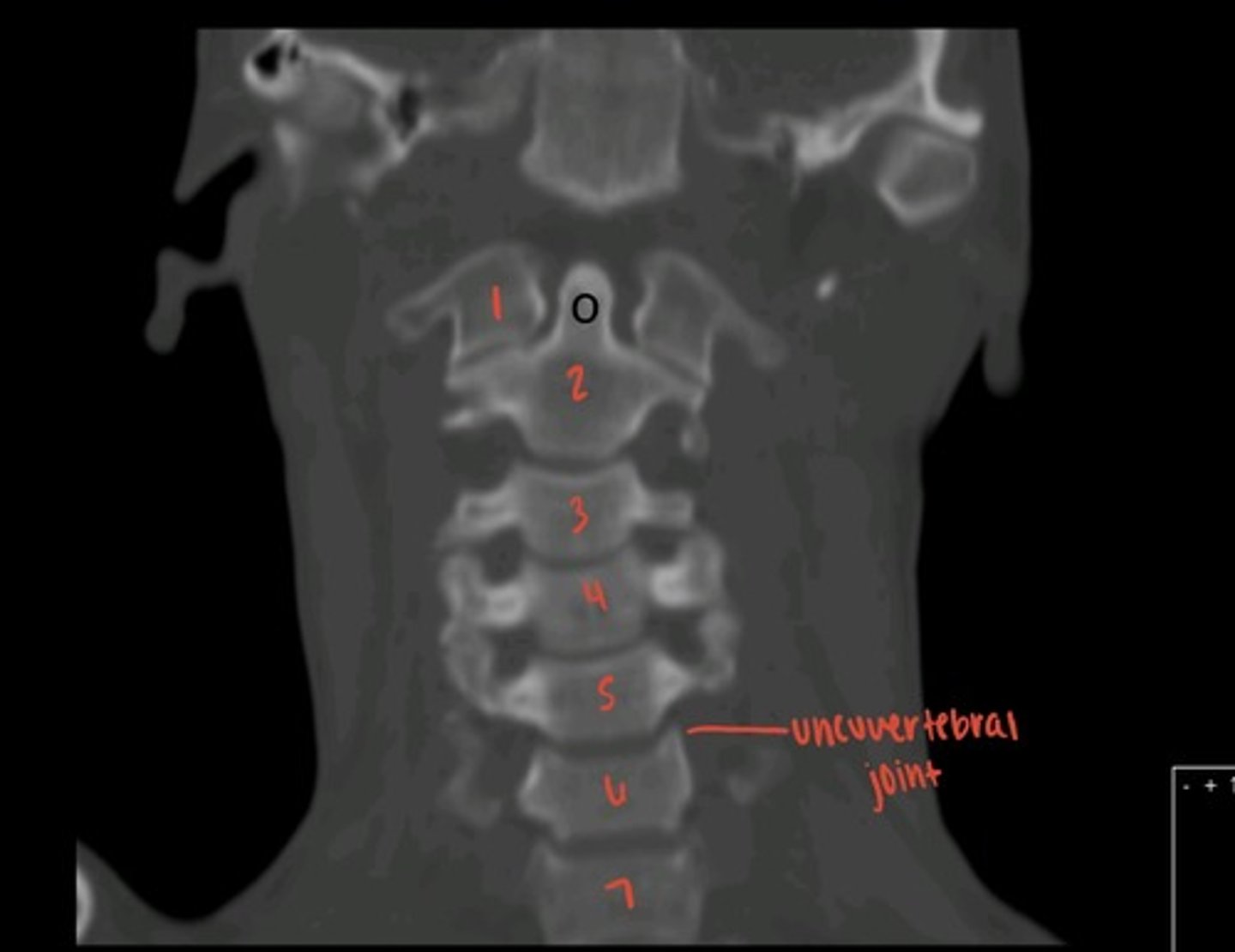
coronal CT c-spine
--> o is dens
what is this image?
when looking for spinal cord abnormalities
when do you get an MRI instead of CT?
CSF is white/bright around cord
why can you see the spinal cord so well in MRI?
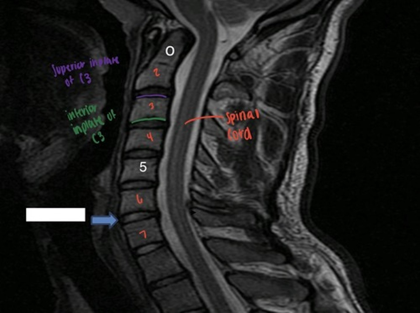
MRI c-spine sagittal
--> blue arrow showing C6-7 disc space
what is this image?
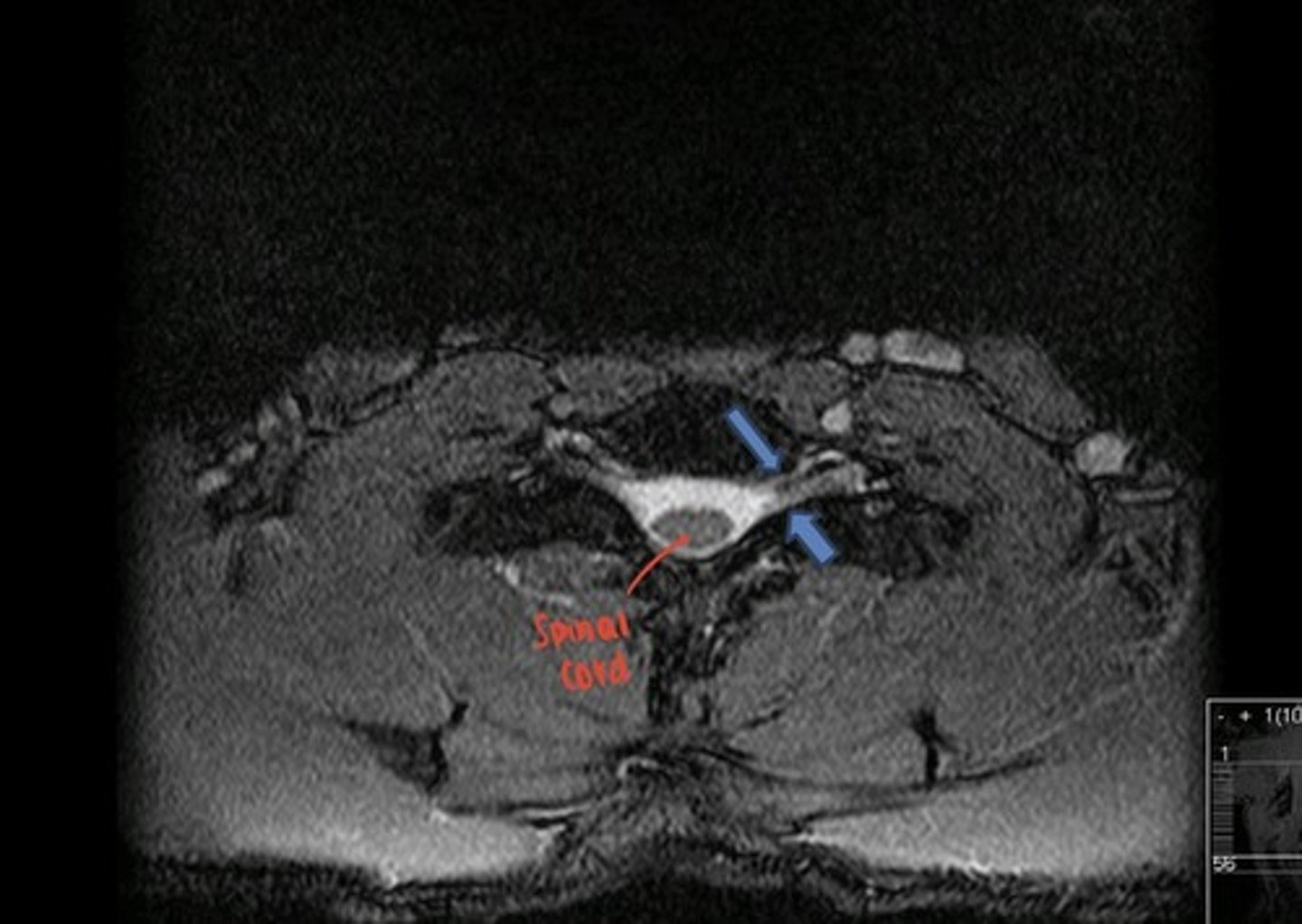
axial MRI c-spine
--> blue arrows showing neural foramina
what is this image?
- AP
- Lateral
- swimmers view
thoracic spine, x-rays
cervicothoracic junction
--> only C7- T5
--> if you cannot see C7, do swimmers
--> arm closest to detector is up
what is the purpose of the swimmers view?
axial, sagittal, coronal
thoracic spine, CT
lots of radiation, going from T1-T12 is a long distance
issue with thoracic spine
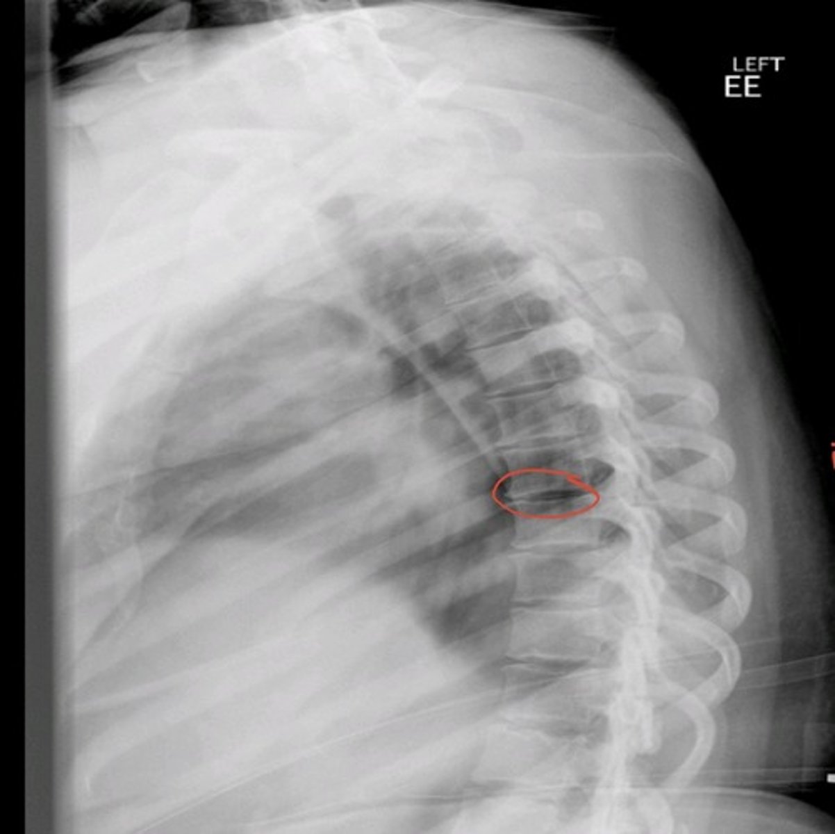
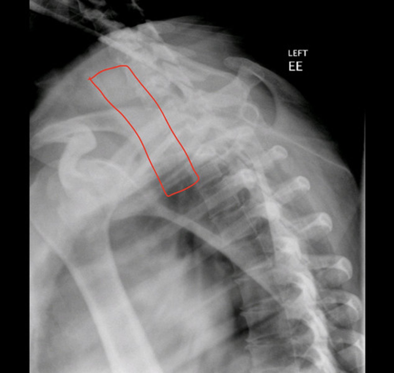
lateral T-spine
--> red circle is indicating intervertebral disc space in thoracic vertebrae, cannot determine level bc ribs are not seen
what is this image?
swimmers view t-spine
what is this image?
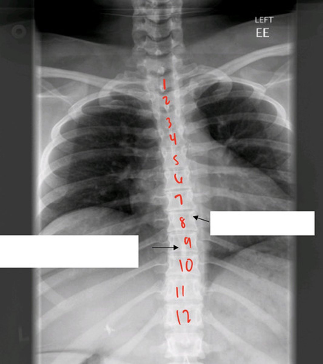
AP T-spine
--> can determine levels bc of ribs
what is this image?

upper arrow: L T-8 pedicle
lower arrow: spinous process of T9
what are the black arrows indicating?
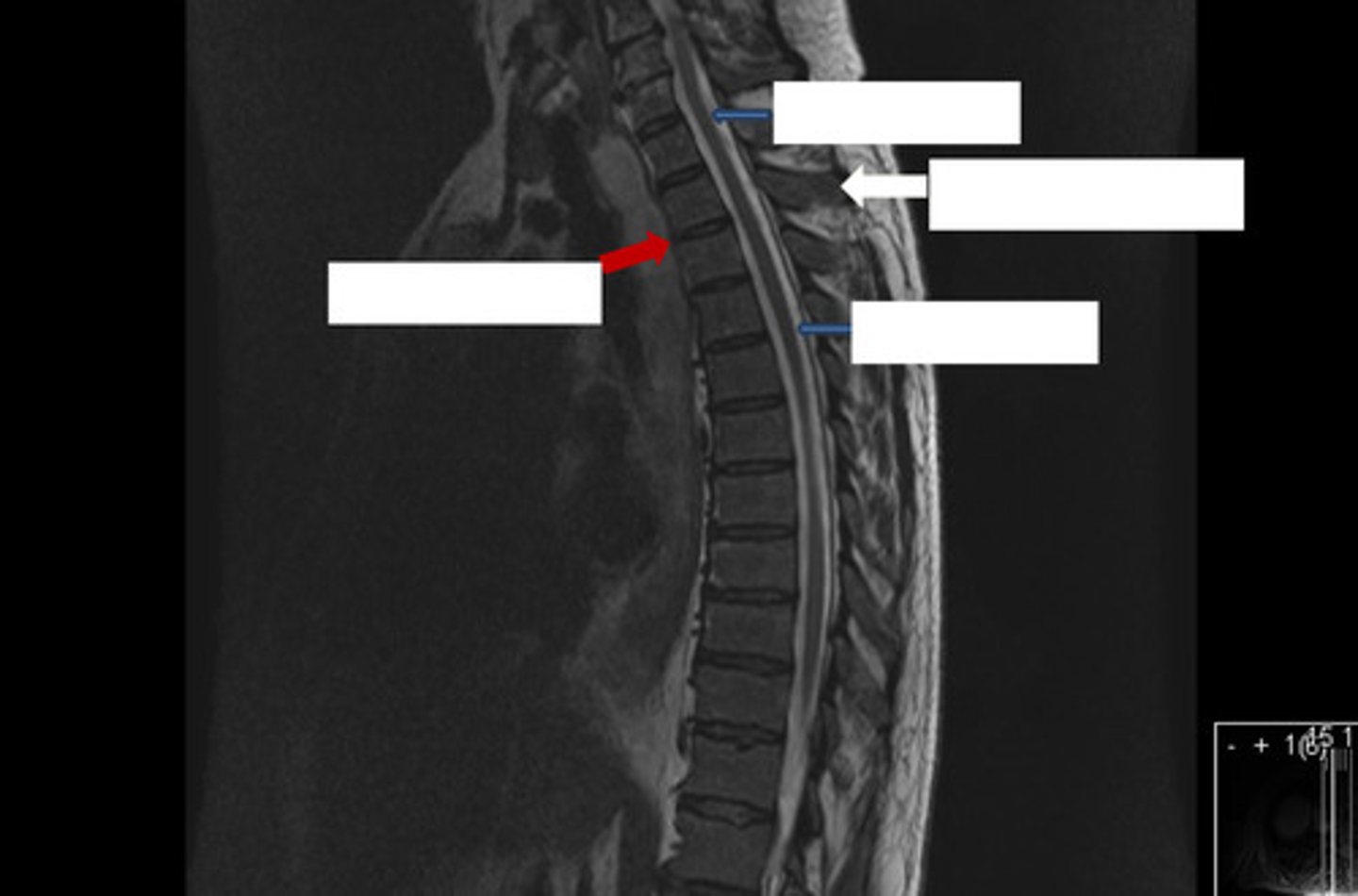
MR T2 sagittal T-spine
--> blue is spinal cord
--> white is spinous process
--> red is disc space
what is this image?
what are the arrows indicating?
- lateral
- AP
- cone down lateral
- obliques
lumbar spine
to see L5/S1 really well
purpose of cone down lateral
"scotty dog", see facet joints
lumbar oblique imaging
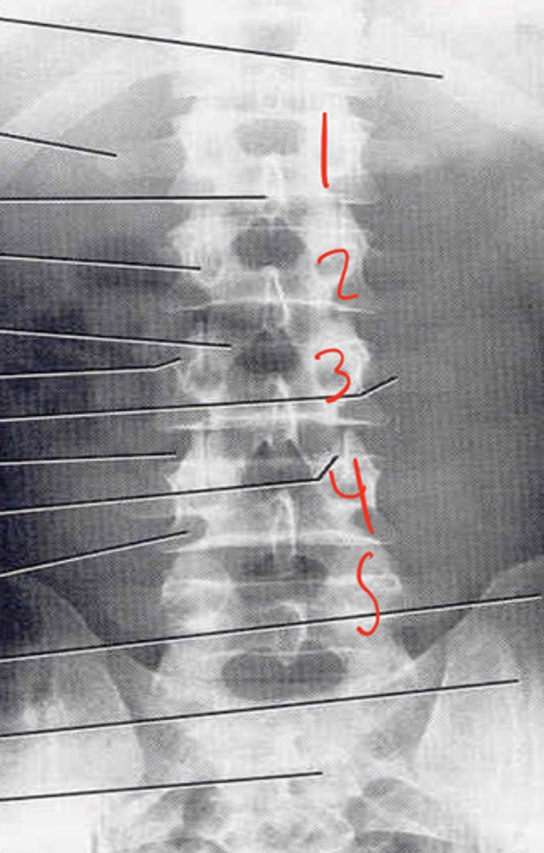
AP lumbar spine
what is this image?
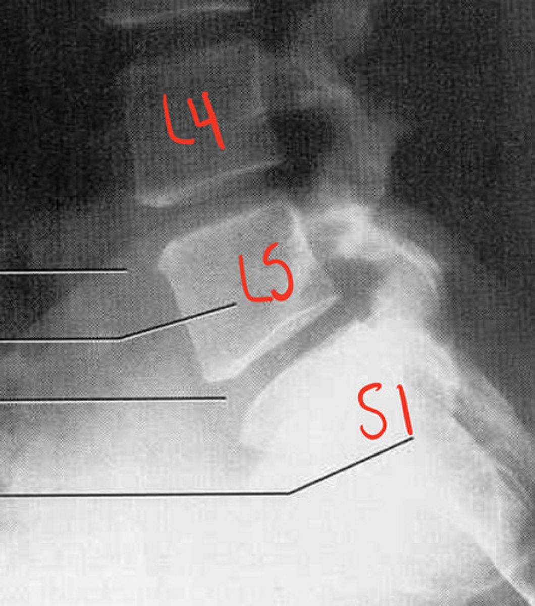
lateral L-spine
--> L3
--> L3-4 disc space
what is this image?
what is the black arrow pointing to?
what is the space below the black arrow?
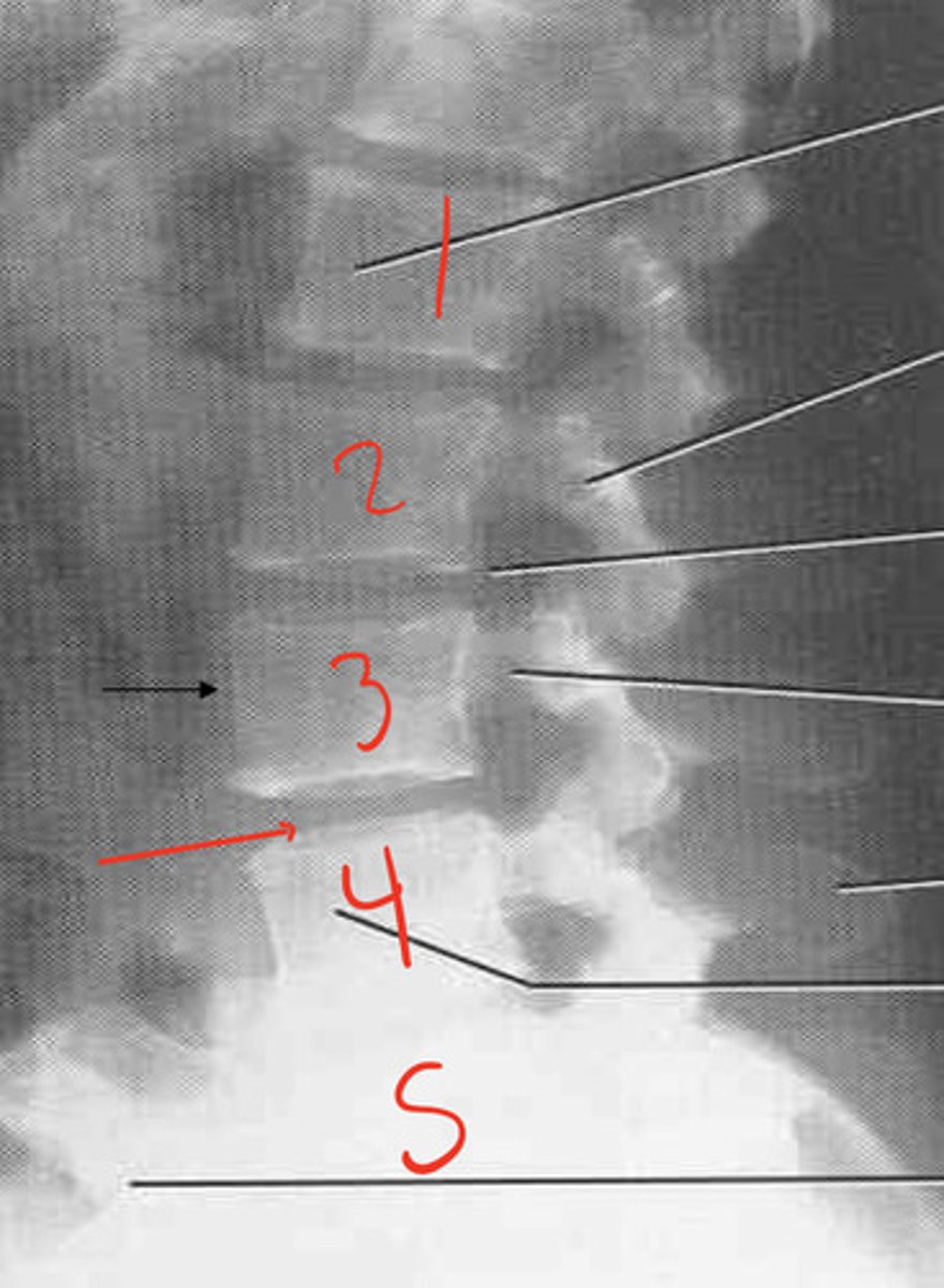
cone down lateral l-spine
what is this image?
oblique l-spine, "scotty"
what is this image?
scotty dog
ear- superior articular process
eye (blue arrow)- pedicle
neck- pars interarticularis
feet- inferior articular process
what is this image?
chronic spondylolysis in L4/L5, very important to find neck**
what is the purpose of the pars interatricularis in scotty?
MR SAG T2 L-spine
what is this image?
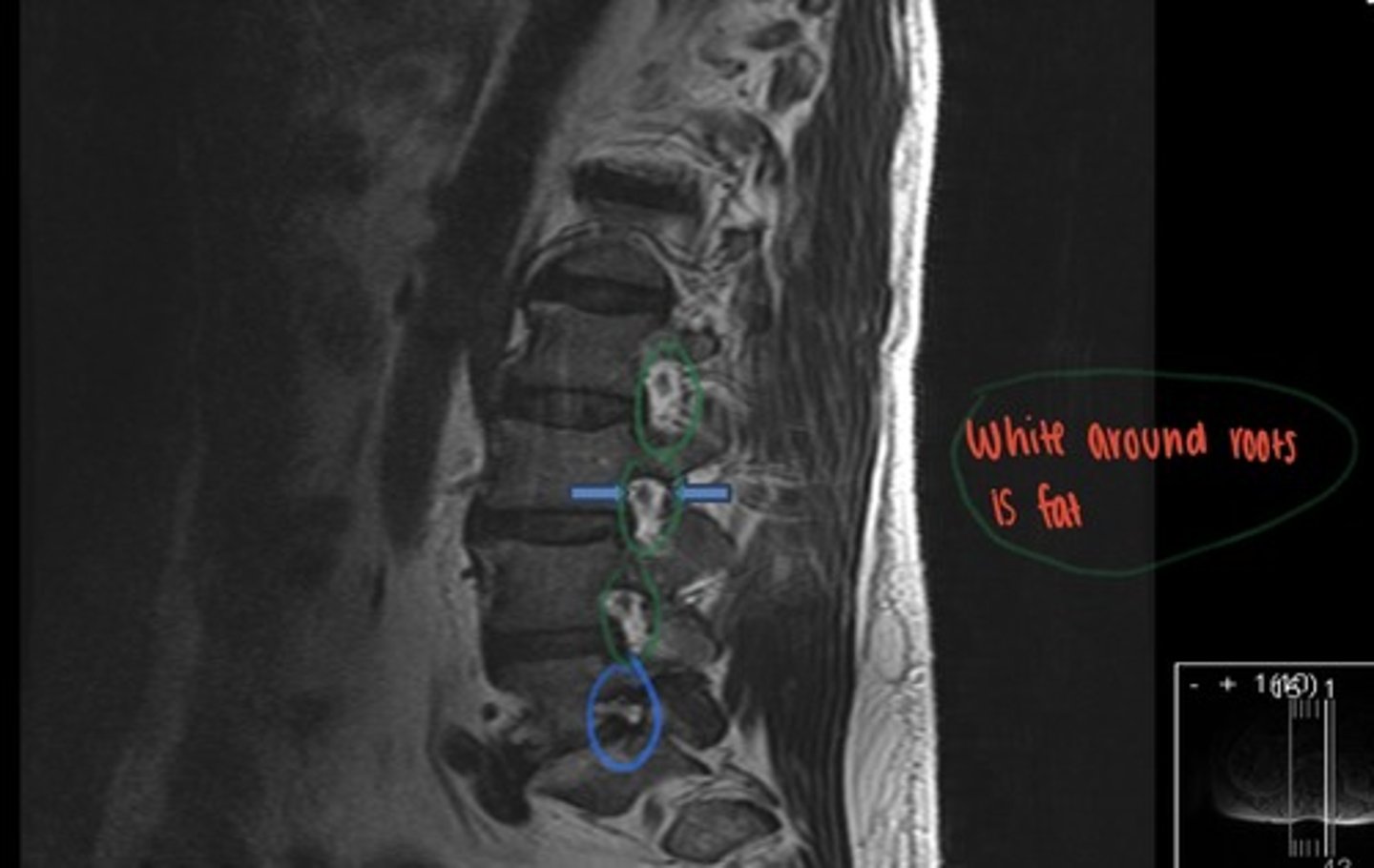
MR L-spine off midline
--> blue arrows showing nerve root
--> L L5SN getting compressed, no fat around it
what is this image?
what is between the blue arrows?
what abnormality is seen on this image?
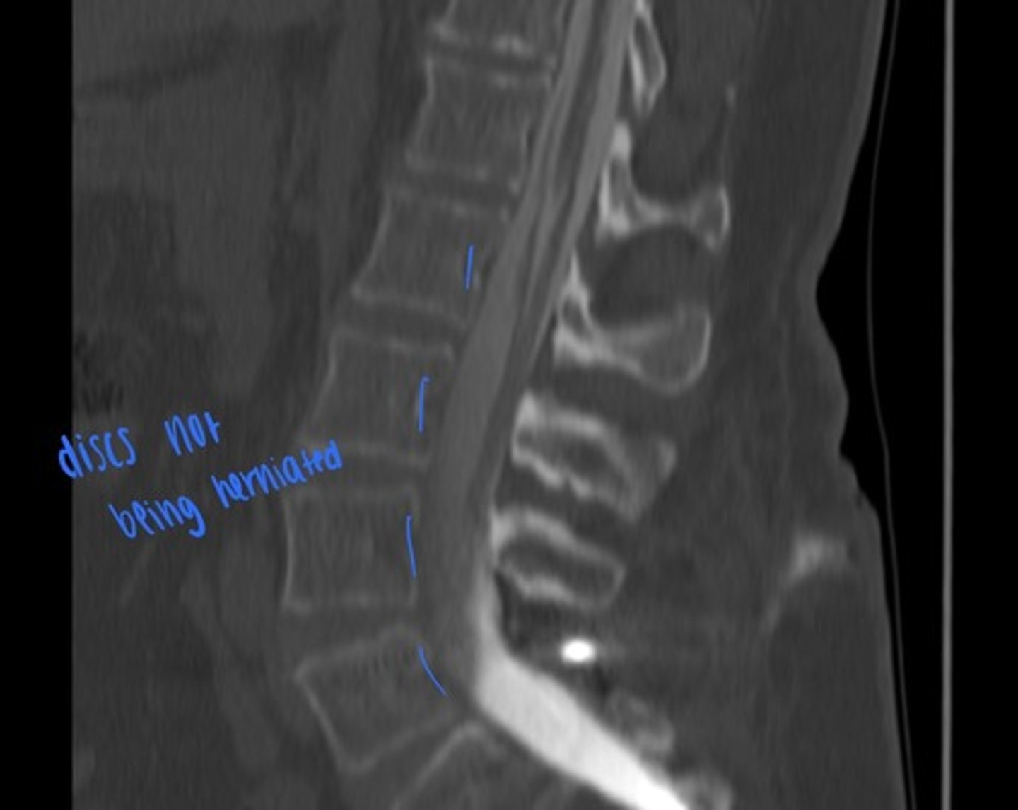
sag L-spine reformat CT
what is this image?
no contrast present

sag L-spine reformat CT
what is this image?
post myelogram, contrast present