Skull
1/105
There's no tags or description
Looks like no tags are added yet.
Name | Mastery | Learn | Test | Matching | Spaced | Call with Kai |
|---|
No analytics yet
Send a link to your students to track their progress
106 Terms
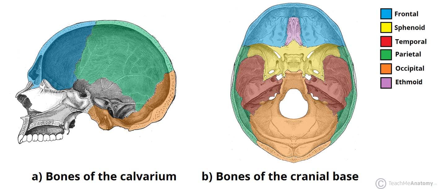
What are the cranial bones?
Frontal
Parietal
Temporal
Sphenoid
Occipital
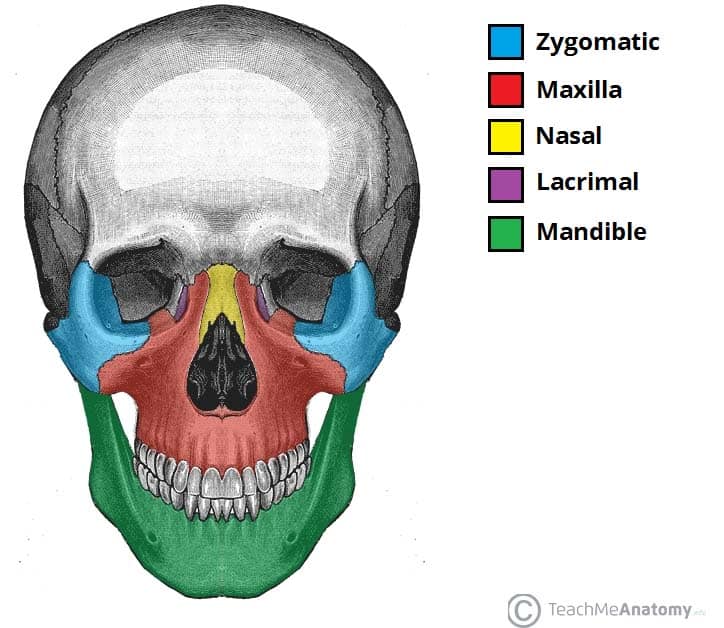
What are the bones of the face?
Maxillae
Zygomatic
Mandible

What bone is #3?
Temporal

What bone is #4?
Mandible

What bone is #5?
Parietal
What bone is #6?
Sphenoid

What bone is #7?
Frontal

What bone is #10?
Zygomatic

What bone is #13?
Maxillae

What bone is #14?
Occipital
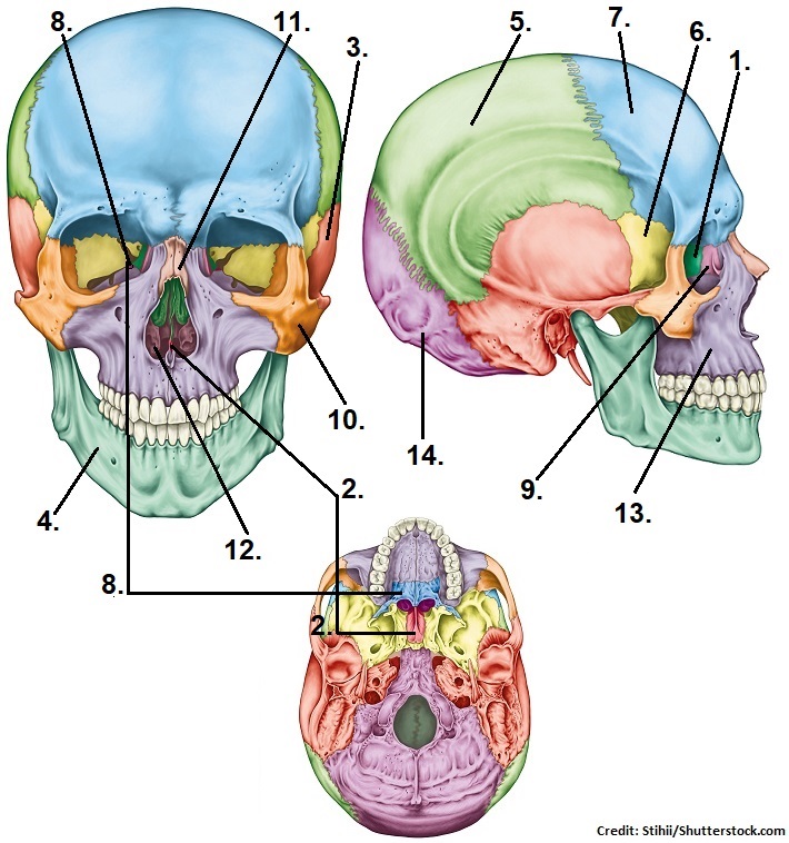
What is the pterion?
An area of weakness on the skull
What 4 bones intersect to make the pterion?
Frontal, parietal, sphenoid, and temporal bones
What is the significance of the pterion?
It is highly vulnerable to fracture; trauma here can cause a epidural hematoma

Label cranial suture #2?
Coronal suture

Label cranial suture #3
Squamous suture

Label cranial suture #4
Sagittal suture
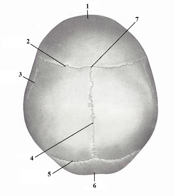
Label cranial suture #5
Lamboid suture

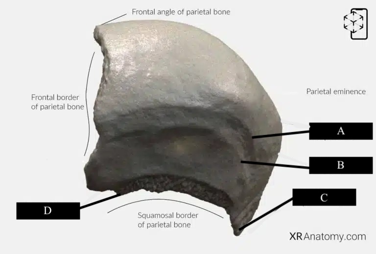
Label A
Frontal eminence

Label C
Frontal surface

Label D
Superciliar arch

Label E
Temporal surface

Label F
Zygomatic process

Label G
Supraorbital margin

Label H
Frontal notch

Label I
Glabella
Label J
Superciliary arch

Label K
Supraorbital foramen

Label L
Temporal line
Label A
Superior temporal line

Label B
Inferior temporal line

Label A
External auditory meatus

Label B
Squamous portion of temporal bone

Label C
Zygomatic process

Label D
Mandibular fossa
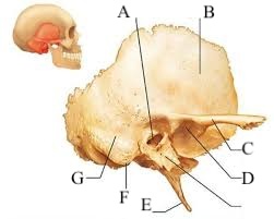
Label E
Styloid process

Label F
Mastoid process
What is significant about the mandibular fossa?
It articulates with the mandibular condyle to create the temporal mandibular joint (TMJ)
What does the TMJ do?
Allows for up-and-down, side-to-side, and forward-and-back motions of the jaw
What is significant of the styloid process?
It is the site of a lot of muscle attachments
What is significant of the zygomatic arch?
It is the joining of the zygomatic process, temporal bone, and temporal process of zygomatic bone
Where is the inferior limit of the cerebral hemisphere of the brain?
Zygomatic arch

Label 1
Optic canal

Label 2
Chiasmatic sulcus

Label 3
Hypophysial fossa

Label 4
Lesser wing

Label 5
Greater wing

Label 6
Anterior clinoid process
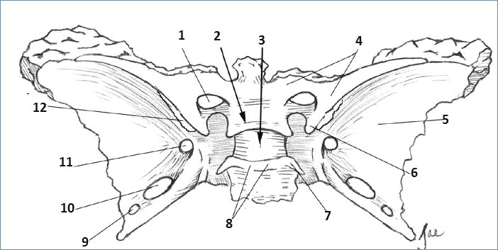
Label 7
Posterior clinoid process

Label 8
Dorsum sellae

Label 9
Foramen spinosum

Label 10
Foramen ovale

Label 11
Foramen rotundum

Label 12
Superior orbital fissure

Label A
Body of sphenoid

Label B
Tuberculum sellae

Label C
Lateral plate

Label D
Medial plate
Label E
Pterygoid process
What are the 3 parts of the sphenoid body?
Sella turcia
Hypophysial fossa
Dorsum sallae
What is significant about the hypophysial fossa?
It holds the pituitary gland
What borders the sella turcia?
Anterior and posterior clinoid processes

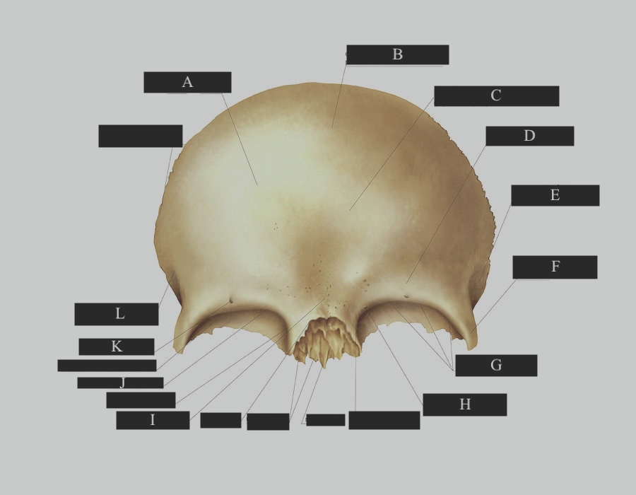
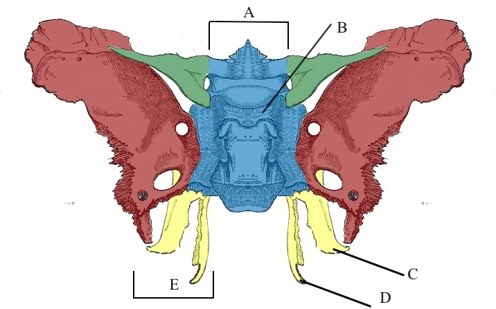
Label A
External occipital protuberance

Label B
Squamous part of occipital bone

Label C
Mastoid border

Label D
Jugular process

Label E
Jugular tubercle

Label F
Lateral part

Label G
Basion

Label H
Basilar part

Label I
Occipital condyle

Label J
Hypoglossal duct

Label K
Condylar duct

Label L
Condylar fossa

Label M
Foramen magnum

Label N
Nuchal plane

Label O
Inferior nuchal line

Label P
External occipital crest

Label Q
Superior nuchal line
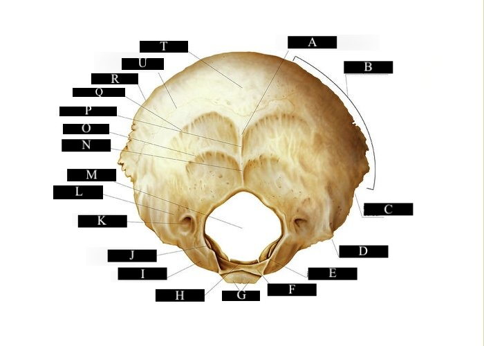
Label R
Lambdoid border

Label U
Highest nuchal line

Label T
Occipital plane

Label 1
Condylar process

Label 2
Head of condyle

Label 3
Pterygoid fovea

Label 4
Neck of condyle

Label 5
Mandibular notch
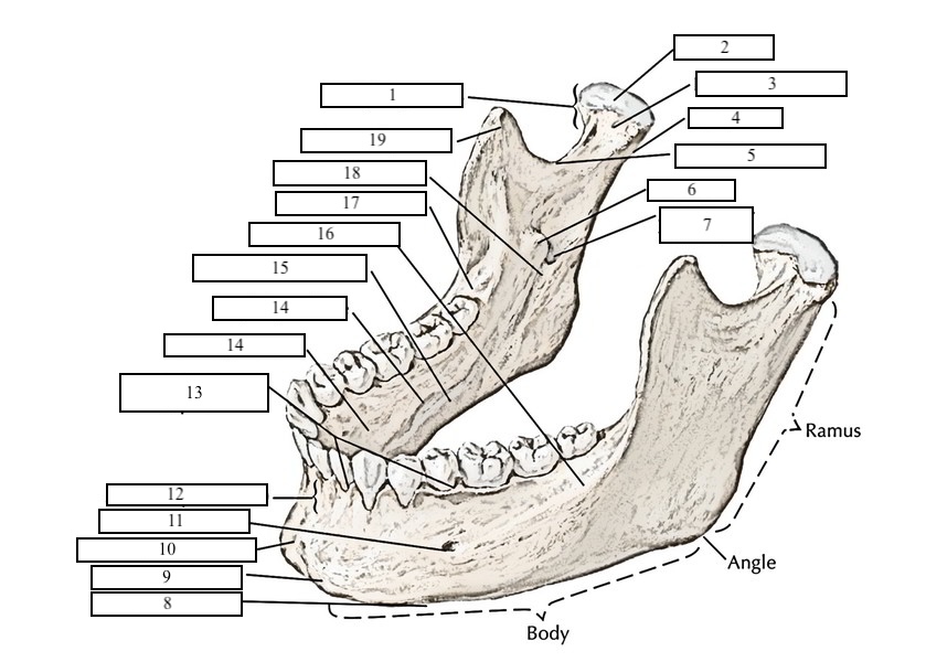
Label 6
Lingula

Label 7
Manidbular foramen

Label 8
Base of mandible

Label 9
Mental tubercle

Label 10
Mental protuberance

Label 11
Mental foramen

Label 12
Alveolar process

Label 13
Interalveolar septa

Label 14
Sublingual fossa

Label 15
Mylohyoid line

Label 16
Submandible fossa

Label 18
Mylohyoid groove

Label 19
Coronoid process