MicroBiology Mod. 2
1/57
There's no tags or description
Looks like no tags are added yet.
Name | Mastery | Learn | Test | Matching | Spaced | Call with Kai |
|---|
No analytics yet
Send a link to your students to track their progress
58 Terms
Prokaryotes vs Eukaryotes
Prokaryotes:
one circular chromosome, not in a membrane
No histones (except Archaea)
No organelles
Bacteria: peptidoglycan cell walls
Binary Fission
Eukaryotes:
Paired chromosomes in nuclear membrane
Histones
Organelles
Polysaccharide cell walls
Mitotic Spindle
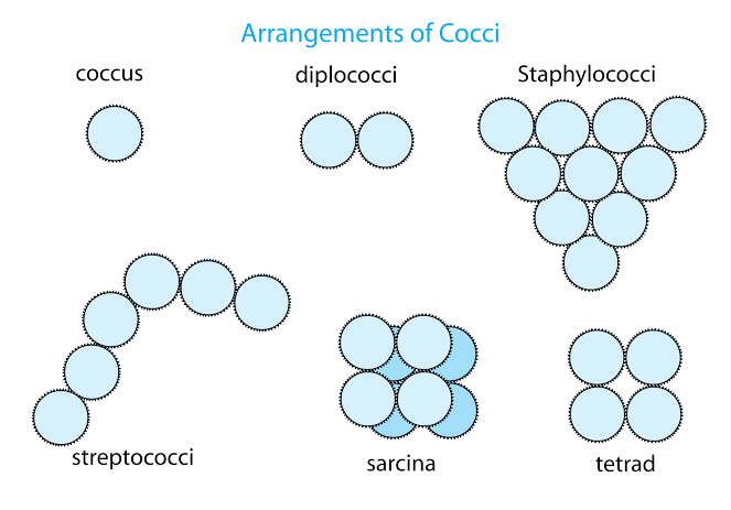
Coccus
Round bacteria
Bacillus
Rod-like bacteria
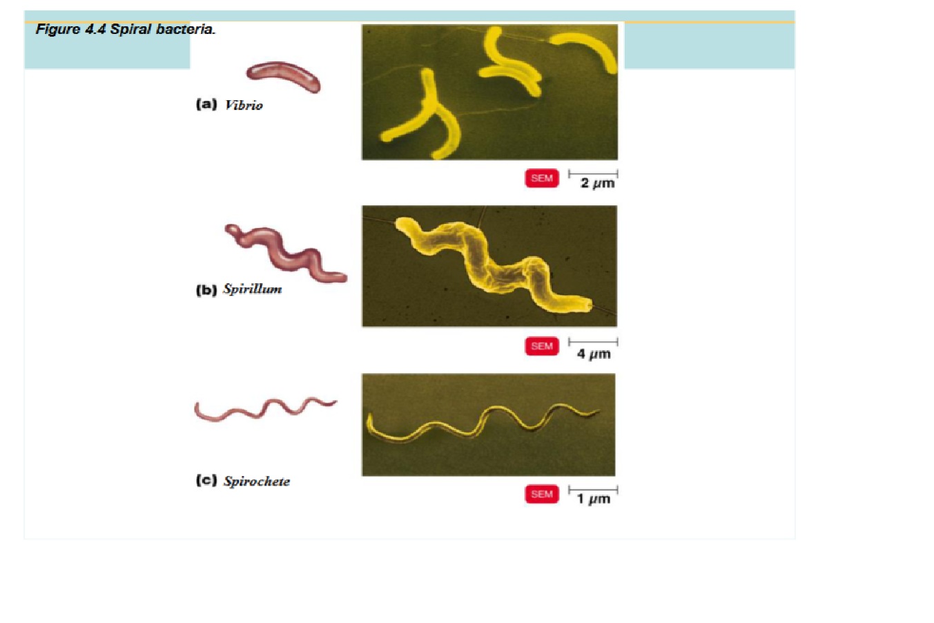
Spiral
Spiral shaped bacteria
Vibrio - bent or C-shaped
Spirillum - rigid cell wall, use external flagella to move
Spirochete - flexible cell wall, internal flagella (axium fillament) for movement
Pleomorphic
Multiple shapes at once
e.g. coccobacilli
Arrangements: (due to how they divide)
Pairs:
diplococci (pair and round)
diplobacilli (pair and rod-like)
Chains:
Streptococci (chain and round)
Streptobacilli (chain and rod-like)
Clusters:
Staphylococci (cluster and round)
Cocci Specific:
Sarcina:
cube-shaped
Tetrad:
square shaped
Glycocalyx
Not every bacteria cell has one
pod/husk outside the cell wall
a layer of material containing substantial amounts of sticky carbohydrates (allows it to stick to surfaces or a host)
capsule - neatly organized glycocalyx that prevents phagocytosis (when specialized cells engulf, internalize, and destroy large particles, like bacteria and viruses)
slime layer - loose and unorganized glycocalyx where the extracellular polysaccharide allows cells to attach
helps induce disease/ increase virulence factor
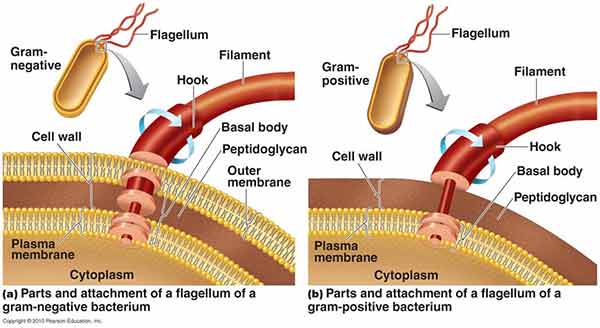
Flagella
Usually on bacilli and spirillum
Outside the cell wall
made of chains of flagellin (a protein in prokaryotes)
3 Basic Parts:
Filament - the long, outermost region that contains the protein flagellin arranged in several chains that form a helix around a hollow core
Hook - consisting of a different protein, where the filament is attached to
Basal Body - anchors the flagellum to the cell wall and plasma membrane (composed of a small central rod inserted into a series of rings: 2 pairs, outer and inner, of rings if gram negative and 1 inner pair of rings if gram positive)
Flagella proteins are H. antigens
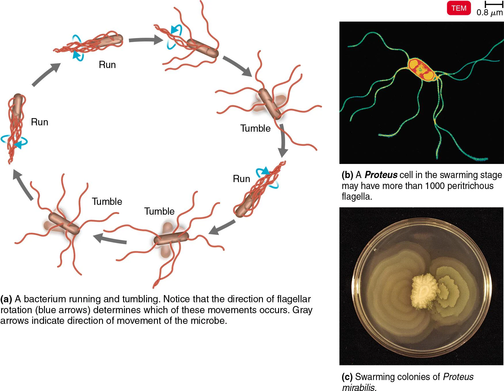
How Flagella help the cell move
Rotates from the basal body
Run - swim in a direction (counterclockwise)
Tumble - switch direction (clockwise)
Allow the cell to move toward or away from stimuli (taxis)
Stimuli include chemicals (chemotaxis) and light (phototaxis)
Listen to chemotactic signals: if positive (attractant), they go towards with many runs and few tumbles. If negative (repellent), they move away with more tumbles than before.
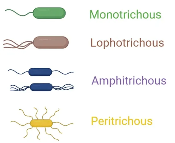
Flagella Arrangement
Atrichous - without flagella
Monotrichous - single flagellum at one end (polar)
Amphitrichous - one flagellum at both ends (polar)
Lophotrichous - a tuft of flagella at one end (polar)
Peritrichous - distributed all around the cell
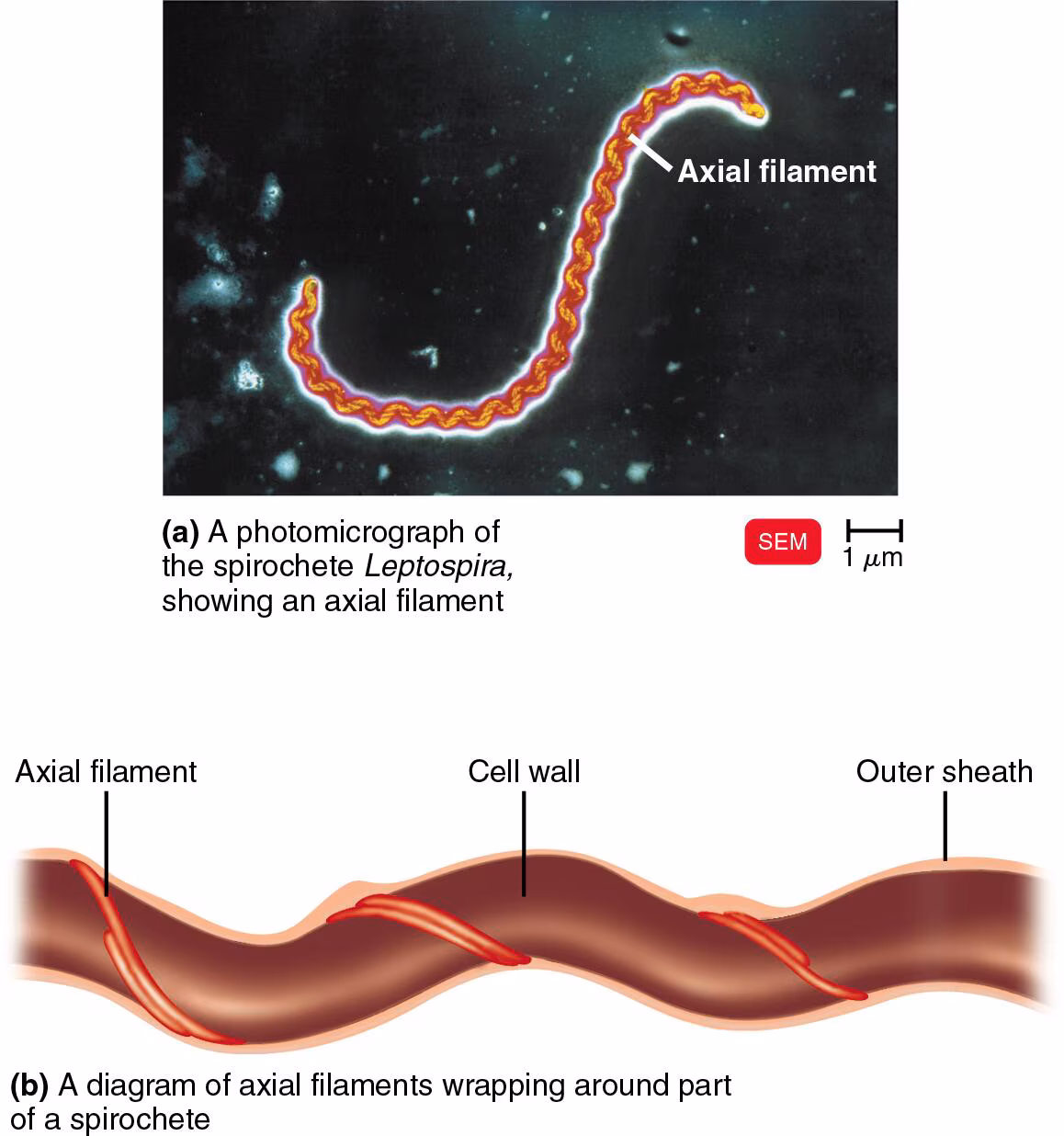
Axial Filaments
bundles of fibrils that arise at the ends of the cell beneath an outer sheath and spiral around the cell
in spirochetes
called “endoflagella”
anchored at one end of the cell
rotation causes the cell to move
Fimbriae
allow attachment (involved in forming biofilms)
Pili
Used for motility
gliding motility
twitching motility
Transfer DNA
Cell Wall
Prevents osmotic lysis (changes in osmotic pressure causing bursting)
made of peptidoglycan (a repeating sugar molecule - polymer of disaccharide - linked by polypeptides, or proteins)
Gram positive cell wall
thick layer of peptidoglycan (on the exterior surface of the cell)
teichoic acid, integrated for support and rigidity, consist of alcohol and phosphate
lipoteichoic acid in the periplasmic space

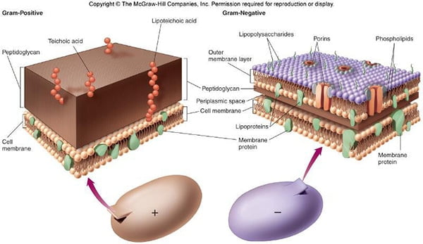
Gram negative cell wall
two plasma membranes
thin peptidoglycan layer in the periplasmic space (makes it susceptible to breakage)
lipopolysaccharide (LPS) in the outer/secondary membrane (phospholipids on the inside facing the periplasm and LPS on the outside acting as a physical and chemical barrier)
Gram stain mechanism
Crystal violet-iodine crystals form in the cell
In gram positive:
alcohol dehydrates peptidoglycan
CV-1 crystals do not leave, staining it purple
In gram negative:
alcohol dissolves the outer membrane and leaves holes in the peptidoglycan
CV-1 washes out
stained pink/red after w/ another stain
acid fast cell wall
Mycobacterium
These bacteria contain high concentrations (60%) mycolic acid in their cell wall that prevents the uptake of dyes, including those used in the Gram stain
the mycolic acid forms a layer outside of a thin layer of peptidoglycan
the hydrophobic lipid cell wall allows mycobacterium to clump together and stick to the walls of a flask
acid fast stain
used to identify bacteria of the genus Mycobacterium
stained with carbolfuchsin, which penetrates bacteria more effectively when heated.
carbolfuchsin penetrates the cell wall, binds to the cytoplasm, and resists removal by washing with acid-alcohol.
Acid-fast bacteria retain the red color of carbolfuchsin because it’s more soluble in the cell wall’s mycolic acid than in the acid-alcohol.
If the mycolic acid layer is removed from the cell wall of acid-fast bacteria, they will stain gram-positive with the Gram stain.
Lysozyme
found in human sweat and tears)
digests the disaccharide in peptidoglycan
This act is analogous to cutting the steel supports of a bridge with a cutting torch: the gram-positive cell wall is almost completely destroyed by lysozyme.
Protoplast
wall-less gram positive cell
cell wall destroyed by lysozyme
susceptible to osmotic lysis when placed in a hypotonic environment (cell bursting when solute concentration is higher inside the cell and water rushes in to dilute the concentration)
Spheroplast
Have lost most of the cell wall but retain portions of the outer membrane
when lysozyme is applied to Gram negative bacteria
consists of the cellular contents, plasma membrane, and remaining outer wall layer
susceptible to osmotic lysis when placed in a hypotonic environment (cell bursting when solute concentration is higher inside the cell and water rushes in to dilute the concentration)
L forms
of the genus proteus
partially or completely lose cell walls and can sometimes revert to the normal form once damaging condition of removed
swells into irregular shapes
develop in response to penicillin (which inhibits cell wall formation) or lysozyme (which digests peptidoglycan in the cell wall)
susceptible to osmotic lysis when placed in a hypotonic environment (cell bursting when solute concentration is higher inside the cell and water rushes in to dilute the concentration)
Mycoplasma
Naturally lack a cell wall and are not produced by lysozyme treatment
smallest form of bacteria
Damage to cell wall summary
Protoplast = Purely no wall (cell wall completely removed)
Spheroplast = Some wall remains (Gram-negative, outer membrane retained)
L-form = Lost wall (partially or completely, often reversible)
Mycoplasma = Missing wall naturally (no cell wall to begin with)
Penicillin (in regards to cell wall damage)
targets cell wall construction and inhibits peptide bridges in peptidoglycan
dos not affect gram negative cell walls
Plasma membrane structure
Phospholipid bilayer - the polar hydrophilic heads are on the two surfaces of the lipid bilayer, and the nonpolar hydrophobic tails are in the interior of the bilayer.
• 3 Carbon glycerol
• 2 Fatty acid chains
• Phosphate group
Peripheral proteins - one side or the other
Integral proteins - permanently embedded into the membrane
Transmembrane proteins - spans the entire plasma membrane
Fluid mosaic model of the plasma membrane
Fluid mosaic model
the fatty acid tails cling together, so phospholipids in the presence of water form a self-sealing bilayer allowing breaks and tears in the membrane to heal themselves
Membrane is viscous, allowing proteins to move freely enough to perform their functions without destroying the structure of the membrane.
Proteins move to function
Phospholipids rotate and move laterally
Plasma membrane functions
Selective permeability allows passage of some molecules
Enzymes for ATP production
Photosynthetic pigments on infoldings called chromatophores or thylakoids
Damage to the membrane by alcohols, quaternary ammonium (detergents) and polymyxin antibiotics causes leakage of cell contents
Semi-permeable membrane
some substances are able to cross while others are not
Simple diffusion
a form of passive transport
movement of a solute from an area of high concentration to an area of low concentration
transports small molecules, such as oxygen and carbon dioxide, across the cell membrane
Fascilitated diffusion
a form of passive transport
solute combines with a transporter protein in the membrane
movement of ions or large molecules across the plasma membrane
does NOT expend energy
Active transport
requires a transporter protein and ATP
it expends energy
low concentration to a high concentration, so it goes AGAINST the concentration gradient
Osmosis
movement of water across a selectively permeable membrane from an area of high water concentration to an area of low water concentration
Osmosis: isotonic solution
no net movement of water occurs
Osmosis: hypotonic solution
water moves into the cell
high solute concentration inside the cell
low solute concentration outside the cell
if the cell wall is strong, it contains the swelling
if the cell wall is weak/damaged, the cell bursts (osmotic lysis)
Osmosis: hypertonic solution
water moves out of the cell
low solute concentration inside the cell
high solute concentration outside the cell
causes the cytoplasm to shrink when water rushes out of the cell to dilute the high concentration outside (called plasmolysis)
plasmolysis
the shrinkage of a cell's cytoplasm away from the cell wall due to water loss by osmosis when the cell is placed in a hypertonic solution
In bacteria:
Water moves out of the cell into the surrounding environment.
The cytoplasmic membrane pulls away from the cell wall.
Cell growth and metabolism may stop, although the cell is not necessarily killed.
This is one reason why high concentrations of salt or sugar can help preserve food—they create a hypertonic environment that causes plasmolysis in many microorganisms.
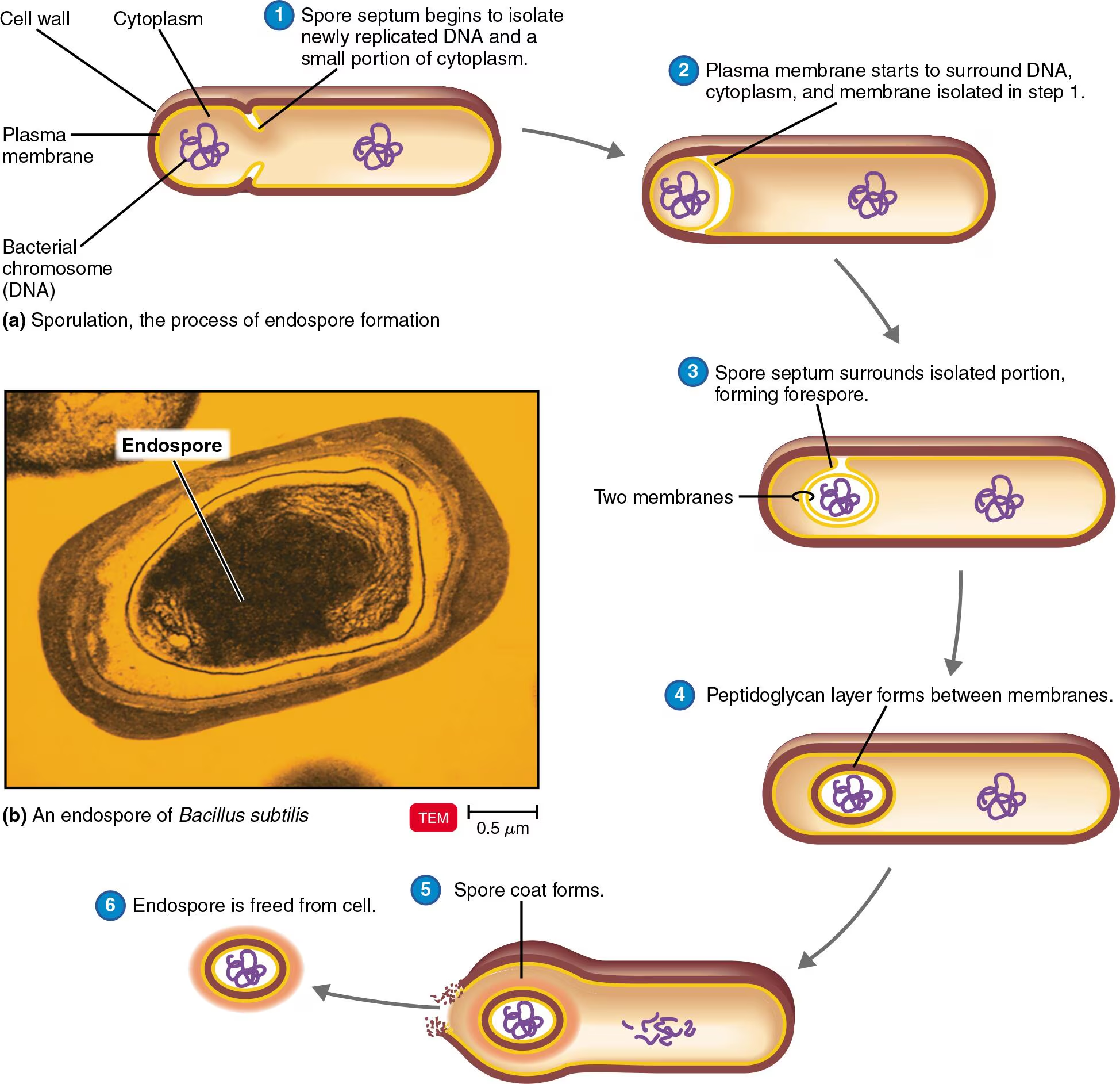
Endospores
Resting (Dormant) cells
• Metabolically inactive
• Resistant to desiccation, heat, chemicals
• Sporulation occurs during stress (unfavorable environment)
• Bacillus, Clostridium
• Sporulation: Endospore formation
• Germination: Return to vegetative state (occurs when spore finds a favorable environment)
Inside a bacteria cell
Nuclear area with DNA (nucleoid)
Ribosomes for protein synthesis
Cytoplasm is the substance inside the plasma membrane
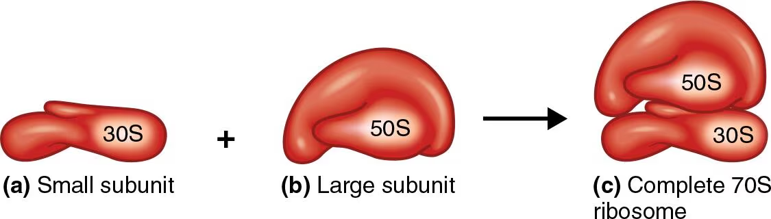
Prokaryotic Ribosomes
Protein synthesis
• 70S (Composed of over 30 proteins and rRNA)
• S = Svedberg Units (Centrifuge sedimentation)
• 50S + 30S subunits
Inclusions:
Cells may accumulate certain nutrients when they are plentiful and use them when the environment is deficient.
(reserve deposits)
Metachromatic granules (volutin) - phosphate reserves
Polysaccharide granules - energy reserves
lipid inclusions - energy reserves
sulfur granules - energy reserves
carboxysomes - Ribulose 1,5- diphosphate carboxylase for CO2 fixation
gas vacuoles - protein-covered cylinders
magnetosomes - iron oxide (destroys H2O2)
Eukaryote: nucleus
largest structure in the cell, and contains almost all of the cell’s hereditary information (DNA)
contains chromosomes wrapped around proteins called histones
genetic material is a threadlike mass called chromatin until it gets ready to divide where it condenses into chromosomes
Eukaryote: Rough Endoplasmic reticulum
responsible for synthesis, folding, and initial modification of proteins
contains ribosomes
Eukaryote: Smooth Endoplasmic reticulum
synthesizes phospholipids, fats, and steroids
does not have ribosomes
Eukaryote: golgi apparatus
modifies, sorts, packages, and ships proteins and lipids to their final destinations inside or outside the cell
Eukaryote: lysosome
digestive enzymes
found only in animal cells
Eukaryote: vacuole
brings food into cells and provides support
derived from the golgi apparatus
some serve as temporary storage organelles for substances such as proteins, sugars, organic acids, and inorganic ions
others form during endocytosis to help bring food into the cell
store metabolic wastes and poisons
take up and excrete excess water to prevent osmotic lysis
Eukaryote: cytoplasm
where all the organelles are stored
Eukaryote: flagella and cilia
projections are few and are long in relation to the size of the cell, they are called flagella (algae)
projections are numerous and short, they are called cilia (protozoa)
Eukaroyte: ribosomes
free ribosomes - unattached to any structure in the cytoplasm.
synthesize proteins used inside the cell.
membrane-bound ribosomes - attach to the endoplasmic reticulum.
synthesize proteins destined for insertion in the plasma membrane or for export from the cell.
80S ribosomes
large 60S sub unit containing three molecules of rRNA
smaller 40S subunit with one molecule of rRNA.
Eukaryote: mitochondria and chloroplasts
produce ATP
perform photosynthesis
Eukaryote: peroxisome
oxidizes various organic substances
protect others parts of the cell from toxic effects of H2O2.
Molds
The fungal thallus (growth) consists of hyphae (long strands of cells)
a mass of hyphae is a mycelium
Yeasts
Unicellular fungi
Fission yeasts divide symmetrically
Budding yeasts divide asymmetrically
Fungal Diseases (Mycoses)
Systemic mycoses - Deep within body
Subcutaneous mycoses - Beneath the skin
Cutaneous mycoses - Affect hair, skin, nails
Superficial mycoses - Localized, e.g., hair shafts
Opportunistic mycoses - Caused by normal microbiota or fungi that are usually not pathogenic
Viruses
Don’t fit our definition of a living organism
They are composed of Nucleic Acid & Protein
Viruses are obligate intracellular ‘parasites’
No metabolic ability
They infect every type of life form: animals to algea
Each has a “Host Range”
Evolve quickly
Genome consists of singe or double stranded DNA or RNA
capsid - protein shell enclosing the viral genome (built from proteins called capsomeres)
Some have lipid membranes:
viral envelope (contains capsid and aids in entry to the cell)
Prions
Prions - proteins that cause brain diseases in
mammals
• Infectious proteins that build up in nervous tissue
• Propagate by converting normal proteins into the prion
version
• Creutzfeldt-Jakob disease – acquired (vCJD) or
inherited
• PRNP gene PrP normal protein
• Scrapie; Mad Cow disease (BSE)