Bio Topic 2 -> DNA
 Call Kai
 Learn
Learn Practice Test
 Spaced Repetition
 Match
Match Flashcards
 Knowt Play
1/22

There's no tags or description
Looks like no tags are added yet.
Name | Mastery | Learn | Test | Matching | Spaced | Call with Kai |
|---|
No analytics yet
Send a link to your students to track their progress
23 Terms
Structure of mononucleotides
DNA nucleotide → deoxyribose sugar + a phosphate group + nitrogenous base (ATCG)
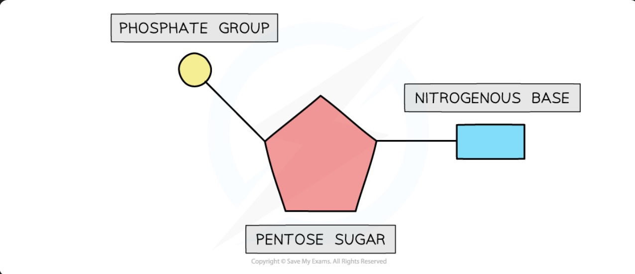
RNA nucleotide → ribose sugar + a phosphate group + nitrogenous base (AUCG)
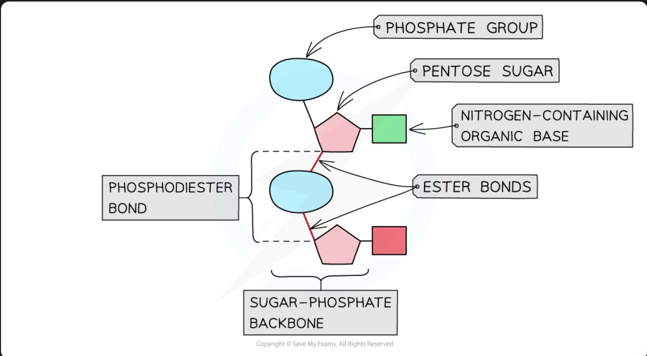
Phosphodiester bond
Nucleotides join via condensation reactions and make a phosphodiester bond to form polynucleotides like DNA and RNA
Phosphate group of one nucleotide binds to the OH group of the pentose sugar of the next
Ester bond → between an OH group and an acid group (phosphate group acts as the acid)
What is the structure of DNA
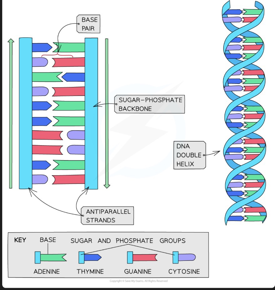
DNA molecule → made of 2 polynucleotide strands running in opposite directions → each strand is held tg by phosphodiester bonds
The 2 strands are held tg by hydrogen bonding between the complimentary nitrogenous bases (A → T and C → G)
What is the structure of RNA
RNA molecule → 1 polynucleotide strand made of a ribose sugar, a phosphate group and nitrogenous base (AUCG) → shorter than DNA
What is a gene
A sequence of nucleotide bases that codes for the production of a specific sequence of amino acids that makes up a protein
How is a protein formed
Transcription → DNA is transcribed and an mRNA molecule is produced
Translation → mRNA is translated and an amino acid sequence is produced
transCription before transLation
C is before L
What is the process of transcription
Part of the DNA molecule unwinds and the Hydrogen bonds between the complimentary base pairs break
Exposes the gene to be transcribed
A complimentary copy of the code from the gene is made called the mRNA → Free RNA molecules pair up via hydrogen bonds with their complementary bases on the template strand → phosphodiester bonds form between the RNA molecules to form the sugar - phosphate backbone of the mRNA which is catalysed by RNA polymerase
When the gene has been transcribed → the mRNA and DNA template strands’ hydrogen bonds break
Double stranded DNA molecule reforms
mRNA molecule leaves the nucleus via a pore in the nuclear envolope
What is the process of translation
After leaving the nucleus via a pore in the nuclear envolope, mRNA attaches to a ribosome
There are free tRNA molecules in the cytoplasm, each with a complementary anticodon to the codon on the mRNA (triplet of bases) and a region for a specific amino acid to bind to → tRNA is already binded to the specific amino acid and when it reads the complementary codon on the mRNA it attaches to the ribosome
At the beginning of the mRNA, there is a start codon which codes for a specific amino acid → starts translation
2 tRNA molecules fit onto the ribosome at one time → amino acids form a peptide bond
Repeats until the stop codon is read
Polypeptide chain folds to from a protein
What is the nature of the genetic code
Triplet code → A triplet of bases code for one amino acid
Non overlapping → Each base is only read once so the adjacent codons dont overlap → the same base is not used to make another codon
Degenerate → There are 4 bases on the mRNA (AUCG) → 4³ (64) combinations of codons possible → but only 20 amino acids exist → so multiple codons can code for the same amino acid → limits the effect of mutations
What is a peptide bond
Forms between amino acids
What is the primary structure of a protein
Specific to each protein
The sequence of amino acids is the primary structure of a protein
Gene codes for the primary structure of a protein
Primary structure → affects the shape + function of the protein
What is the secondary structure of a protein
Secondary structure refers to the hydrogen bonds that form between peptide bonds
Hydrogen bonds can form these 2 shapes :
Alpha helix → hydrogen bonds form between every 4th peptide bond
Beta pleated sheet → forms when the protein folds so that 2 parts of the polypeptide chain are parallel so hydrogen bonds form between parallel peptide bonds
What is the tertiary structure of a protein
Aditional bonds forming between the R groups in the amino acids after secondary structure → creates a 3D structure
What is the quarternary structure of a protein
Proteins with more than 1 polypeptide chain
What is the structure of a globular protein
Compact
Spherical → non polar hydrophobic R groups are in the centre of the protein away from the aqeuous surroundings + polar hydrophillic R groups are on the outside of the protein
Have specific shapes
What is the function of a globular protein
Due to the polar hydrophillic R groups on the outside → globular proteins are soluble in water → can be easily transported around the body
What is an example of a globular protein
Haemoglobin → has a quarternary structure (4 polypeptide chains) → each polypeptide chain contains a haem group
If there are changes to the sequence of amino acids (primary structure) → protein would fold differently → sickle cell anaemia
The haem group in each subunit contains an Fe 2+ ion → binds to oxygen reversibly → useful because oxygen is not soluble in water so transported around the body in haemoglobins
What is the structure of a fibrous protein
Long polypeptide chains that have cross linkages due to hydrogen bonding
→ repeafting structure → creates an organised structure
What is the function of a fibrous protein
Due to large no of hydrophobic R groups → not soluble in water
Strong + insoluble → structural proteins eg collagen
What is the structure of collagen
Formed from 3 polypeptide chains held tg by hydrogen bonding to form a triple helix
Covalent bonds form cross links between R groups of amino acids → holds collagen molecules tg to form fibrils → have staggered ends → not in a perfect line → stronger
What is the function of collagen
Structural protein
Many hydrogen bonds within the triple helix → strong
The fibrils have staggered ends → strong
Insoluble → high no of hydrophobic R groups
What is the role of an enzyme
Biological catalyst → speeds up the rate of reaction by finding an alternate pathway with a lower activation energy for the reaction to occur
Structure of enzymes
Globular protein → highly specific → active site has a specific shape for a specific substrate
Enzymes 3D structure is determined by the tertiary structure → if altered → active site changes shape → substrate no longer fits → enzyme cant carry out its function
High pH or temp → denatures the enzyme by changing the shape of the active site