BIO 167 Exam 6 Learning Targets
1/111
There's no tags or description
Looks like no tags are added yet.
Name | Mastery | Learn | Test | Matching | Spaced | Call with Kai |
|---|
No analytics yet
Send a link to your students to track their progress
112 Terms
Describe the gross anatomy of the spinal cord
31 segments
each giving rise to a pair of spinal nerves
protected by bone, meninges, and CSF
Describe the location of the spinal cord
located w/in the vertebral canal
foramen magnum → 1st/2nd lumbar vertebra
Explain how specific spinal nerves pass enter/exit the vertebral canal
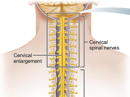
cervical enlargement
where nerves serving upper extremities arise
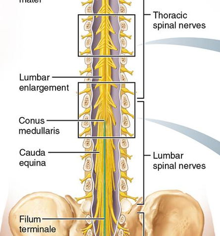
lumbar enlargement
where nerves serving lower extremities arise

conus medullaris
tapered end of the spinal cord

filum terminate
pia mater extension to coccyx
holds spinal cord in position
contains no neurons, only connective tissue
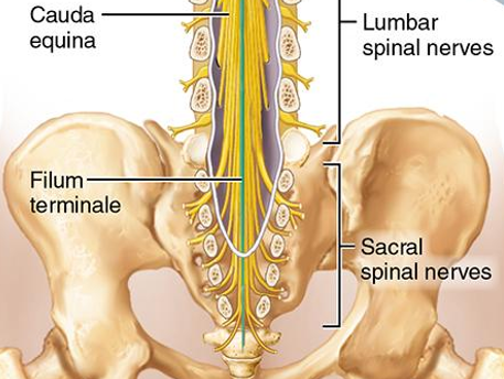
cauda equina
lower spinal nerve roots that “chase” their exits
spinal segment
spinal nerve
Describe the structure, location and extent of the spinal meninges.
composed of dura mater, arachnoid mater, and pia mater
foramen magnum to the coccyx, separating the spinal cord from the vertebral canal
List in order the structures passed from the bone of the vertebra to the surface of the spinal cord.
epidural space
dura mater
subdural space
arachnoid mater
subarachnoid space
pia mater
denticulate ligament
lumbar cistern

epidural space
contains fat

subdural space
deep to dura mater

subarachnoid space
contains CSF
deep to arachnoid mater
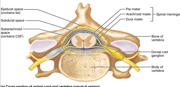
denticulate ligament
extensions connecting spinal cord to dura mater meninx
lumbar cistern
space for lumbar puncture
subdural space functional significance
potential space acts as a structural buffer
allows for minor movement between meningeal layers
houses "bridging veins" that drain blood to venous sinuses
acts as part of the lymphatic system around the spinal cord
subarachnoid space functional significance
protecting and nourishing CNS
cushions spinal cord
denticulate ligament functional significance
stabilize the spinal cord within the dura mater, suspending it within the cerebrospinal fluid
anchors spinal cord laterally to prevent excessive side-to-side motion
lumbar cistern functional significance
safe, accessible reservoir of CSF for lumbar puncture
anterior median fissure
deeper groove running length of spinal cord

posterior median sulcus
shallower groove

gray commissure
bridge connecting masses of gray matter
encloses central canal

central canal
continuous w/in chamber in brain
contains CSF
runs length of spinal cord
ventral horns
have some interneurons
mainly house cell bodies of somatic motor neurons
axons exit cord via ventral rootlets, which fuse into ventral roots of spinal cord
amt of ventral gray matter = amt of skeletal muscle innervation at that level = spinal cord enlargements

dorsal horns
consist of interneurons

lateral horns
thoracis and superior lumbar segments
mainly consist of cell bodies of autonomic motor neurons to visceral effectors (sympathetic division)
neurons exit cord through ventral root

anterior columns
ventral
composed of myelinated and nonmyelinated fibers

posterior columns
dorsal
composed of myelinated and nonmyelinated fibers
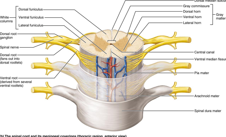
lateral columns
lateral
composed of myelinated and nonmyelinated fibers

functional neuron associated w/ anterior horn
cell bodies of somatic motor neurons
some interneurons
functional neuron associated w/ lateral horn
cell bodies of autonomic motor neurons
functional neuron associated w/ posterior horn
interneurons
general fxn of the spinal cord
provides 2-way conduction pathway to and from brain
major reflex center
list the number and name of spinal nerves
cervical spinal nerves: C1 - C8
thoracic spinal nerves: T1 - T12
lumbar spinal nerves: L1 - L5
sacral spinal nerves: S1 - S5
coccygeal nerves: Co1
describe if the nerves are mixed, sensory, or motor
describe the length of a spinal nerve
name the types of fibers that travel through the dorsal root and ventral root
dorsal root: carries sensory info
ventral root: carries motor info
name the branches of a typical spinal nerve and describe the regions or structures innervated by these branches
spinal nerve plexus
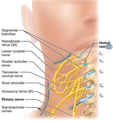
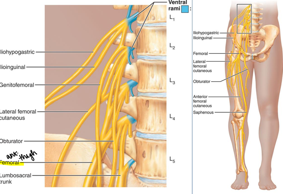
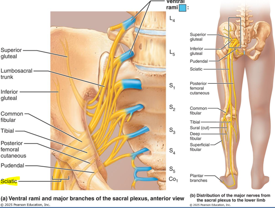
name the 4 plexuses of the body
cervical plexus
brachial plexus
lumbar plexus
sacral plexus
cervical plexus location
deep to sternocleidomastoid
brachial plexus location
located in neck and axilla

lumbar plexus location
w/in psoas major
sacral plexus location
along the posterolateral wall of pelvis
ventral rami of cervical plexus
C1 - C4 (C5)

ventral rami of brachial plexus
C5 - T1
often receiving fibers from C4 and T2
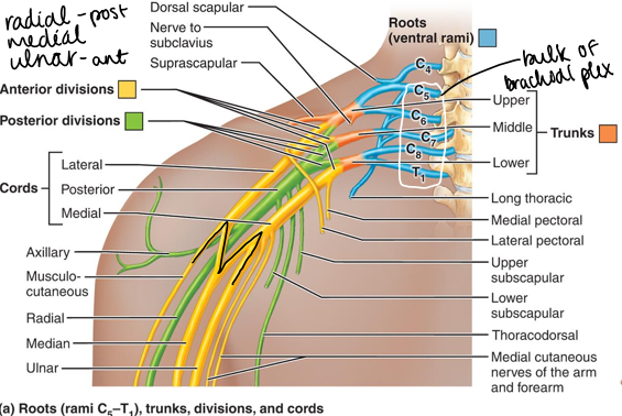
ventral rami of lumbar plexus
L1 - L4

ventral rami of sacral plexus
L4 - S4
“lumbosacral trunk” = L4 + L5 contributions

cutaneous branches of cervical plexus
skin over back of head, ear region, anterior neck, and shoulder region
cutaneous branches of brachial plexus
skin over shoulder and all parts of upper extremity
cutaneous branches of lumbar plexus
skin over lower abdomen, buttock, external genitalia, many thigh regions, medial leg and foot
cutaneous branches of sacral plexus
skin over gluteal region, external genitalia, and lower extremity
innervations of intercostal nerves
cervical plexus motor branch
muscles on back of neck: trapezius, sternocleidomastoid
diaphragm: phrenic nerve - fibers from C3 - C5
brachial plexus motor branch
muscles of shoulder and upper extremity
lumbar plexus motor branch
muscles of lower abdomen, medial and anterior thigh regions
sacral plexus motor branch
muscles of lower extremity
what is a dermatome and its clinical significance
area of skin supplied by cutaneous branches of a single spinal nerve
damage to the spinal cord can be determine by which dermatomes are affected (skin poke)
reflex
rapid, automatic response to stimuli
reflex arc
conduction pathway involving two or more neurons
reflex center
identify and describe the components of a simple reflex arc
receptor
sensory neuron
integration center
motor neuron
effector
Rod Stewart Is My Everything
give an example of how a reflex helps to maintain homeostasis
visceral reflexes help maintain heart rate, respiration, digestion, and urination
functional classification of reflexes
somatic
visceral
intrinsic reflex
born with
unlearned, unpremeditated, involuntary
ex: help maintain posture, avoid pain and injury, control visceral activities
acquired reflex
results from practice and repetition
ex: driving skills, playing a musical instrument
somatic reflex
activate skeletal muscles
maintain homeostasis w/ skeletal muscle contractions
visceral reflex
activate visceral effectors
cardiac and smooth muscles
glandular secretion
help maintain homeostasis associated w/ heart rate, respiration, digestion, and urination
types of spinal reflexes
somatic reflexes mediated by the spinal cord
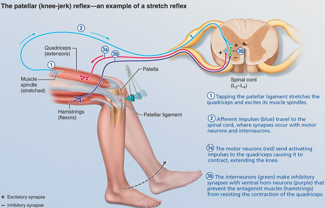
stretch reflex
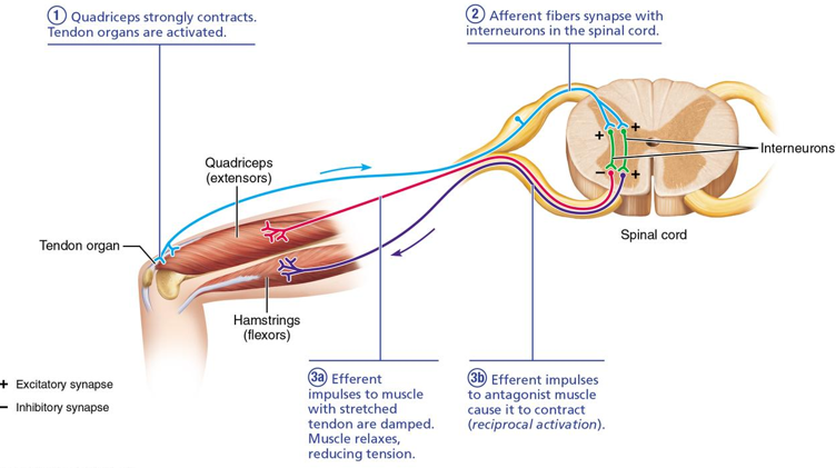
tendon reflex
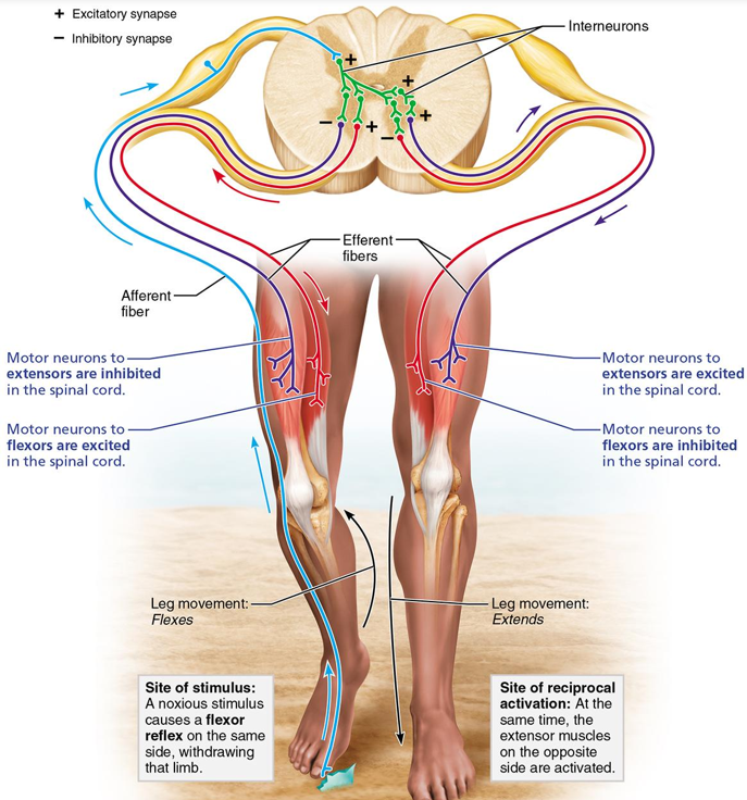
flexor (withdrawal) reflex
crossed-extensor reflex
stretch reflex
muscle tightens in response (patellar reflex)
antagonist relaxes
tendon reflex
muscle relaxes in response
flexor (withdrawal) reflex
finger pricked w/ needle
crossed-extensor reflex
ipsilateral withdrawal reflex and contralateral extensor reflex
describe the ANS
system of motor neurons innervating smooth and cardiac muscle, and glands
name some visceral functions controlled by the ANS
speeds or slows heart rate
shunts blood to areas in need
adjust blood pressure and body temperature
increases or decreases stomach secretions
give examples of visceral effectors
smooth and cardiac muscle, glands
2 principle divisions of the ANS
parasympathetic
sympathetic
parasympathetic division
rest and digest
promotes maintenance functions and conserves body energy
sympathetic division
fight/flight system
mobilizes body during activity or threatening situations
describe the general effects of each division
parasympathetic effects
energy use by body is low
directs activities like digestion, pooping, and peeing
ex: after eating
blood pressure drops
heart rate drops
eyes accommodate for close reading
GI tract is actively digesting food
sympathetic effects
exercise, embarrassment, excitement, emergency
increase in heart rate, respirations
dry mouth, cold and sweaty skin, dilated pupils
visceral blood vessels constrict
bronchioles in lungs dilate
liver releases more glucose into blood (more energy)
nonessential actions (digestion) are slowed
describe the “two-neuron hook-up” of the ANS
preganglionic neuron - autonomic ganglia - postganglionic neuron
uses chain to reach effector
describe the origin and termination point of a preganglionic and postganglionic neuron in the parasympathetic and sympathetic nervous system
give the general name of the “autonomic ganglion” of the parasympathetic nerveous system and describe its location
terminal ganglia
near or w/in walls of visceral effector organ
give the general names of the “autonomic ganglia” of the sympathetic nerveous system and describe their locations
sympathetic chain ganglia: paravertebral ganglia
collateral ganglia: close to large abdominal arteries
lie close to the spinal cord
describe the general trends with respect to the location of ganglia and the length of the postganglionic fiber in the parasympathetic and sympathetic systems
describe the specific organization of the sympathetic division of the ANS
identify the origin of the preganglionic sympathetic fibers and describe the path taken by these fibers to enter the sympathetic trunk
describe the 3 possible routes taken by the preganglionic fibers once in the sympathetic trunk
describe the three possible routes taken by the preganglionic ribers once in the sympathetic trunk
describe how the sympathetic fiber leaves the sympathetic trunk to rejoin the spinal nerve
describe the origin and termination point of the postganglionic sympathetic fiber
describe the sympathetic trunk
explain the relationship b/t chromaffin cells in the adrenal medulla and the sympathetic division
white ramus communicans
splanchnic nerve
gray ramus communicans