A-Level AQA Psychology - Biopsychology
1/199
Earn XP
Description and Tags
Name | Mastery | Learn | Test | Matching | Spaced | Call with Kai | Chat |
|---|
No analytics yet
Send a link to your students to track their progress
200 Terms
What is the nervous system?
The nervous system is a specialised network of cells in the human body and our primary communication system via electrical signals.
What are the 2 main functions of the nervous system?
1.Collect, process, and respond to information.
2.Coordinate the working of different organs and cells in the body.
What is the nervous system made up of?
It is made up of the central nervous system (CNS) and the peripheral nervous system (PNS).
What 2 things make up the CNS?
Brain.
Spinal cord.
What makes up the PNS?
Millions of neurons (nerve cells) to and from the CNS.
What is the brain?
Centre of all conscious awareness.
Cerebral cortex (outer brain layer) is highly developed in humans.
Divided into 2 hemispheres by the corpus callosum.
What is the spinal cord?
Extension of the brain.
Responsible for reflex actions [e.g. pulling away from a hot plate]: without conscious awareness.
The spinal cord passes messages to and from the brain and connects nerves to the PNS.
What does the PNS do?
Sends information via millions of neurons to the CNS from the outside world and transmits messages from the CNS to muscles and glands.
What 2 systems is the PNS subdivided into?
Somatic Nervous System (SNS)
Autonomic Nervous System (ANS)
What does the SNS do?
Controls voluntary muscle movement and receives information from sensory receptors.
What does the ANS do?
Governs vital involuntary (without conscious awareness) functions such as breathing, heart rate, digestion, stress responses.
What are the 2 divisions of the ANS?
Sympathetic Nervous System.
Parasympathetic Nervous System.
What does the Sympathetic Nervous System do?
Gets the body ready for fight or flight.
What does the Parasympathetic Nervous System do?
Calms the body down: rest and digest.
What are 6 features of the sympathetic state?
Increases heart rate.
Increases breathing rate.
Dilates pupils.
Inhibits digestion.
Inhibits saliva production.
Contracts rectum.
What are 6 features of the parasympathetic state?
Decreases heart rate.
Decreases breathing rate.
Constricts pupils.
Stimulates digestion.
Stimulates saliva production.
Relaxes rectum.
What is a neuron?
The basic building blocks of the nervous system, neurons are nerve cells that process and transmit messages through electrical and chemical signals.
What do neurons enable?
Communication within the nervous system.
What do neurons contain?
Cell body (soma): this contains the nucleus.
Dendrites.
An axon.
How do dendrites communicate?
They receive information from other neurons and pass these to the cell body (the control centre).
How do axons communicate?
It then carries this message (as an electrical signal known as action potential) to the axon terminals (the end of the axon).
What do the terminal buttons then do?
They then make synaptic connections via neurotransmitters and communicate with the next neuron in the chain.
What is the axon covered by?
The myelin sheath.
What does the myelin sheath do?
Speeds up electrical transmission.
What is the myelin sheath segmented by?
Nodes of Ranvier.
What do the nodes of Ranvier do?
Forces signals to ‘jump’ the gaps.
When does the inside of a neuron become positively charged?
When the inside of a neuron is activated by a stimulus.
What occurs in a positively charged neuron?
Action potential.
What are the 3 types of neuron?
Sensory.
Relay.
Motor.
What is the structure of a sensory neuron?
Long dendrites and short axons.
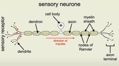
What is the function of a sensory neuron?
These carry messages from the CNS to the PNS.
Where are sensory neurons located?
Located outside the CNS in the PNS in clusters known as ganglia.
What is the structure of a relay neuron?
Short dendrites and short axons
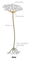
What is the function of a relay neuron?
These connect the sensory neurons to the motor or other relay neurons. They make up 97% of all neurons.
Where are relay neurons located?
Most are found within the brain and the visual system.
What is the structure of motor neurons?
Short dendrites and long axons
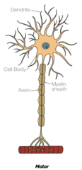
What is the function of motor neurons?
These connect the CNS to effectors such as muscles and glands.
Where are motor neurons located?
The cell bodies are in the CNS, but the axons are in the PNS.
What is synaptic transmission?
Signals within neurons are transmitted electrically.
Signals between neurons are transmitted chemically across the synapse.
Once message reaches the postsynaptic sites, the message is transformed into an electrical signal again.
What is synaptic transmission: chemical transmission?
The process by which neighbouring neurons communicate with each other by sending chemical messages across the gap that separates them. Within a neuron, the signal is transmitted electrically. When the electrical impulse reaches the end of the neuron (the presynaptic terminal) it triggers the release of neurotransmitter from the synaptic vesicles.
What are neurotransmitters?
Brain chemicals released from the synaptic vesicles that relay signals across the synapse from one neuron to another. Neurotransmitters can be broadly divided into those that perform an excitatory function and those the perform an inhibitory function.
Synaptic transmission: neurotransmitters. How do they work?
1. Neurotransmitter crosses the gap from the presynaptic neuron terminal to the postsynaptic neuron receptor site.
2. It is taken up by a postsynaptic receptor site on the dendrites of the next neuron.
3. The chemical message is converted back into an electrical impulse and the transmission process begins again.
What is excitation in synaptic transmission?
When a neurotransmitter increases the positive charge of the postsynaptic neuron. This increases the likelihood that the postsynaptic neuron will pass on the electrical impulse. An example of this type of neuron would be adrenaline.
What is inhibition in synaptic transmission?
When a neurotransmitter increases the negative charge of the postsynaptic neuron. This decreases the likelihood that the postsynaptic neuron will pass on the electrical impulse. An example of this type of neuron would be serotonin.
What is summation in synaptic transmission?
Whether a postsynaptic neuron fires or not. The excitatory and inhibitory influences are summed and the net effect decides whether the neuron fires or not (what charge it has: positive or negative). The action potential of the postsynaptic neuron is only triggered if the net effect reaches the threshold.
What is the endocrine system?
One of the body's major information systems that instructs glands to release hormones directly into the bloodstream. These hormones are carried towards the target organs in the body. Communicates via chemicals.
What is the function of the endocrine system?
The endocrine system works alongside the nervous system to control vital functions in the body. It instructs glands to release hormones into the bloodstream that are carried towards target organs. The endocrine system acts more slowly but with powerful effects as it provides a chemical system of communication.
What is a gland?
An organ in the body that synthesises substances such as hormones.
What is a hormone?
A biochemical substance that circulates in the blood but only affects target organs. They are produced in large quantities but disappear quickly. Their effects are very powerful.
What are 8 examples of glands and hormones produced?
Hypothalamus → corticotropin-releasing hormone (CRH)
PITUITARY gland → 'master gland': controls the release of hormones from all other endocrine glands
THYROID gland → thyroxine
Parathyroid gland → parathyroid hormone (PTH)
ADRENAL gland → adrenaline
Pancreas → insulin and glucagon
Ovaries → oestrogen
Testes → testosterone
How do the endocrine and autonomic nervous systems work together during a stressful event? (fight or flight response)
1. A stressful event is perceived.
2. The hypothalamus activates the pituitary gland.
3. The pituitary gland triggers the sympathetic nervous system because adrenaline is released from the adrenal medulla (part of the adrenal gland).
4. The ANS changes from the parasympathetic to the sympathetic state.
Evaluate the fight or flight response: Limitation - Gray (1988)
P: One limitation to the fight-or-flight explanation is that human behaviour is not limited to only two responses.
EE: Gray (1988) suggests that the first response to danger is to avoid it altogether, which is demonstrated by a ‘freeze’ response. During the freeze response, humans are hyper-vigilant while they appraise the situation to decide the best course of action.
L: This suggests that the fight-or-flight explanation of behaviour is limited and doesn’t fully explain the complex cognitive and biological factors that underpin the human responses to stress/danger.
Evaluate the fight or flight response: Limitation - Taylor et al (2002)
P: One issue with the fight or flight response is that it doesn’t fully explain the stress response in females.
E: Taylor et al. (2002) suggest that females adopt a ‘tend and befriend’ response in stressful/dangerous situations: Women are more likely to protect their offspring (tending) and form alliances with other women (befriending), rather than fight an adversary or flee. This highlights a beta bias within this area of psychology: psychologists assumed that females responded in the same way as males until Taylor suggested otherwise.
L: Therefore, while the original fight-or-flight explanation may have been limited in its application to females, it has prompted more recent research which has provided an alternative explanation more relevant to females, which is actually a strength.
What is hemispheric lateralisation?
•Lateralisation is the idea that the two halves (hemispheres) of the brain are functionally different/specialised.
•If a function is dealt with by one hemisphere it is lateralised.
•Therefore, the division of functions between the two hemispheres is called hemispheric lateralisation.
How is language lateralised?
Left hemisphere: verbal (the analyser)
Language processing (for most people):
Broca’s area: Left frontal lobe
Wernicke’s area: left temporal lobe
Language is lateralised as it is performed by one hemisphere.
Right hemisphere: non-verbal (the synthesiser)
Can only produce rudimentary words and phrases but contributes to the emotional context of what is being said and visual-motor tasks..
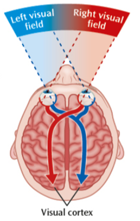
Why are vision, motor, sensory not lateralised?
•Vision, motor and somatosensory appear in both hemispheres.
•Motor: Cross-wiring/contralateral wiring.
•Vision: both contralateral and ipsilateral (opposite and same-sided).
- Each eye receives light from the left visual field (LVF) and RVF.
- The LVF of both eyes is connected to the right hemisphere (RH).
- The RVF of both eyes is connected to the LH.
What is the right hemisphere like with recognising?
•Emotions in others (Narumoto et al., 2001).
•Participants shown photos of a face that is split (e.g. half smiling/half neutral).
•Found that the emotion displayed on the left hand side of the picture is the emotion recognised by the participant (Heller & Levy, 1981).
What is the right hemisphere like with spacial relationships?
•Clarke, Assal & de Tribolet (1993).
•Case study of a woman with damage to the right hemisphere.
•She often got lost, even in familiar situations, unless she had verbal instructions that contained a distinguishable visual feature (“turn right at the red house”).
What is the most contrasting thing about left and right hemisphere?
The left focuses on detail whereas the right processes overall patterns.
What is split-brain research?
A series of studies which began in the 1960s (and still ongoing) involving people with epilepsy who had experienced a surgical separation of the hemispheres of their brains to reduce severity of their epilepsy.
What were the aims of Sperry’s (1968) research?
To investigate how the hemispheres function when they can’t communicate with each other to determine if each hemisphere is specialised.
What was Sperry’s (1968) procedure?
Sperry and Gazzaniga used 11 patients whose corpus callosum had been severed, meaning that their hemispheres did not communicate. Patients were presented with images to either the left or the right visual field. A range of tasks was used, and patients had to respond with either their left hand (right hemisphere) or their right hand (left hemisphere), or verbally. The word is flashed on the screen for no more than 1/10 second or else the participants eyes will move.
What were Sperry’s (1968) findings?
It was found that the patient could say what they saw in their right visual field but not in their left. This is because the left hemisphere processes information in the right visual field: Because this hemisphere is responsible for language, the patient can say what they saw. However, they could not say what they saw in the left visual field because that information is processed in the right hemisphere which cannot process language. The patient could, however, draw this image with their left hand as this task did not require language. Sperry argued that his studies give considerable support to his argument for lateralisation of function: the left hemisphere specialises in language while the right hemisphere specialises in tasks involving spatial analysis.
What were Sperry’s (1968) conclusions?
These observations show how certain functions are lateralised in the brain and support the view that the left hemisphere is verbal and the right hemisphere is ‘silent’ but emotional.
What is a limitation of research investigating hemispheric lateralisation? scope for generalisation
P: The primary limitation of the research investigating hemispheric lateralisation is its limited scope for generalisation.
EE: Split-brain procedures are rarely carried out in modern healthcare which means that split-brain patients are very difficult to recruit. This leads to studies with very limited samples: in some cases, only one person and research that often takes an idiographic approach.
Sperry's split-brain patients were compared to a neurotypical control group but none of the participants in the control group had epilepsy. Therefore, epilepsy may act as a confounding variable – differences in hemispheres may be the result of their condition/surgery rather than split-brain. The extent of disconnection between hemispheres also varied between patient.
L: This suggests any conclusions drawn are only representative of those individuals and cannot be generalised to the wider population.
( /CA) P: However, there is research showing that even in “connected” (neurotypical) brains, the 2 hemispheres process information differently.
•EE: Fink et al (1996) used PET scans to identify which brain areas were active during a visual processing task. In a task where non-split brain participants were asked to attend to global elements of an image (e.g. looking at a picture of a whole forest) it was found that regions of the right hemisphere were more active. When required to focus on finer details (e.g individual trees in the picture), the left hemisphere dominated.
•L: This suggests that hemispheric lateralisation is a feature of both connected and split-brain individuals (supporting Sperry’s conclusions that the hemispheres have distinct roles).
What is another limitation of Sperry’s research? oversimplified
P: One limitation is that the differences in function between the two hemispheres are oversimplified and exaggerated (especially now in pop culture: artist vs mathematician brain)
EE: The difference is a lot less clear-cut as many behaviours associated with one hemisphere can be effectively performed by the other when the situation requires it. For example, language may not be restricted to the left hemisphere. Turk et al. (2002) discovered a patient that suffered damage to the left hemisphere but developed the capacity to speak in the right hemisphere.
L: The hemispheres’ flexibility suggests that lateralisation is not fixed and is more complex than Sperry’s research advocates.
What is a strength of lateralisation? provides an adaptive function
P: One strength of lateralisation is that it provides an adaptive function - it would not have evolved and been maintained if it didn’t have some value.
EE: It is assumed that it increases neural processing capacity - allowing animals to be able to perform two tasks simultaneously with greater efficiency (multitask). Research by Lesley Rogers et al. (2004) showed that chickens with a lateralised brain could find food while watching for predators whereas chickens reared in the dark, whose brain was not lateralised, could not do this.
L: This provides evidence for the advantages of brain lateralisation and demonstrates how it can enhance brain efficiency in cognitive tasks.
C/A: However, this research was carried out on animals, so it is difficult to generalize the findings to humans.
What is a strength of split-brain specifically? scientific
P: Experiments on split-brain patients were scientific.
EE: All visual stimuli was presented for only 0.1 seconds. This was another control as it is too quick for eye movements to cause visual information to enter both visual fields. Luck et all (1989): Gazzaniga showed that split-brain patients perform better than controls on certain tasks – they were faster at identifying "the odd one out" in an array of similar objects
In ‘normal’ brains the LH's better cognitive strategies are "watered down" by the inferior RH
L: The scientific nature of ‘split-brain’ research has further enabled discoveries of lateralisation of function
What is localisation of function?
The theory that different areas of the brain are responsible for specific behaviours, processes or activities.
What did Broca and Wernicke propose?
Broca and Wernicke proposed that different parts of the brain perform different tasks and control different parts of the body (CORTICAL SPECIALISATION)
If a certain area of the brain becomes damaged through illness or injury, the function associated with that area will be affected
What is the curious case of Phineas Gage?
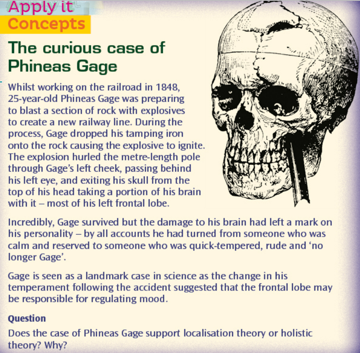
How does Phineas Gage support localisation of function?
The specific changes observed in his behaviour support theories about the localisation of brain function, or the idea that certain functions are associated with specific areas of the brain. The part of the brain damaged in the accident was the area in the frontal cortex associated with planning, reasoning and control – Gage’s personality changed from him being mild-mannered to rude and hostile. Today, scientists better understand the role that the frontal cortex has to play in important higher order functions such as reasoning, language and social cognition. In those years, while neurology was in its infancy, Gage's extraordinary story served as one of the first sources of evidence that the frontal lobe was involved in personality.
How many brain hemispheres are there?
2
What are the 2 hemispheres joined by?
A bundle of fibres called the corpus collosum.
What happens because brain function is generally lateralised?
Brain function is generally lateralised. Left side of body is controlled by the right hemisphere and vice versa.
What is the outer layer of both hemispheres called?
The cerebral cortex.
What does the cerebral cortex do?
Provides our higher functioning.
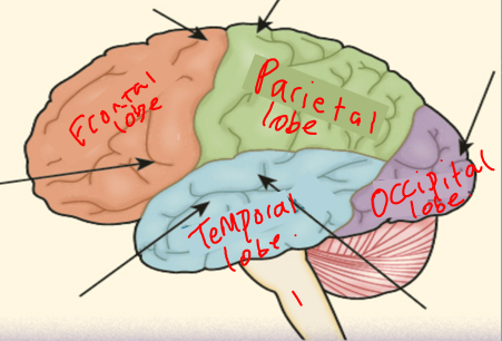
What are the 4 different lobes of our brain?
Frontal lobe
Parietal lobe
Occipital lobe
Temporal lobe
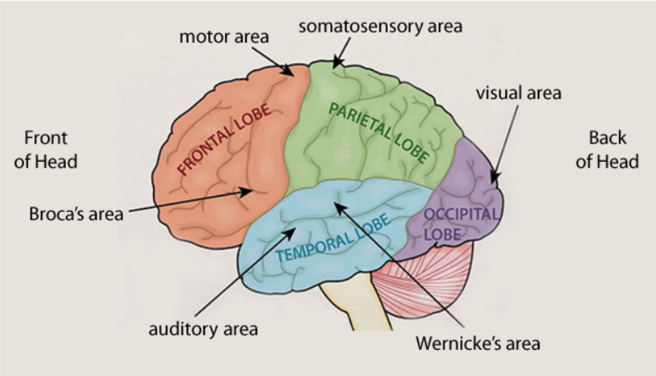
What cortex is associated with the frontal lobe?
The motor cortex
Where is the motor cortex located?
Back of frontal lobes.
In both hemispheres (L-R R-L)
What is the function of the motor cortex?
Controls voluntary movement on the opposite side of the body (left hemisphere - right side). Damage to this area might result in loss of control over fine movements.
What cortex is associated with the parietal lobes?
The somatosensory cortex.
What is the somatosensory cortex located?
Parietal lobes.
In both hemispheres (L-R R-L)
The somatosensory cortex is separated from the frontal lobe/ motor cortex by a ‘valley’ called central sulcus.
What is the function of the somatosensory cortex?
Where sensory information from skin produces sensations like touch, pressure, pain, temp
Amount of somatosensory area for a body part shows it’s sensitivity (need more neural connections)
E.g. the face requires greater sensitivity than the trunk and so has a larger proportion of the somatosensory cortex
What cortex is associated with the occipital lobe?
The visual cortex.
Where is the visual cortex located?
Occipital lobe (back of brain)
The brain has 2 visual cortices, 1 in each hemisphere
What is the function of the visual cortex?
Main visual centre, referred to as Area V1.
Each eye sends information from the left visual field to the right visual cortex/ the right visual field to the left visual cortex.
Nerve impulses are transferred from the retina to the visual cortex via optic nerves.
Damage to LEFT hemisphere: blindness in part of the RIGHT visual field in BOTH eyes
Different parts process different info e.g colour
What cortex is associated with the temporal lobe?
The auditory cortex.
Where is the auditory cortex located?
Temporal lobes
The brain has 2 auditory cortices, 1 in each hemisphere (left ear goes to right hemisphere and right ear goes to left hemisphere)
What is the function of the auditory cortex?
Analyses speech-based information
The process starts at the cochlea which detects sound and then transports messages to the brain stem for basic processing, then onto the auditory cortex
Damage does not necessarily result in total deafness
The more damage, the more hearing loss
Which hemisphere is language located in?
The left hemisphere.
Where is Broca’s Area located?
Left frontal lobe.
What is the function of Broca’s Area?
Responsible for speech production. Damage to the area results in:
Slow speech
Laborious speech
Speech lacking fluency (Patient “Tan” – only say Tan)
People with Broca's Aphasia have difficulty with prepositions and conjunctions (a, the, and)
Where is Wernicke’s Area located?
Left temporal lobe.
What is the function of Wernicke’s Area?
This area is important for understanding language and accessing words
Found that patients who had damage had specific language impairments e.g. inability to comprehend language; Anomia (struggle to find the word you need)
Fluent speech (when they could access the words quickly) but struggled to produce meaningful language (neologism – nonsense words)due to lack of understanding.
Where are the cortexes and lobes located?
How do modern brain scan studies, such as Peterson et al.'s research, provide stronger evidence for localisation than Phineas Gage’s case study?
Brain scan studies like Peterson et al.'s provide more reliable, controlled evidence of brain function localisation, as they allow researchers to observe brain activity in real-time and under standardized conditions.
Case studies like Phineas Gage’s offer valuable insights but are limited by their uniqueness and lack of control, making it difficult to generalize findings or establish definitive cause-and-effect relationships.
What is a strength of localisation of function? Brain scan evidence.
P: Although Phineas Gage’s case provided one of the first sources of evidence for localisation theory (showing the frontal lobe was involved in personality), neurology at that time was in its infancy. There is now more recent and robust evidence from brain scans that supports the idea that many everyday brain functions are localised.
EE: In 1988, Peterson et al used brain scans to demonstrate how Wernicke’s area, found in the left temporal lobe and associated with language comprehension, was active during a listening task and Broca’s area, found in the left frontal lobe and associated with speech production, was active during a reading task.
In addition, a review of long-term memory studies by Peterson in 1996 revealed that semantic and episodic memories reside in different parts of the prefrontal cortex.
L: This objective and scientific evidence supports localisation theory as it shows that some specific areas of the brain activate and control certain functions.
What is a limitation of localisation of function? contradictory research
P: Lashley’s work challenges the idea that all cognitive functions are strictly localised.
EE: He suggests that while basic motor and sensory functions are localised, higher mental functions like learning are not. In his study, he removed between 10% and 50% of the cortex in rats learning a maze. No specific area was found to be more important than another, suggesting that learning involved the whole cortex rather than a single region.
L: This supports the view that complex cognitive functions are distributed more holistically.
In addition, Lashley’s law of equipotentiality—where other parts of the brain can take over functions if one area is damaged—further contradicts localisation theory, as it supports the idea of plasticity and shows how functions can be re-distributed rather than fixed in one area.
What is a strength of localisation of function? Neurosurgery.
P: Evidence from neurosurgery
EE: Brain damage has been linked to mental disorders, supporting localisation. Neurosurgery is used as a last resort to treat some disorders by targeting specific brain areas.
For example, cingulotomy targets the cingulate gyrus, which has been implicated in OCD. Dougherty et al. (2002) studied 44 people with OCD who had undergone a cingulotomy. After 32 weeks, 30% showed a successful response to surgery, and 14% showed a partial response.
L: This suggests that behaviours linked to mental disorders may be localised.