rp2 -> Preparation of stained squashes of cells from plant root tips
1/10
There's no tags or description
Looks like no tags are added yet.
Name | Mastery | Learn | Test | Matching | Spaced | Call with Kai |
|---|
No analytics yet
Send a link to your students to track their progress
11 Terms
how to prepare squashes of cells from plant root tips
Cut a thin slice of root tip (5mm from end) using scalpel and mount onto a slide
Soak root tip in hydrochloric acid then rinse
Stain for DNA eg. toluidine blue
Lower coverslip using a mounted needle at 45 degrees without trapping air bubbles
Squash by firmly pressing down on glass slip but do not push sideways
Why not push cover slip sideways?
Avoid rolling cells together / breaking chromosomes
Why squash / press down on cover
slip?
● (Spreads out cells) to create a single layer of cells
● So light passes through to make chromosomes visible
Why soak roots in acid?
● Separate cells / cell walls
● To allow stain to diffuse into cells
● To allow cells to be more easily squashed
● To stop mitosis
Why is a stain used?
To distinguish chromosomes
Why are root tips used?
where mitosis occurs
Describe how to set-up and use an optical microscope
1. Clip slide onto stage and turn on light
2. Select lowest power objective lens (usually x 4)
3a. Use coarse focusing dial to move stage close to lens
3b. Turn coarse focusing dial to move stage away from lens until image comes into focus
4. Adjust fine focusing dial to get clear image
Swap to higher power objective lens, then refocus

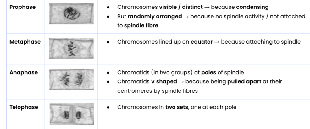
Explain how the stages of mitosis can be identified
In interphase (not mitosis), chromosomes aren’t visible but nuclei are. In mitosis, chromosomes are visible.
mitotic index?
● Proportion of cells undergoing mitosis (with visible chromosomes)
● Mitotic index = number of cells undergoing mitosis / total number of cells in sample
how to determine a reliable MI from observed squashes
● Count cells in mitosis in field of view
● Count only whole cells / only cells on top and right edges → standardise counting
● Divide this by total number of cells in field of view
● Repeat with many / at least 5 fields of view selected randomly → representative sample
● Calculate a reliable mean
how to calculate the time cells are in a certain phase of mitosis
Identify proportion of cells in named phase at any one time
● Number of cells in that phase / total number of cells observed
Multiply by length of cell cycle