Microscopic Evaluation of urine
1/71
There's no tags or description
Looks like no tags are added yet.
Name | Mastery | Learn | Test | Matching | Spaced | Call with Kai |
|---|
No analytics yet
Send a link to your students to track their progress
72 Terms
What does the microscopic tell us?
- evidence of renal disease as opposed to lower urinary tract infection
- indication of the type and activity of a renal lesion or disease condition.
What are microscopes that can be used for microscopic evaluation of urine?
bright field, phase contrast, and interference contrast.
What stains can be used for the microscopic evaluation of urine?
supravital stains and acetic acid
Where do casts like to go on a slide at 10x?
They tend to congregated near the edges of the coverslips
What is considered abnormal condition for RBC in urine?
>3/hpf
What does RBC cells, proteinuria, and RBC casts indicate?
glomerular or tubular bleeding
What does it indicate if there are RBC cells without proteinuria and RBC casts?
indicate bleeding below the kidney or contamination.
What is happening in hypotonic dilute urine with RBCs?
high pH, low specific gravity- cells swells up and release their hemoglobin (Ghost cells)
What do RBCs look like in a hypertonic culture?
Cells will be crenated
What causes RBCs to lyse with urine?
Cells will lyse with the addition of acetic acid
What does a large number of lymphocytes indicate?
early tissue rejection in kidney transplant patients
What is the limit of WBC that should not be exceed in urine?
should not exceed 3-4/hpf in normal urine
What is WBC's accompanied proteinuria indicate?
pyelonephritis
What is WBCs without proteinuria?
lower urinary tract infection
What are glitter cells?
It is hypotonic urine degenerated wbc's, cells expand and granules demonstrate Brownian movement.
What do renal tubular epis look like?
- round and slightly larger than leukocytes
- Each contains a single, large nucleus

What do large numbers of renal tubular epis suggest?
suggests renal disease
What do Squamous epithelium look like?
Large flat cells with single nucleus and large cytoplasm. edges can fold/curve making for unusual transformations

What do transitional epithelium look like?
Smaller than squamous cells, usually pear-shaped.
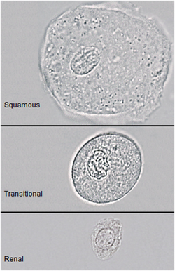
Where is transitional epithelium found?
located in renal pelvis, bladder
What can large numbers of transitional epithelium suggest?
disease of the bladder
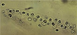
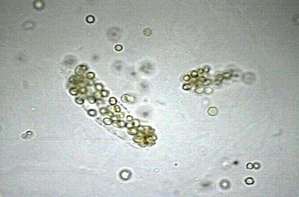
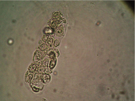
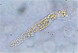
What are casts?
Proteins in urine that congeal in the lumen of the nephron
What do casts look like?
Cylindrical bodies with a uromodulin protein matrix
What is Cast usually associated with and where is it generally formed?
- in distal and collecting tubules
- associated with proteinuria due to casts protein matrix
What are the classifications of homogenous matrix casts?
- Hyaline
- Waxy
What is normal limit of Hyaline cast?
2 or fewer per LPF
What can hyaline casts caused by?
- acute glomerulonephritis
- acute pyelonephritis
- malignant hypertension
- chronic renal failure
What do hyaline casts look like?
- colorless, transparent, and low refractive index.
What does the hyaline casts serve as?
the matrix for which all casts are formed.
What do waxy casts look like under the microscope?
- Highly refractile readily visible
- well defined edges, sharp, blunt, uneven ends
- cracks or fissures from lateral margins
Prolonged stasis and tubular obstruction
What is a waxy cast associated with?
tubular inflammation and degeneration, chronic renal disease
What does a WBC cast look like on a microscope?
Often look the cells are stuck to the outside.
What do WBC accompany and indicate?
they accompany proteinuria, bacteriuria and acute pyelonephritis
What do RBC cast look like on a microscope?
usually bright orange or reddish brown.
How will a RBC cast usually occurs?
diseases to the basement membrane of the glomerulus.
What should RBC casts be regarded as?
renal disease
Where are epithelial cell cast usually seen?
Generally renal tubular epis with in the protein matrix.
What is epithelial cell cast associated with?
- Inflammatory condition of the kidney.
- Suggests intrinsic kidney disease involving renal tubes
What accompanies Epithelial cell casts?
proteinuria almost always accompanies this.
What are mixed casts?
They have at least tow defined and distinct portions.
implies that more than one part of the nephron has been injured.
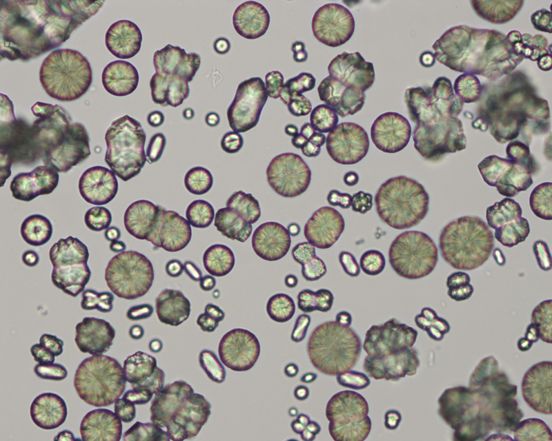
What are granular casts?
composed maidly of uromodulin protein and cast granulation
What do granular casts look like?
often colorless, shades of yellow, has all shapes and sizes. Coarse and finely granular
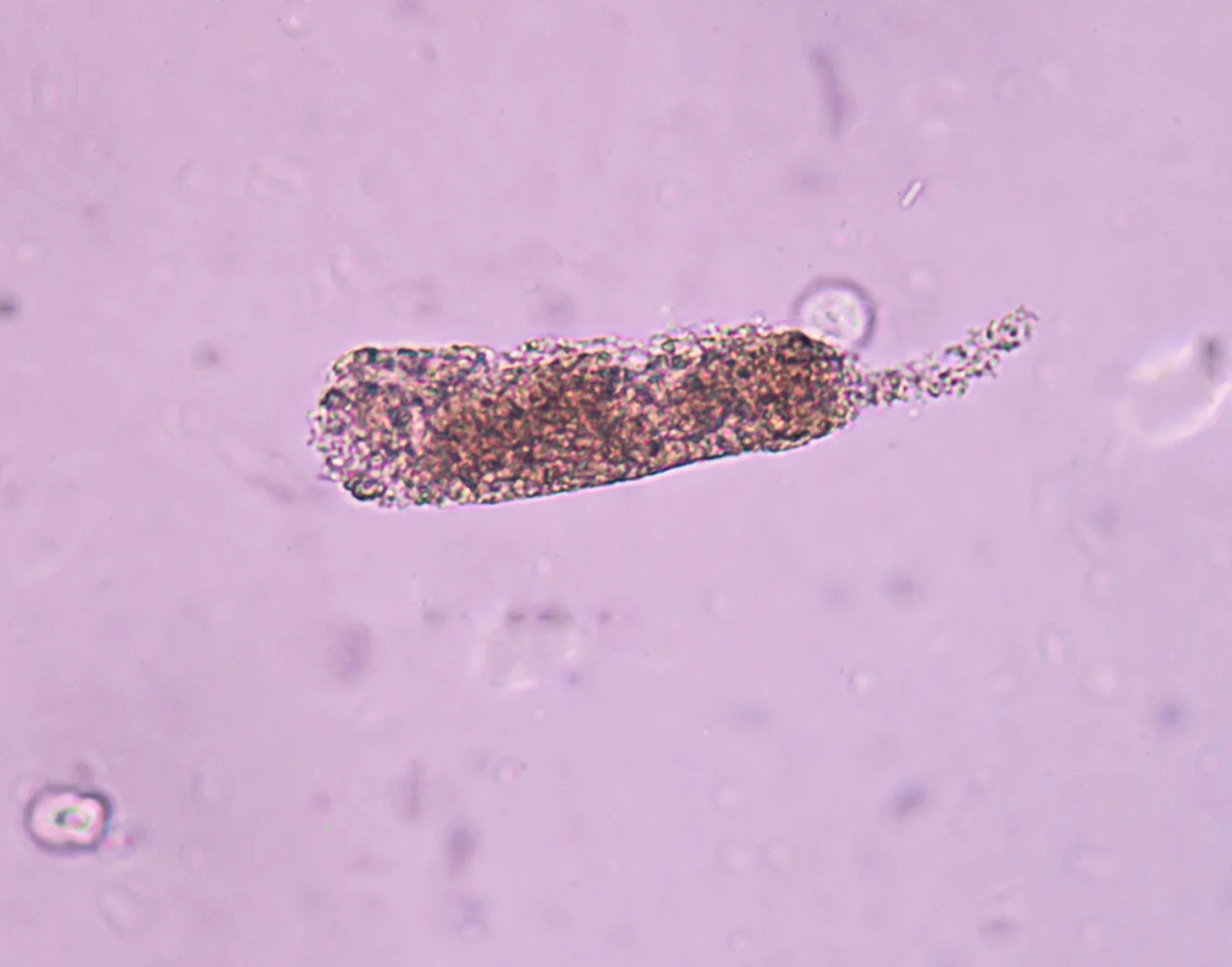
What do granular casts indicate
Pyelonephritis or lead intoxication
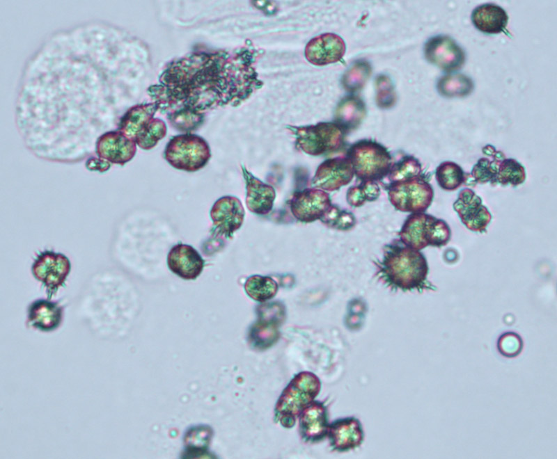
What are fatty a cast?
matrix contains free fat globules, oval fat bodies, and matrix can be hyaline or granular
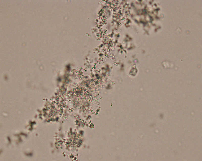
What are bacteria that can be seen in urine?
- bacilli or cocci
- gram negative rods being the most common cause of UTI
What do yeasts most often represent?
a vaginal infection with subsequent contamination of the urine collection.
Who do UTIs usually effect that are caused by yeast?
commonly immunosuppressed patients
What is trichomonas vaginalis?
A contaminant from vaginal secretions
What is enterobius vermuclaris (pinworm)?
fecal contamination
What is mucus known as?
- delicate, ribbon like strands with irregular or serrated ends
- contaminant in the female vaginal epithelium
Why are crystals found in urine?
When chemicals are present in excess of their solubility.
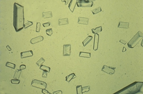
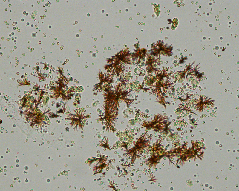
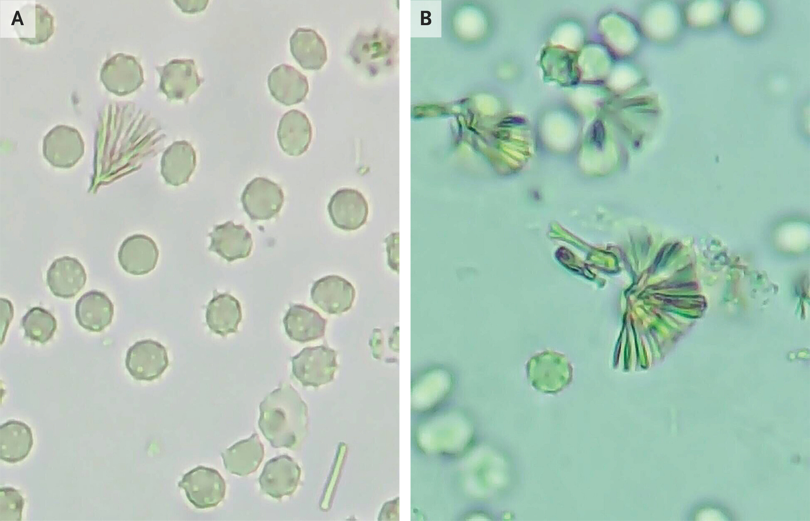
What is the most frequently observed crystal and where is it seen?
Calcium oxalate in acidic urine, but can be seen in alkaline urine.
Dihydrate (normal) and monohydrate (dumbell)
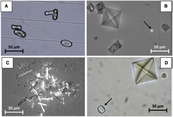
Where are calcium oxalate stones found?
large number of kidney stones or ethylene glycol poisoning.
Why are calcium oxalate stones caused?
ingesting large amounts of oxalate rich foods, tomatoes, asparagus, vitamin C.
What are amorphous urates?
Urate salts
Acidic urine
Refrigeration enhances formation
Pink
uric acid
Colorless to yellow to golden brown in color
Acidic urine
Gout and chemotherapy
Sodium urate
Light yellow slender prisms
Crystals in alkaline urine
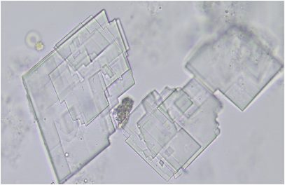
Triple phosphate, amorphous phosphates, ammonium biurate, calcium carbonate, calcium phosphate
All normal
Triple phosphate
Alkaline and neutral urine, coffin lids (3-6 sided prisms)
Enhanced by refrigeration
Urine stasis/chronic UTI (phosphates)
Amorphous phosphates
Common, nonpathologic
White precipitate in alkaline urine
Ammonium biurate
Alkaline urine, nonpathologic. Usually in old urine
Thorny apple
Calcium carbonate
Alkaline/neutral urine. Rare. Shaped like dumbells, rhombi, or needles
Produces carbon dioxide gas with acetic acid
Calcium phosphate
Neutral urine, sometimes acidic.
Colorless prisms/rosettes
Urine statis/chronic UTI (phosphates)
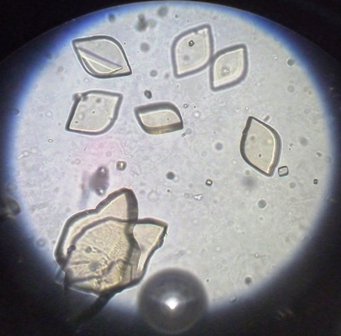
Abnormal crystals
All in acidic urine
Tyrosine and Leucine, cholesterol, cystine, bilirubin, antibiotics, x-ray dyes
Tyrosine
Abnormal (acidic urine). Thin dark needles, colorless or pale yellow-brown
When found alone, suggests rare inherited metabolic disorder
Leucine
Abnormal (acidic urine). Yellow/brown spheres with radial striations
When found alone, suggests rare inherited metabolic disorder
What is suggested when leucine and tyrosine are found together
Severe liver disease
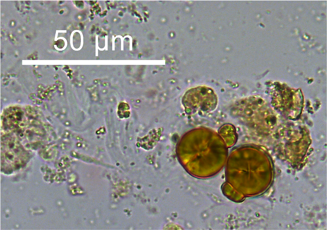
Cholesterol crystals
Abnormal (acidic urine).
Clear, flat, rectangular plates with notched corners
Accompanied by large amounts of fat and protein
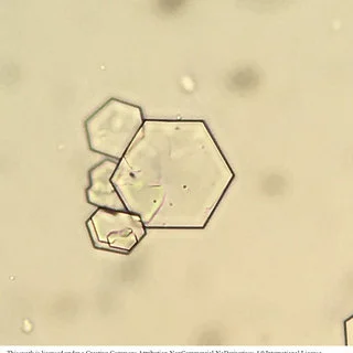
Cystine crystals
Abnormal (acidic urine).
Colorless, hexagonal plate, often layered.
Cystinuria
Bilirubin crystals
Abnormal (acidic urine).
Yellow/brown, fine needles
Positive bilirubin on dipstick, bilirubinemia
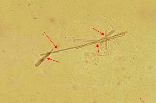
Antibiotic crystals
Abnormal (acidic urine).
Ampicillin - Long, colorless, thin prisms or needles that aggregate
Sulfonamides - Yellow/brown bundles of needles (sheaves of wheat)
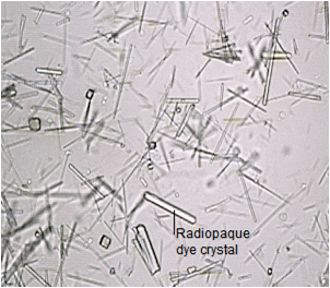
X-ray dye crystals
Abnormal (acidic urine).
Specific gravity >1.040
Long, thin pointed rectangles or flat four-sided notched plates. Similar to cholesterol, but not accompanied by proteinuria or lipiduria