L1 - Light Microscopy
1/33
There's no tags or description
Looks like no tags are added yet.
Name | Mastery | Learn | Test | Matching | Spaced | Call with Kai |
|---|
No analytics yet
Send a link to your students to track their progress
34 Terms
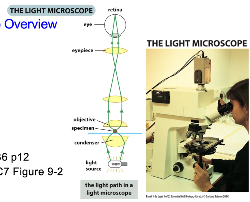
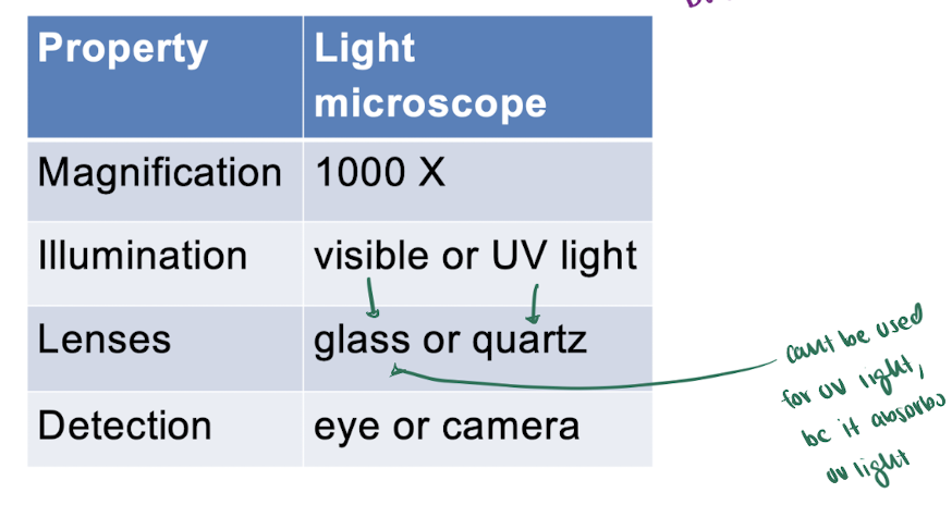
Light Microscope
Type of Light Microscopy
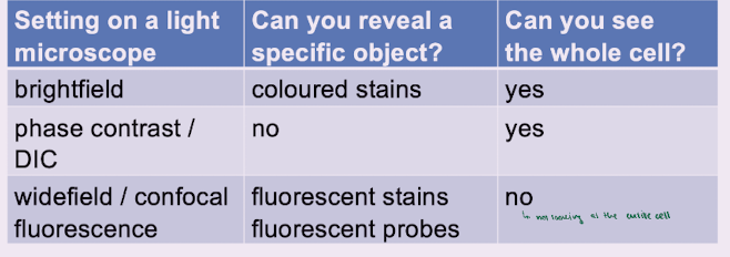
Brightfield = visible light passes thru the specimen
often need stains + living cells
Phase contrast = visible light passes thru the specimen
no stains + a halo + living cells
makes darks darker and lights lighter
DIC = visible light passes thru the specimen
can be alive + no stain + shadows
makes darks darker and lights lighter
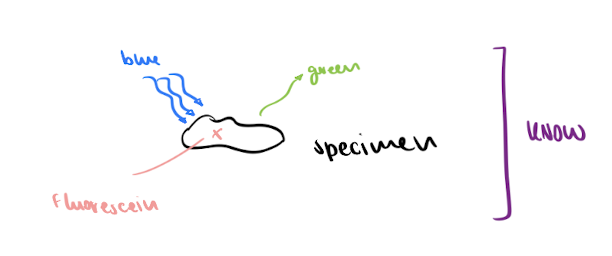
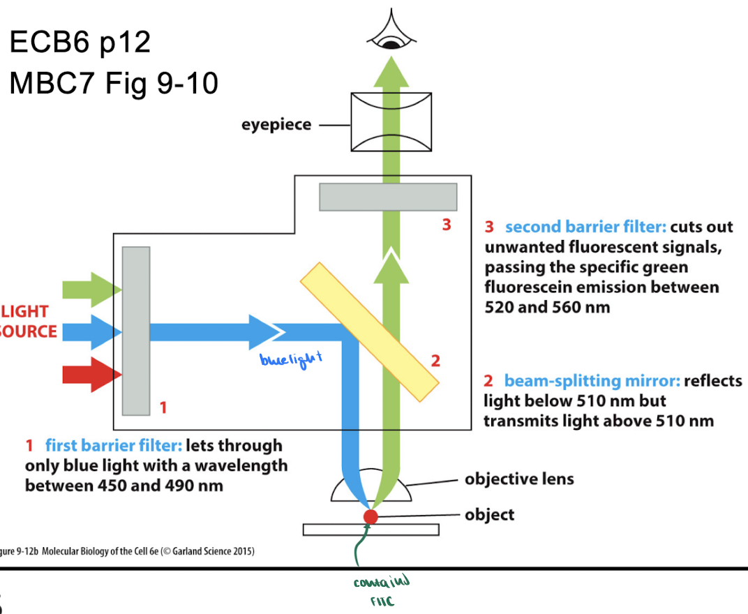
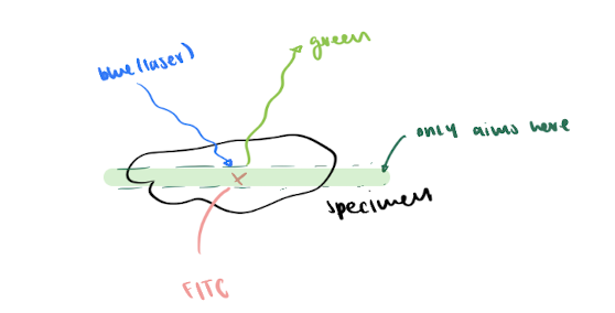
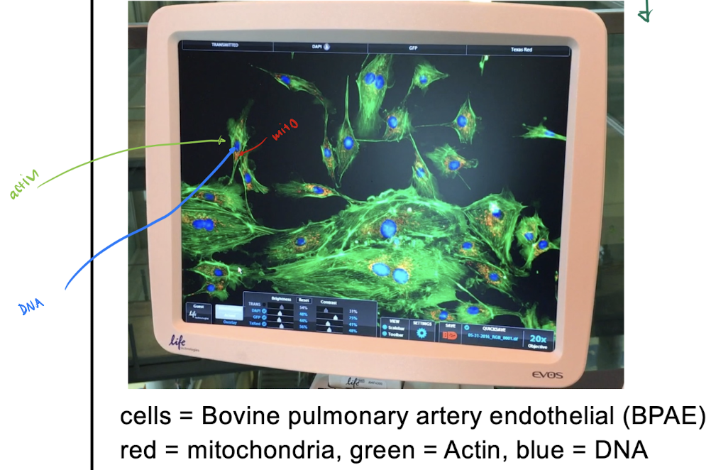
Widefield Fluorescence
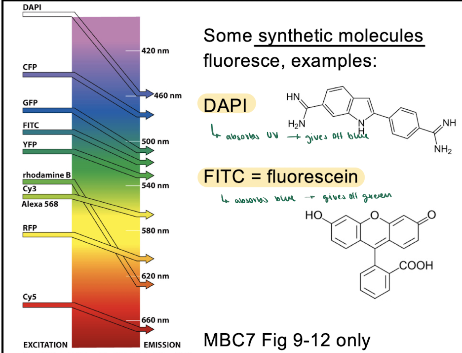
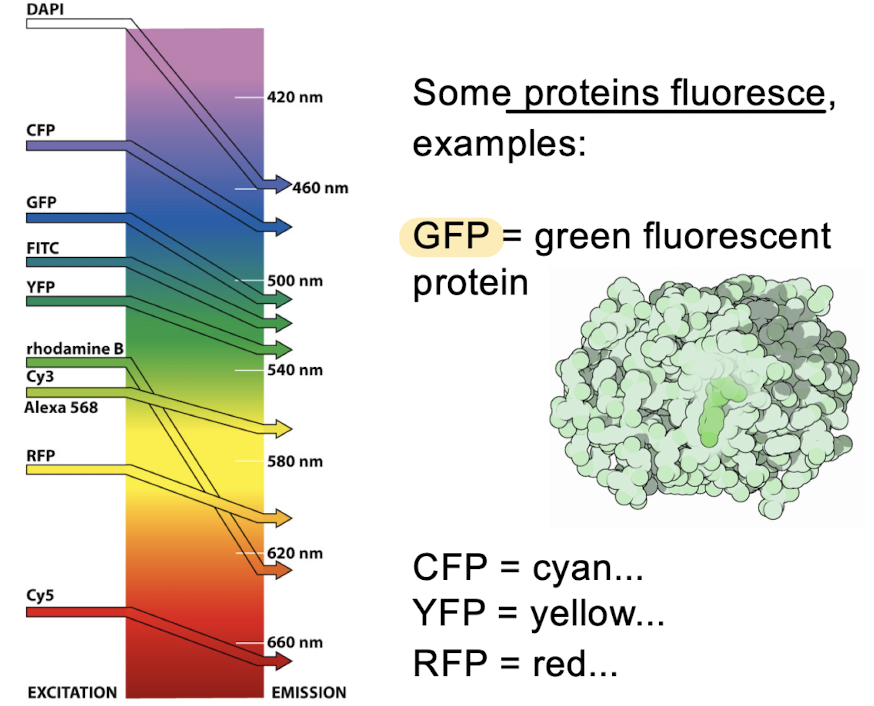
uses fluorescent molecules (DAPI, FITC, rhodamine)
short wavelength (high E) → longer wavelength (low E)
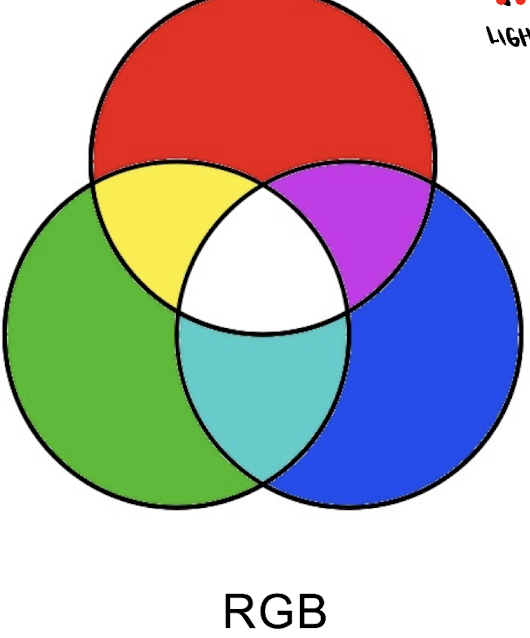
Composite Images = figures made from multiple images
red + green = yellow
purple + green = white
visible light is ADDITIVE (RGB)
= more colours → lighter
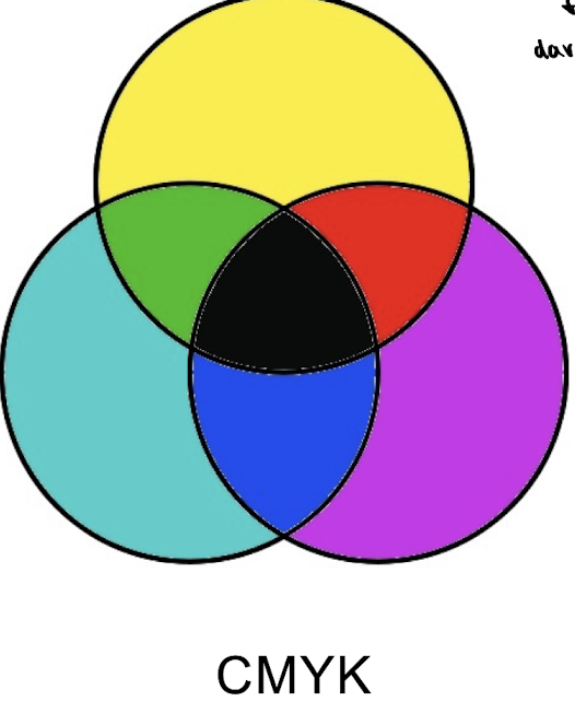
pigments are subtractive (CMYK)
= more colours → darker
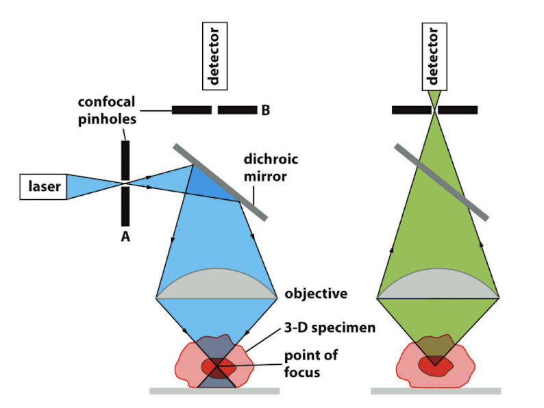
Confocal Fluorescence = shows a 2D optical plane
using a laser to focus on MIDDLE of specimen
shows more detail than wide field
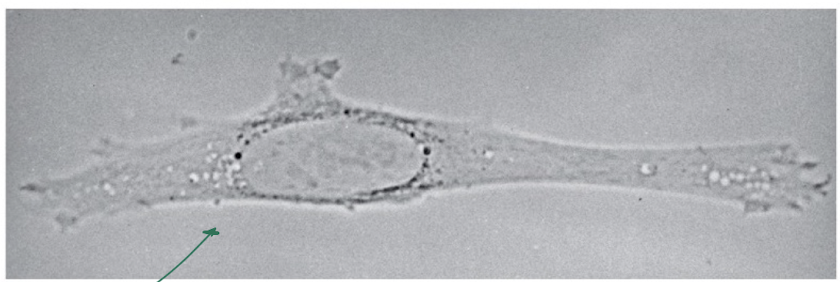
Brightfield microscopy
= visible light passes thru specimen
often need stains
ex. looking at chloroplasts or mito
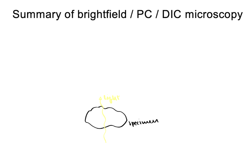
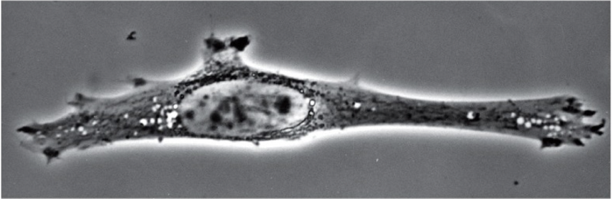
Phase Contrast microscopy
= visible light passes thru specimen
lenses make darks darker + lights lighter
NO stains + get halo
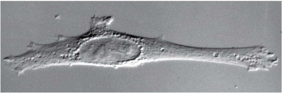
Differential Interference Contrast (DIC) microscopy
= visible light passes thru specimen
can be alive + NO stain + get shadows
lenses make darks darker + lights lighter
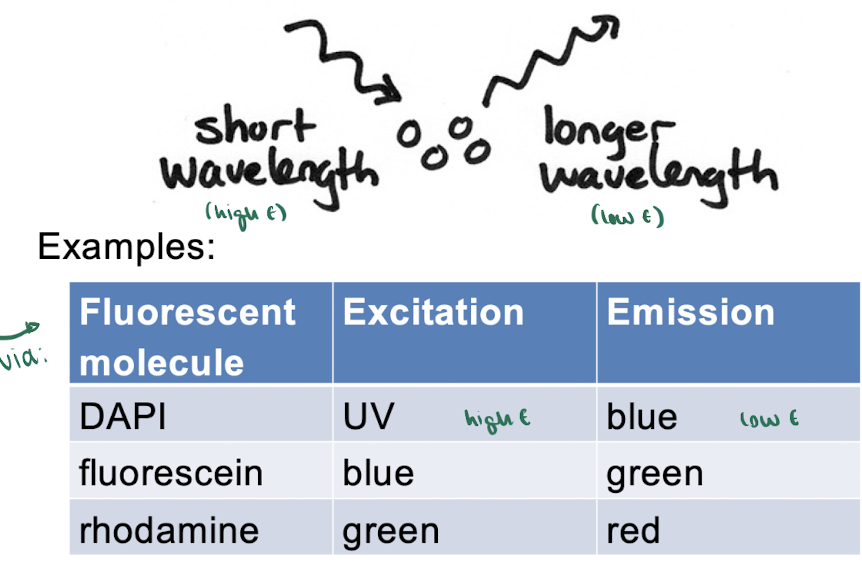
Widefield Fluorescence microscopy
uses fluorescent molecules (DAPI, FITC, rhodamine)
shine short wavelength (high E) on specimen → see longer wavelength (low E)
ex. DAPI absorbs UV → give off blue
ex. FITC absorbs blue → gives off green
(-) : fuzzy images → thus we use confocal fluorescence microscopy to fix issue
Fluorescent Synthetic Molecules
ex. DAPI absorbs UV → give off blue
ex. FITC absorbs blue → gives off green
Fluorescent Proteins
ex. GFP
CFP
YFP
RFP
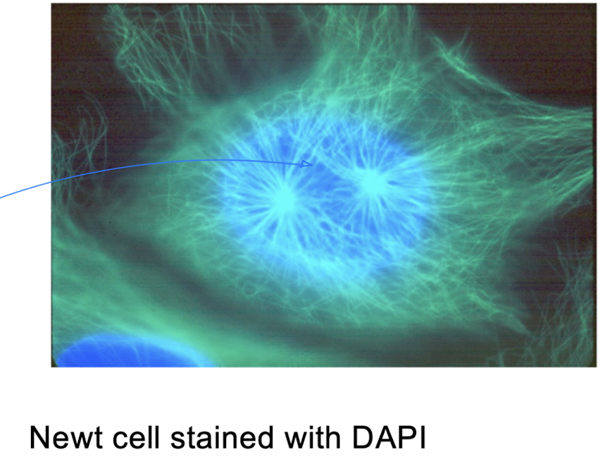
example of wide field
Newt Cell Image
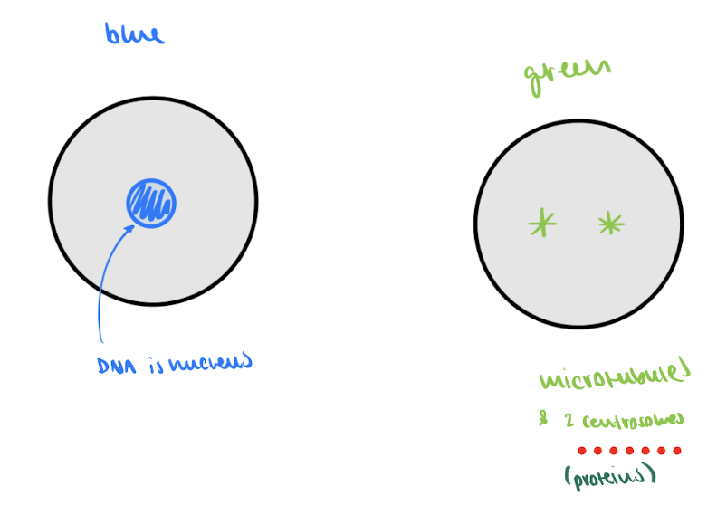
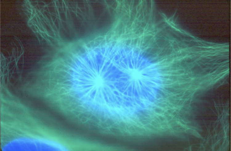
what is the blue?
DNA
what is the green?
Microtubules
Stage of Newt Cell?
G2 or prophase (interphase)
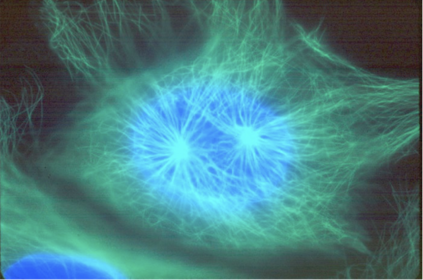
Composite Images
= figures made from multiple images
red + green = yellow
purple + green = white
ex. Newt image = 2 images superimposed
Visible light
additive = more colours → lighter
Pigments
Subtractive = more colours → darker
Red + green =
YELLOW - thus red and green (two proteins) are in the same place
Purple + green =
WHITE
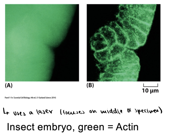
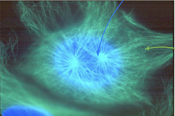
Confocal Fluorescence microscopy
= shows a 2D optical plane within a 3D specimen
uses a laser to focus on MIDDLE of specimen
shows more detail than wide field + more $$$
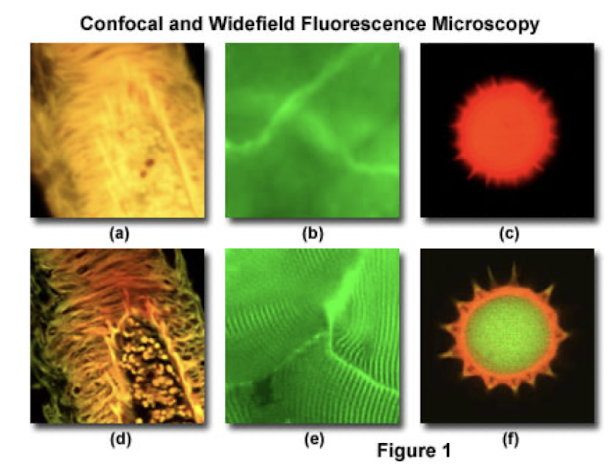
Confocal vs. Widefield microscopy images
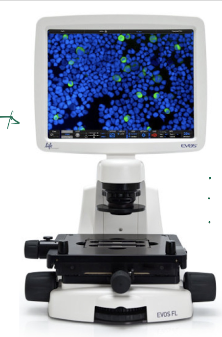
ex. EVOS FL microscope
has: brightfield, phase contrast, and widefield fluorescence
Steps to image cells
Put specimen on slide
whole cells : put 3 microliters of cell culture on slide + coverslip
thin sections of cell/tissue : immobilize in wax/plastic → section w a microtome
Make things visible
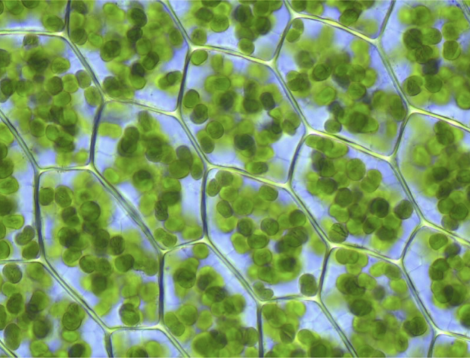
a). via natural colour (ex. chloroplast - green, mitochondria - brown from pigments in ETC)
b). via coloured stains = synthetic molecules with 2 properties: affinity for target + absorbs light
ex. Hematoxylin (nucleic acids) & Eosin (proteins)
c). fluorescent stains = synthetic molecules with 2 properties: affinity for target + absorbs light
ex. DAPI (DNA)
d). fluorescent probes = 2 molecules attached together
used when stains WONT stick
properties : affinity for target, covalent bond, fluoresces light
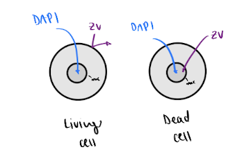
e). specialized fluorescent stains
ex. zombie violent (dead cells purple)
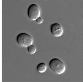
Putting whole cells on slide:
put 3 micrometers of cell culture on slide + add coverslip
ex. yeast
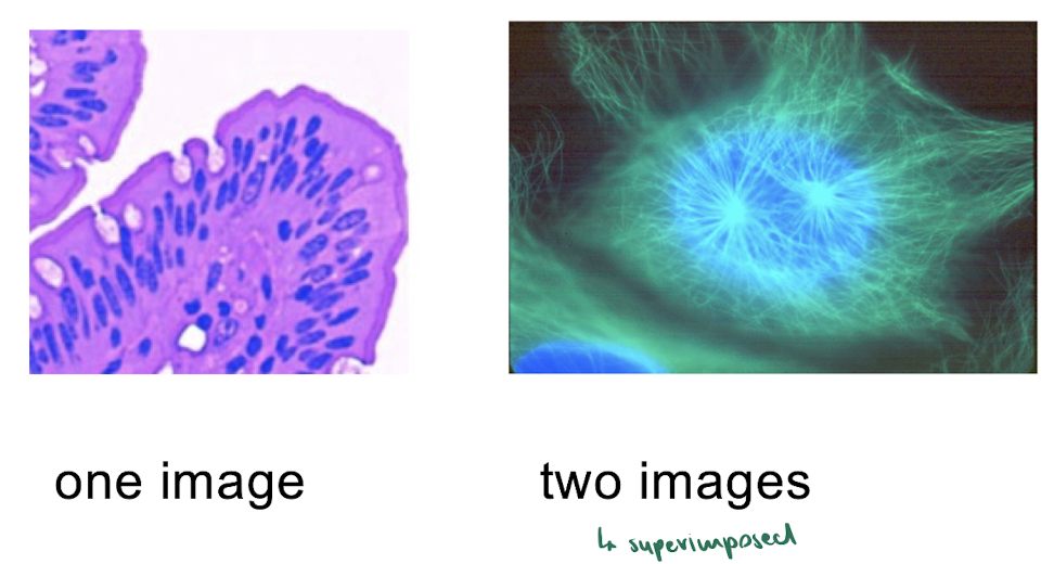
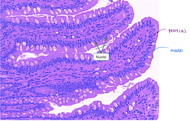
Putting thin section of cells or tissues on slide:
immobilize cells/tissue in wax or plastic → section w a microtome
ex. human intestine biopsy
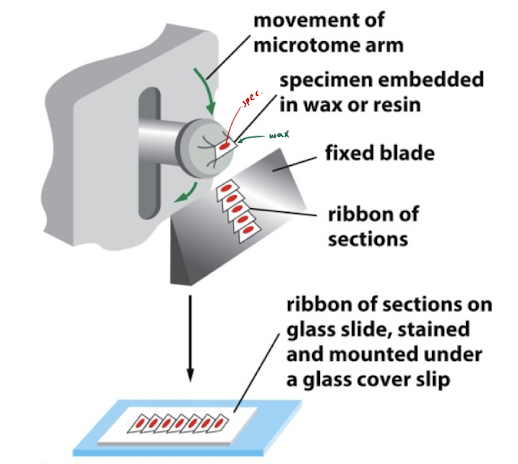
what microscope was used?
DIC
Make things Visible: Natural Colour
some organelles have a natural colour
ex. chloroplasts : green
ex. mitochondria : brown (bc of pigments in ETC)
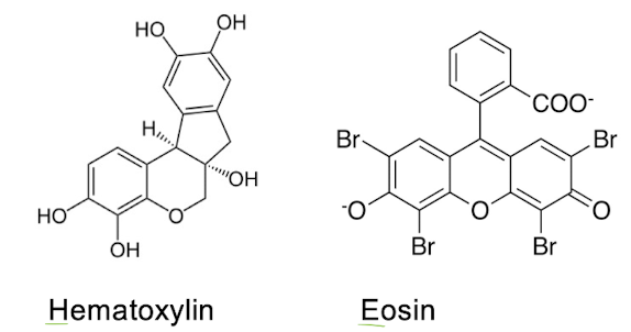
Make things Visible: Coloured stains
= synthetic molecules with 2 properties : affinity for target & absorbs light
ex. Hematoxylin - binds to nucleic acids (DNA/RNA)
ex. Eosin - binds to proteins
→ H&E staining:
by using their natural affinities for staining, it makes nuclei blue & cytoplasm pink (proteins)
H&E Staining : Steps
cell/tissue on slide
add stain #1 (H)
wash
add stain #2 (E)
wash
coverslip
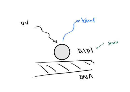
Make things Visible: Fluorescent stains
= synthetic molecules with 2 properties : affinity for target & absorbs light
ex. DAPI (sticks to DNA) → makes nuclei (DNA) fluroesce blue
DAPI is excited by UV light
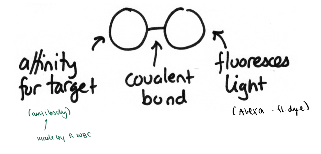
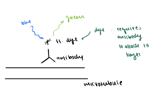
Make things Visible: Fluorescent Probes
= 2 molecules attached together (antibody + fl. dye)
used when stains WONT stick
antibody sticks to target, then fl. dye sticks to antibody via covalent bond
each microtubule is coated with fl. dye (a newt cell would be labelled w anti-microtubule antibodies w fl. dye attached)
Antibody = small proteins made by WBC (b-cells)
high affinity for microtubules → thus used for immunofluorescence
Fluroescent dyes = synthetic molecules that require an antibody to bind to target
ex. Alexa Fluor 488 (green)
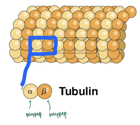
Tubulin proteins are?
heterodimers
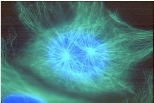
Do you have to use a green fluorescent dye to label a Newt cell
No, bc images are captured in B&W → then coloured
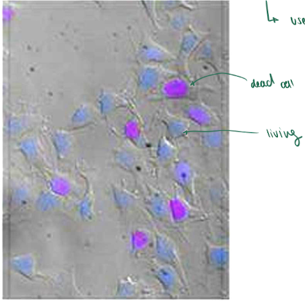
Make things Visible: Specialized Fluorescent stains
ex. Zombie Violet = only stains dead cells purple
used to tell if a cell is dead or alive
L1 Summary
Testable Content:
which method(s) was used?
which method(s) would you use?