Exam 2- Nerves
1/38
There's no tags or description
Looks like no tags are added yet.
Name | Mastery | Learn | Test | Matching | Spaced | Call with Kai |
|---|
No analytics yet
Send a link to your students to track their progress
39 Terms
Cutaneous Innervation Via Cervical Plexus
Skin is innervated by C2, C3, C4 VPR
Lesser occipital nerve (C2), Great auricular nerve (C2, C3), Transverse cervical nerve (C2, C3), supraclavicular nerve (C3, C4)
Emerge at Erb’s Point
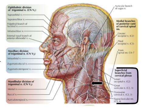
Lesser Occipital Nerve (C2)
Cervical Plexus
Follows posterior border of SCM to reach mastoid process and behind the ear
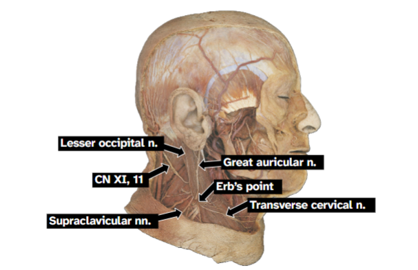
Great Auricular Nerve (C2, C3)
Cervical Plexus
Large
Courses vertically on SCM towards angle of mandible and parotid region

Transverse Cervical Nerve (C2, C3)
Crosses SCM horizontally towards the anterior
triangle of the neck

Supraclavicular nerves (C3, C4)
Crosses the clavicle towards the inferior
posterior triangle and upper chest and thorax

Brachial Plexus branches in posterior triangle
Dorsal scapular nerve (C5). Nerve to subclavius (C5-C6), suprascapular nerve (C5-C6), Long thoracic nerve (C5-C7)
Dorsal Scapular Nerve
Pierces lateral side of middle scalene
Long thoracic nerve
Pierces lateral side of middle scalene
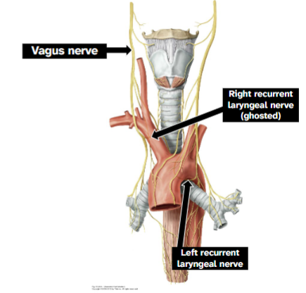
Vagus Nerve (CN X)
Courses inside carotid sheath, anterior to subclavian artery, medial to phrenic
Right vagus nerve gives off right recurrent laryngeal nerve at right subclavian artery
Left vagus nerve gives off left recurrent laryngeal nerve
Gives off superior laryngeal n. branch
▪ Internal laryngeal n. – branch of
sup. laryngeal n. that travels w/
superior laryngeal a.
▪ External laryngeal n. – branch of
sup. laryngeal n. that travels w/
superior thyroid a.
▪ Gives off recurrent laryngeal n. branch
that ascends between trachea &
esophagus
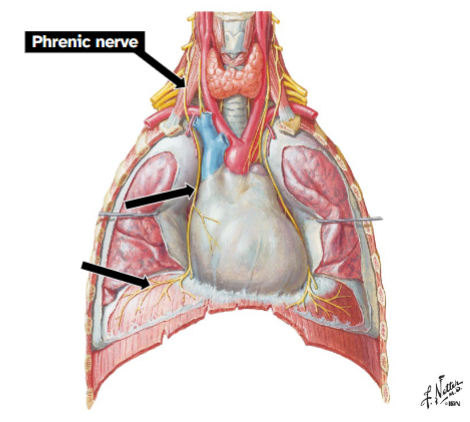
Phrenic Nerve
Formation: VPR C3-C5
Course: Descends on anterior surface of anterior
scalene m., lateral to ascending cervical a.
Enters superior thoracic aperture
Courses along pericardium to the diaphragm
Function: Motor innervation to diaphragm, sensory innervation from parietal peritoneum in right upper quadrant of abdomen
Ansa Cervicalis
Somatic motor innervation to infrahyoid muscles
Formed from C1-C3 VPRs
Loop shape
Superficial to internal jugular v
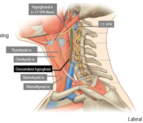
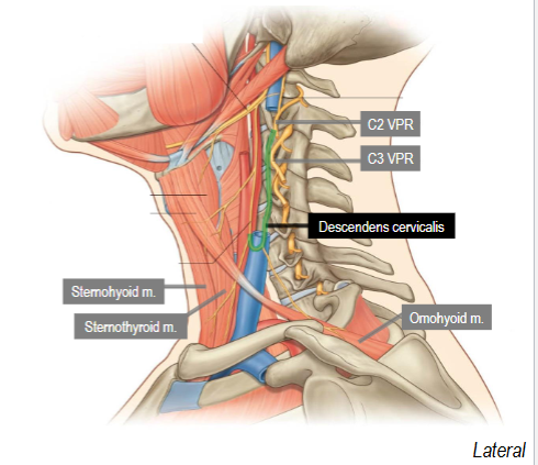
Two components: Descendens hypoglossi, descendens cervicalis
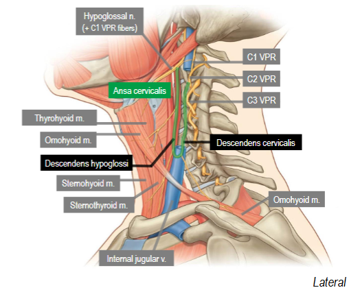
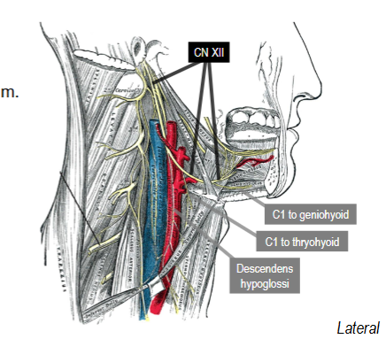
Descendens Hypoglossi
Superior root, Formed from C1 VPR Fibers running with hypoglossal n (CN XII)
Supplies superior belly of omohyoid, upper parts of sternohyoid and sternothyroid
Descendens Cervicalis
Inferior root, Formed from C2 and C3 VPR fibers
Supplies inferior belly of omohyoid, lower parts of sternohyoid and sternothyroid
Trigeminal n (CN V)
Branches into V1, V2, V3
Ophthalmic, maxillary, and mandibular nerves
Sensory nerve of the face
Inferior Alveolar Nerve
Branch of CN V3
Gives off nerve to mylohyoid

Nerve to Mylohyoid
Innervates mylohyoid and anterior belly of digastric
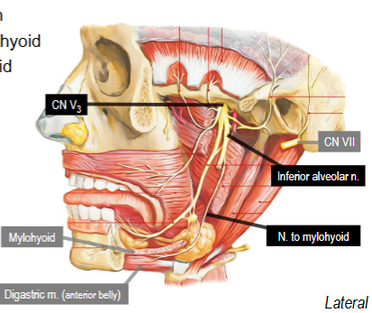
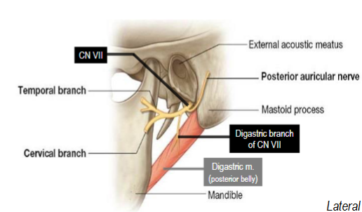
Facial Nerve (CN VII)
Trunk innervates stylohyoid and posterior
Belly of digastric mm
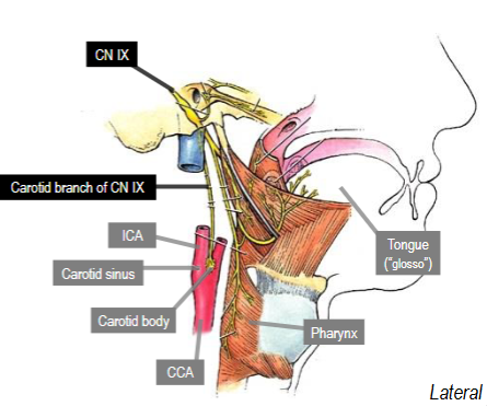
Glossopharyngeal Nerve (CN IX)
Conveys visceral sensory innervation
from carotid sinus (baroreceptors) and
carotid body (chemoreceptors), which
are both located at bifurcation of
common carotid a.
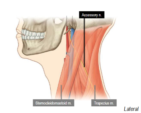
Accessory Nerve (CN XI)
Innervates SCM and Trapezius
Hypoglossal Nerve (CN XII)
- Crosses superficial to carotid arteries
▪ Helps deliver C1 VPR fibers to ansa
cervicalis, thyrohyoid, and geniohyoid mm.
▪ Passes deep to mylohyoid to innervate
tongue muscles
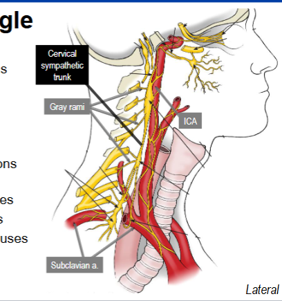
ANS in Anterior Triangle
Superior cervical ganglion: source of internal and external carotid nerves
Middle Cervical: may be absent
Inferior cervical ganglion: may fuse with first thoracic ganglion to form stellate ganglion
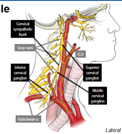
Sympathetic Trunk
Receives preganglionic sympathetic axons
that all:
• Originate at T1-L2 spinal cord levels
• Enter trunk via white rami in thorax
• Fibers ascend to cervical region
Source of postganglionic sympathetic axons
that either:
• Exit via gray rami into cervical spinal nerves
• Supply thoracic viscera via cardiac nerves
• Travel to head & neck via periarterial plexuses
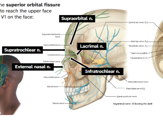
Ophthalmic N, CN V1
Exits cranial cavity via the superior orbital fissure
→ Moves through orbit to reach the upper face
→ Terminal branches of V1 on the face:
→External nasal
→Supratrochlear
→Infratrochlear
→Supraorbital
→Lacrimal
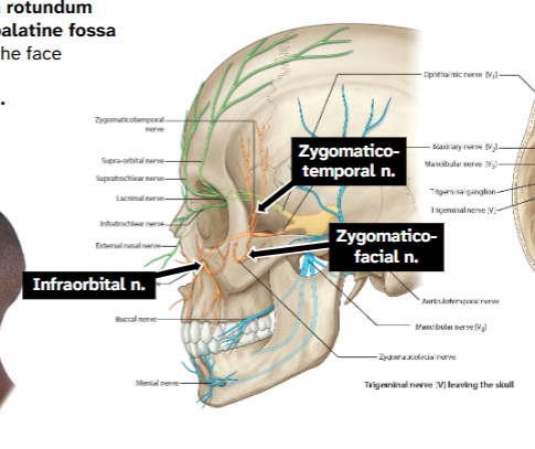
Maxillary Nerve, CN V2
Exits cranial cavity via foramen rotundum
→ Moves through the pterygopalatine fossa
→ Terminal branches of V2 on the face
→Zygomatic-facial n.
→Zygomatico-temporal n.
→Infraorbital n.
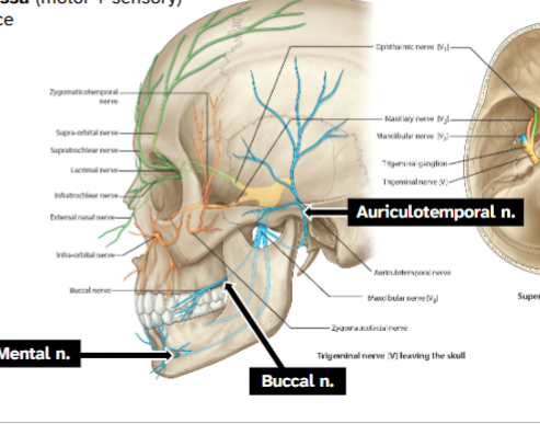
Mandibular Nerve, CN V3
Exits cranial cavity via foramen ovale
→ Courses through infratemporal fossa (motor + sensory)
→ Terminal branches of V3 on the face
→Auriculotemporal n.
→Buccal n.
→Mental n.
CN V3 has BOTH motor & sensory fibers
→ CN V1 and CN V2 are entirely sensory
→ CN V starts in the middle cranial fossa
→ It moves through foramen ovale to the
infratemporal fossa
→ Divides into 3 portions:
→ Main trunk
→ Anterior division
→ Posterior division
Cilary Ganglion
→ Inside the orbit along the lateral side of CN II
→ Receives CN III (pre-gg para); some branches ride CN V1
→ Target: pupillary constrictor muscle
Pterygopalatine Ganglion
→ Housed in the pterygopalatine fossa in the deep midface
→ Receives CN VII’s greater petrosal nerve (pre-gg para)
→ ANS fibers travel with CN V2 branches
→ Target: glandular mucosa of the nasal & oral cavities
Otic Ganglion
Supported by CN V3 just inferior to foramen ovale
→ Receives CN IX’s lesser petrosal nerve (pre-gg para)
→ ANS fibers travel with CN V3’s auriculotemporal nerve
→ Target: parotid gland
- Parasympathetic ganglion containing
postganglionic neurons which receive
the preganglionic fibers from the
glossopharyngeal nerve (CN IX) via its
lesser petrosal nerve branch
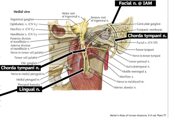
Submandiublar Ganglion
Supported by CN V3’s lingual nerve near back of mouth
→ Receives CN VII’s chorda tympani nerve (pre-gg para)
→ Target: submandibular and sublingual glands
Parasympathetics travel with…
Trigeminal Branches
Sympathetics Travel With…
Arteries
Branches of Mandibular Nerve
Main trunk, anterior division, posterior division
Main Trunk CN V3
Meningeal branch: sensory to meninges, travels with middle meningeal artery
Nerve to medial pterygoid: motor to medial pterygoid
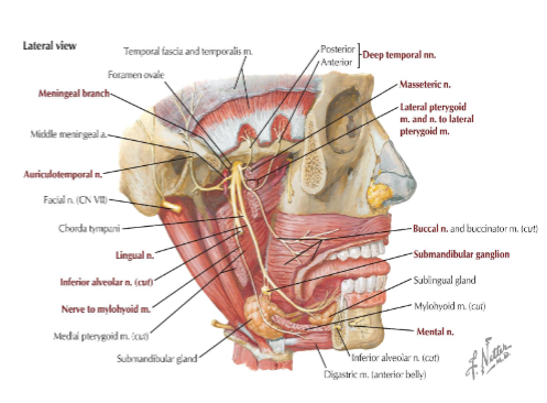
Anterior Division of CN V3
Masseteric nerve
→ Motor to masseter m.
→ Sensory to TMJ
Deep temporal nerves
→ Motor to temporalis m.
Nerve to lateral pterygoid
→ Motor to lateral pterygoid
Buccal nerve
→ Sensory to skin of cheek and nearby
gums
Posterior Division of CN V
Auriculotemporal nerve, lingual, inferior alveolar nerve
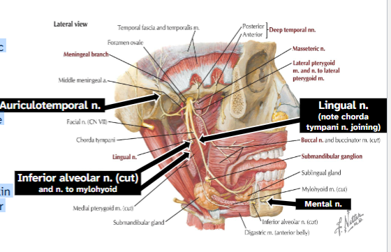
Auriculotemporal Nerve
Sensory to TMJ, skin near ear
→ Surrounds middle meningeal a.
→ Carries postganglionic
parasympathetic fibers from otic
ganglion to parotid gland

Lingual Nerve
Sensory to ant. 2/3 tongue
→ Carries chorda tympani n. to the
submandibular gg. and glands,
and ant. 2/3 tongue (taste)

Inferior Alveolar Nerve
Sensory to mandible teeth & skin
→ Enters mandibular foramen after
giving off n. to mylohyoid
→ Ends as mental n. on the chin

Chorda tympani Nerve
From the facial n. (CN VII)
→ Carries preganglionic
parasympathetic fibers to
the submandibular
ganglion
→ Carries taste fibers from
the anterior two-thirds of
the tongue
→ Leaves the skull through the
petrotympanic fissure
→ Travels with the lingual
nerve of CN V3