exam 1 Caren test
1/81
There's no tags or description
Looks like no tags are added yet.
Name | Mastery | Learn | Test | Matching | Spaced | Call with Kai |
|---|
No analytics yet
Send a link to your students to track their progress
82 Terms
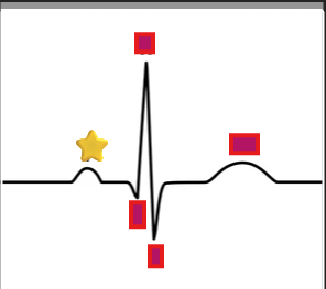
What does the P wave represent on an EKG?
A: Atrial depolarization.
depolarization
The ability to respond with pumping action.
What is the conductivity in Cardiac Cells?
The ability of each cell to receive an electrical stimulus and transmit to an adjacent cells like dominos.
What is contractility?
The mechanical result of depolarization the ability to respond with pumping action.
What is the resting state of a cell during polarization?
No membrane potential and no electrical activity.
What event occurs when a cardiac muscle cell is stimulated during depolarization?
It contracts.
Lead 1
Records differences in electrical potential between RA and LA.
Lead 2
Records the difference in electrical potential between RA and LL.
Lead 3
Records the difference in electrical potential between LA and LL.
What is Frank starlings law also on
handout
What law states that the greater the stretch of the heart muscle, the stronger the contraction?
Frank‑Starling law
What is the amount of blood pumped by the heart in one minute called?
Cardiac output
Is ekg either unipolar or polar?
both
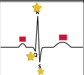
What does the QRS complex represent?
A: Ventricular depolarization.
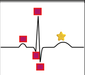
What does the T wave represent?
A: Ventricular recovery (repolarization).
How should lead wires be positioned across a patient?
Loosely, so they don’t pull or lift the electrodes.
Why is it important to position lead wire loosely?
To prevent tension that could lift electrodes off the skin.
What must be done to lead wires each use?
Properly clean and decontaminate them.
What is the purpose of cleaning and decontaminating lead wires?
To maintain hygiene and prevent cross contamination.
What does the ecg stylus inscribe on the moving paper?
Waveforms representing the heart electrical activity.
What do bipolar leads record?
The flow of the electrical impulse between two electrodes (one positive, one negative).
What do unipolar lead use?
One positive electrode and reference point calculated by ECG machine.
How are impulses traveling toward a positive electrode recorded on the ECG?
As upward deflections.
How are impulses traveling away from a positive electrode (or toward a negative electrode) recorded?
As a downward deflections.
What do the vertical lines on ECG paper represent?
Amplitude in electrical voltage.
What do the horizontal lines on ECG paper represent?
time or duration.
Normal heartbeat is
60 to 100 beats for minute
Precordial leads are
the chest lead V1, V2, V3, V4, V5, V6
Each small square equals to
0.04 second
Why should lead wires be kept separated from each other?
to avoid tangling.
What does lead aVR record?
Heart’s voltage between right arm electrode and central point between left arm and left leg. (LA & LL to RA)
What does lead aVF record?
Heart’s voltage between left leg electrode and central point between right arm and left arm. (RA & LA to LL)
what is the central point and where is it going?
is Wilson’s Central Terminal and its going downward and to the left toward the left ventricle. ( they are right arm (RA), left arm (LA), and left leg (LL) )
What does lead aVL record?
A: Heart’s voltage between left arm electrode and central point between right arm and left leg. (RA & LL to LA)
Name the three fascicles of the left bundle branch.
A: Anterior, Posterior, and Septal branches.
Name the branches of the internodal pathway.
A: Anterior, Middle, and Posterior branches.
Where is the Bundle of His located?
A: Right side of the interventricular septum above the ventricles.
How does the electrical impulse spread through the heart?
A: From endocardium to myocardium.
Which fascicle of the left bundle branch is longest and thinnest?
A: The anterior fascicle.
What is the correct conduction pathway of the heart?
A: SA node → AV node → Bundle of His → Bundle branches → Purkinje fibers.
What is the pacemaker of the heart?
A: The SA node.
What is the normal rate of the SA node?
A: 60–100 bpm.
What is the normal rate of the AV node?
A: 40–60 bpm.
What is the normal rate of Purkinje fibers?
A: 20–40 bpm.
Where do impulses from the AV node travel next?
Through the Bundle of His to the Bundle Branches.
Which fascicle of the left bundle branch is shorter and thicker?
A: The posterior fascicle.
What are the three types of EKG leads?
A: Precordial, Augmented, and Standard.
Name the four chambers of the heart.
A: Right atrium, Right ventricle, Left atrium, Left ventricle.
What are the two types of heart valves?
A: Semilunar (SL) and Atrioventricular (AV).
Where is the Thebesian valve located?
A: Near the entrance of the coronary sinus.
Where is the Eustachian valve located?
A: Near the entrance of the inferior vena cava (IVC).
What is the function of heart valves?
To control and maintain blood flow through the body.
Which chamber receives deoxygenated blood?
A: Right atrium.
Which chamber receives oxygenated blood?
A: Left atrium.
Name the three layers of the heart.
A: Epicardium, Myocardium, Endocardium.
What guards the coronary sinus?
A: The Thebesian valve.
Where does the coronary sinus drain?
A: Into the right atrium.
Describe the blood flow through the heart.
A: Superior Vena Cava → Inferior Vena Cava → Coronary Sinus → Right Atrium → Tricuspid Valve → Right Ventricle→ Pulmonic Valve → Main Pulmonary Artery → Lungs → the four Pulmonary veins → Left Atrium → Mitral Valve) → Left Ventricle → Aortic Valve (Aov) → Body.
What surrounds the heart?
A: The pericardium.
How much fluid does the pericardium contain?
A: 10–50 mL.
What can a dilated coronary sinus be mistaken for?
A: The descending aorta.
Name the branches of the aortic arch.
A: Brachiocephalic, Left common carotid, Left subclavian.
Which ventricle is a higher pressure chamber?
A: The left ventricle.
What is the time value of one small box on EKG paper?
A: 0.04 seconds.
What are the functions of the pericardium?
A: Prevents friction, trauma, infection, and provides lubrication.
What is the cardiac cycle?
A: One complete contraction and relaxation of the heart.
Define stroke volume.
A: Amount of blood ejected from the heart each beat.
Define cardiac output.
A: Amount of blood pumped into the aorta each minute.
What does Frank-Starling’s law state?
A: Greater stretch = greater contraction (length-tension relationship).
Where is the SA node located?
A: In the anterior portion of the right atrium.
Which arteries feed the Bundle of His?
A: Anterior and posterior descending coronary arteries.
Which artery usually supplies the AV node?
A: Right coronary artery.
Define excitability.
A: Each cell’s ability to respond to an electrical stimulus.
Define automaticity.
A: Ability to generate an electrical impulse independently.
What gases are transported through the circulatory system?
A: Oxygen and carbon dioxide.
Describe myocardial cells.
A: Branched, single nucleus, connected by intercalated discs.
What is the plasma membrane of myocardial cells called?
A: Sarcolemma.
What protein filaments make up myocardial cells?
A: Actin and myosin.
Where is the tricuspid valve located?
Between the right atrium and right ventricle.
Where is the mitral (bicuspid) valve located?
Between the left atrium and left ventricle.
Where is the pulmonic valve located?
Between the right ventricle and the pulmonary artery.
Where is the aortic valve located?
A: Between the left ventricle and the aorta.