Bio of Cancer Exam 4 (Ch.11-13,J.C.7/8)
 Call Kai
 Learn
Learn Practice Test
 Spaced Repetition
 Match
Match Flashcards
 Knowt Play
1/79

There's no tags or description
Looks like no tags are added yet.
Name | Mastery | Learn | Test | Matching | Spaced | Call with Kai | Chat |
|---|
No analytics yet
Send a link to your students to track their progress
80 Terms
Glucose
Glycolysis:
-Starts with Glucose (6 carbons) → Glycolysis = 2 ATP produced per glucose
-Ends with 2 x Pyruvate ( 3 carbons)
Energy produced per cycle (two cycles per glucose):
• Production of one ATP per cycle
• Production of three NADH per cycle
• Production of one FADH2 per cycle
* Gluconeogenesis can generate glucose as a precursor for synthesizing other carbohydrates
• Therefore, elevated glucose utilization can be used to drive pathways other than oxidative ATP generation
>Electron transport = ~32 ATP produced per glucose
• GLUT1: A membrane transport protein for glucose
Pyruvate
Glycolysis:
-Starts with Glucose (6 carbons)
-Ends with 2 x Pyruvate ( 3 carbons)→ Pyruvate = Output of glycolysis
Krebs Cycle:
Eight step circular enzymatic cascade
• Initial Substrate = Acetyl-CoA (produced from pyruvate by pyruvate dehydrogenase)
• Final Product = Oxaloacetate (recombined with AcetylCoA to continue cycle
- Pyruvate dehydrogenase (PDH) is the enzyme that catalyzes the conversion of pyruvate to Acetyl-CoA
• The availability of oxygen has a major impact on the fate of pyruvate
-During anaerobic respiration pyruvate is converted into lactate by the enzyme lactate dehydrogenase
• LDHA: The lactose dehydrogenase enzyme that forms lactate from pyruvate
• PDK1: An enzyme that phosphorylates/inactivates PDH to prevent the formation of Acetyl-CoA from pyruvate
Acetyl-CoA
Krebs Cycle:
Eight step circular enzymatic cascade
• Initial Substrate = Acetyl-CoA (produced from pyruvate by pyruvate dehydrogenase)
• Final Product = Oxaloacetate (recombined with AcetylCoA to continue cycle)
* Pyruvate dehydrogenase (PDH) is the enzyme that catalyzes the conversion of pyruvate to Acetyl-CoA
* = Input of Krebs cycle (and many other processes)
Metabolic enzyme regulation: Pyruvate Dehydrogenase
For example,
• When the cells is in need of Acetyl-CoA, the expression of PDKs is reduced and their activity is inhibited
• When the cell does not want to produce Acetyl-CoA, the expression of PDKs is increased, and their activity is increased
-Dietary fats are metabolized by the process of β-oxidation
• Converts lipids into Acetyl-CoA
• Aerobic respiration: In the presence of O2 pyruvate is converted to Acetyl-CoA by pyruvate dehydrogenase
Lipogenesis generates phospholipids from Glyceraldehyde-3-phosphoate (glycolysis) and Acetyl-CoA
• PDK1: An enzyme that phosphorylates/inactivates PDH to prevent the formation of Acetyl-CoA from pyruvate
Lactate
• Anaerobic respiration: In the absence of O2 , pyruvate is converted to lactate by the enzyme lactate dehydrogenase
The importance of lactate production is one of four explanations for the importance of the Warburg Effect
The Warburg Effect: The importance of lactate
-During anaerobic respiration pyruvate is converted into lactate by the enzyme lactate dehydrogenase-
1.) Regeneration of NAD+
• Continual glycolytic flux requires a constant supply of NAD+
2.)Acidification of the extracellular space
• Lactate export also exports H+ ions
• Acidification aids metastasis by facilitating ECM breakdown
3.)Lactate induces angiogenesis
• Similar to VEGF, extracellular lactate can signal to endothelial cells to undergo angiogenesis
• LDHA: The lactose dehydrogenase enzyme that forms lactate from pyruvate
Pyruvate Dehydrogenase
Krebs Cycle:
Eight step circular enzymatic cascade
• Initial Substrate = Acetyl-CoA (produced from pyruvate by pyruvate dehydrogenase)
• Final Product = Oxaloacetate (recombined with AcetylCoA to continue cycle
- Pyruvate dehydrogenase (PDH) is the enzyme that catalyzes the conversion of pyruvate to Acetyl-CoA
Metabolic enzyme regulation: Pyruvate Dehydrogenase
PDH activity is tightly regulated by two types of proteins:
1.) PDH Kinases (PDKs) phosphorylate and inactivate PDH
2.) PDH Phosphatases (PDPs) dephosphorylate and activate PDH
• The expression and activity of PDKs and PDPs are highly regulated
For example,
• When the cells is in need of Acetyl-CoA, the expression of PDKs is reduced and their activity is inhibited
• When the cell does not want to produce Acetyl-CoA, the expression of PDKs is increased, and their activity is increased
• Aerobic respiration: In the presence of O2 pyruvate is converted to Acetyl-CoA by pyruvate dehydrogenase
PDKs/PDPs
• PDH Kinases (PDKs) phosphorylate and inactivate PDH
• PDH Phosphatases (PDPs) dephosphorylate and activate PDH
• The expression and activity of PDKs and PDPs are highly regulated
Aerobic metabolism
Glycolysis→Kerbs→ ETC = ~32 ATP
uses oxygen
Anaerobic metabolism
Which pathways generates ATP faster: aerobic or anaerobic metabolism? Anaerobic (glycolysis)
glycolysis in the cytosol= ~2 ATP
Warburg Effect
• In the 1920s, Otto Warburg discovered the tumor cells utilize anaerobic metabolic pathways even in the presence of ample oxygen availability!
• This phenomenon is now known as the Warburg effect
Four explanations for the importance of the Warburg Effect
1) The rate of ATP production
2) Metabolic intermediates are used for biosynthesis
3) The dangers of oxygen metabolism
4) The importance of lactate production
Four explanations for the importance of the Warburg Effect
1) The rate of ATP production
2) Metabolic intermediates are used for biosynthesis
The Warburg Effect: The rate of ATP production
• Provided ample supply of glucose, glycolysis can generate ATP faster than glycolysis + Krebs + ETC
• Tumor cells still use Krebs/ETC, but it is not as essential as you might expect
Metabolic intermediates are used for biosynthesis
Elevated glycolytic flux drives anabolic pathways
• Most anabolic pathways begin with chemical intermediates from glycolysis/Krebs cycle
Anabolic pathways to know:
- The pentose phosphate pathway (PPP) generates nucleotides for DNA synthesis
-Lipogenesis generates phospholipids from Glyceraldehyde-3-phosphoate (glycolysis) and Acetyl-CoA
-Gluconeogenesis can generate glucose as a precursor for synthesizing other carbohydrates
• Therefore, elevated glucose utilization can be used to drive pathways other than oxidative ATP generation
Four explanations for the importance of the Warburg Effect
3) The dangers of oxygen metabolism
4) The importance of lactate production
The dangers of oxygen metabolism
Oxygen: With great power comes great responsibility
• During aerobic respiration, oxygen serves as the final electron acceptor of the ETC
- Due to the high electronegativity of oxygen (loves to “steal” electrons)
• Oxygen’s high electronegativity makes oxygen very reactive
- Often “steals” electrons from other chemicals
• Reactive oxygen species (ROS) refers to a class of oxygen-based chemicals that can cause severe damage to cells
> DNA damage is a major target of ROS
The importance of lactate production
The Warburg Effect: The importance of lactate
During anaerobic respiration pyruvate is converted into lactate by the enzyme lactate dehydrogenase
1) Regeneration of NAD+
• Continual glycolytic flux requires a constant supply of NAD+ During anaerobic respiration pyruvate is converted into lactate by the enzyme lactate dehydrogenase
2) Acidification of the extracellular space
• Lactate export also exports H+ ions
• Acidification aids metastasis by facilitating ECM breakdown
3) Lactate induces angiogenesis
• Similar to VEGF, extracellular lactate can signal to endothelial cells to undergo angiogenesis
Gluconeogenesis
• Gluconeogenesis can generate glucose as a precursor for synthesizing other carbohydrates
Pentose Phosphate Pathway
• The pentose phosphate pathway (PPP) generates nucleotides for DNA synthesis
Lipogenesis
• Lipogenesis generates phospholipids from Glyceraldehyde-3-phosphoate (glycolysis) and Acetyl-CoA
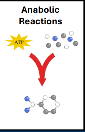
Anabolism
• Anabolic reactions
• Builds complex molecules using smaller molecules
• Requires energetic input (ATP)
- Continuous cell division of cancer cells requires a substantial amount of anabolism
- Anabolism requires a substantial amount of energy (e.g. catabolism)
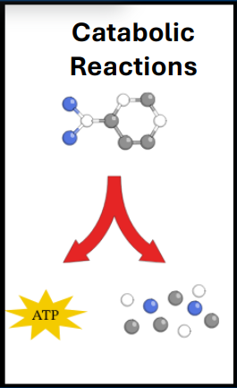
Catabolism
• Catabolic reactions
• Breaks down complex molecules into small molecules
• Releases energy that can be stored at ATP
-Anabolism requires a substantial amount of energy (e.g. catabolism)
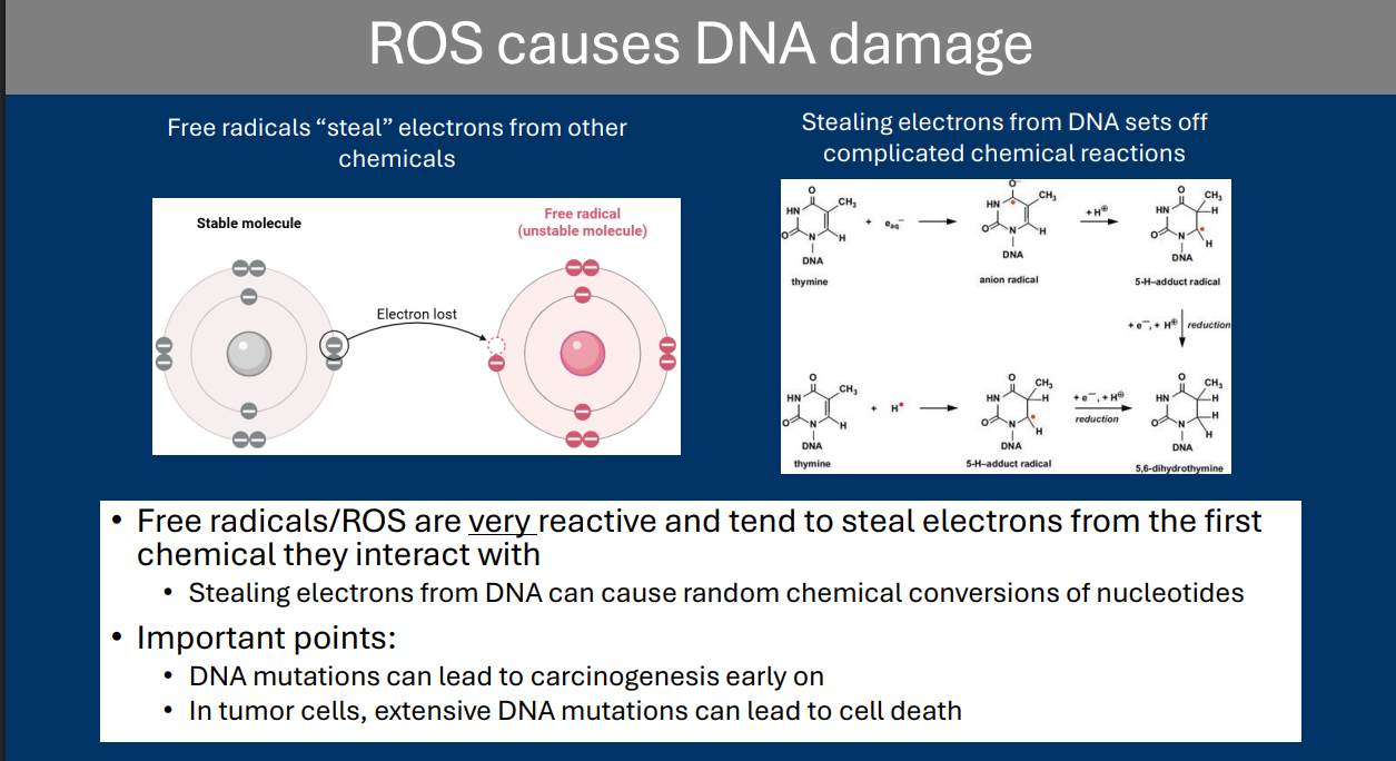
Reactive Oxygen Species
Reactive oxygen species (ROS) refers to a class of oxygen-based chemicals that can cause severe damage to cells
• DNA damage is a major target of ROS
Nrf2
The Nrf2 pathways is a common pathway that regulates ROS scavenging
Pathway overview:
• Nrf2: Transcription factor, with expression regulated by ROS
• Cul3: Ubiquitin ligase that ubiquitinates Nrf2
• Keap1: Adaptor protein that bridges an interaction between Cul3 and Nrf2
The Nrf2 Signaling Pathway
• Under “normal” conditions:
• Nrf2 interacts with Keap1 which acts as a bridge between Nrf2 and Cul3
• Cul3 is a ubiquitin ligase that polyubiquitinates Nrf2 leading to its degradation
• End result: In the absence of ROS, Nrf2 expression remains low
• Under conditions of high ROS:
• ROS molecules modify the structure of Keap1
• Keap1 releases Nrf2
• Nrf2 is no longer poly-ubiquitinated/degraded by Cul3
• Nrf2 translocates into the nucleus and transcribes genes needed for the removal of ROS
> Example genes = Catalase, SOD1, SOD2, Glutathione
End result: In the presence of ROS, Nrf2 expression increases and protective genes are expressed
Keap1
The Nrf2 Signaling Pathway
• Keap1: Adaptor protein that bridges an interaction between Cul3 and Nrf2
• Under “normal” conditions:
• Nrf2 interacts with Keap1 which acts as a bridge between Nrf2 and Cul3
• Under conditions of high ROS:
• ROS molecules modify the structure of Keap1
• Keap1 releases Nrf2
Cul3
The Nrf2 Signaling Pathway
• Cul3: Ubiquitin ligase that ubiquitinates Nrf2
• Under “normal” conditions:
• Nrf2 interacts with Keap1 which acts as a bridge between Nrf2 and Cul3
• Cul3 is a ubiquitin ligase that polyubiquitinates Nrf2 leading to its degradation
• Under conditions of high ROS:
• Nrf2 is no longer poly-ubiquitinated/degraded by Cul3
GLUT1
• HIF-1α promotes the expression of:
• GLUT1: A membrane transport protein for glucose
mTOR
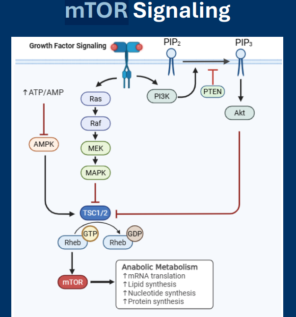
• The mTOR signaling pathways is a master regulator of anabolic metabolism
• Revolves around activation of mTOR, a protein kinase with many regulatory targets in the cell
• mTOR activation increases nearly all anabolic pathways
• Pathway activity is upregulated in >50% of cancers
• mTOR: Protein kinase that promotes anabolic metabolism when activated
• Rheb is an activator of mTOR (Inactive GDP-Rheb can no longer activate mTOR)
• mTOR signaling is repressed by AMPK
TSC1/2
Regulation of metabolic pathways: mTOR
• TSC1/2: GTPase-activating protein that induces Rheb to convert GTP (active) to GDP (inactive).
RheB
Regulation of metabolic pathways: mTOR
• Rheb: G-protein that activates mTOR
• TSC1/2: GTPase-activating protein that induces Rheb to convert GTP (active) to GDP (inactive).
• Rheb is an activator of mTOR
Akt
• Activation PI3K pathway via activation of AKT
AMPK
Known pathways that inactive mTOR:
• AMPK
- mTOR signaling is repressed by AMPK
• AMPK is an “energy sensor” kinase:
- Activated by high AMP/ATP ratio
- Inactivated by high ATP/AMP ratio
• Low energy availability (i.e. high AMP/ATP) activates AMPK
• AMPK activates TSC1/2
Critical thinking questions
1.) How would the regulation of the enzymatic activities of Pyruvate Dehydrogenase and Lactate Dehydrogenase influence a cell’s tendency to utilize oxygen for metabolism?
2.) What is the difference between anabolic and catabolic pathways? Why are each important for carcinogenesis?
3.) What is the Warburg effect? What are some possible reasons why cancer cell metabolism follows this phenomenon?
1.)PDH activation increases the utilization of oxygen by directing pyruvate into the mitochondria, while LDH inhibition forces pyruvate toward mitochondrial oxidation, thus increasing oxygen consumption. Conversely, PDH inhibition and LDH activation (common in cancer/hypoxia) reduce oxygen utilization and increase lactate production.
(The regulation of Pyruvate Dehydrogenase (PDH) and Lactate Dehydrogenase (LDH) acts as a metabolic switch determining whether a cell uses oxygen to generate energy (oxidative phosphorylation) or relies on anaerobic fermentation (glycolysis).)
2.) Anabolic pathways need energy (ATP) and they use small molecules to make larger complex molecules (like bricks making houses). Catabolic pathways Break down complex larger molecules into smaller molecules and release energy (ATP)( taking a house and turning it into bricks). Continuous cell division of cancer cells requires a substantial amount of anabolism .Anabolism requires a substantial amount of energy (e.g. catabolism)
3.) The Warburg effect is the phenomenon where even in the Prescence of plenty of oxygen tumor cells will still use anaerobic metabolic pathways. There are four possible reasons for this : The rate of ATP ( anaerobic metabolism makes ATP faster), Metabolic intermediates are used for biosynthesis, the dangers of oxygen metabolism, and the importance of lactate production
Critical thinking questions
4.) In what ways does HIF-1α signaling help activate the metabolic pathways responsible for the Warburg effect?
5.) What is the function of Nrf2? How can we explain the observation that Nrf2 can function as either a tumor suppressor or a proto-oncogene depending on context?
6.) What is mTOR signaling and how does it affect cellular metabolism? What is the rationale for the suppression of mTOR signaling by the activation of AMPK?
7.) What are some ways in which our diet can either increase or decrease our chances of developing cancer?
4.) HIF-1α signaling helps activate the metabolic pathways responsible for the Warburg effect by acting as a major transcription factor which upregulates glucose intake and glycolysis while inhibiting mitochondrial respiration ( aerobic respiration) It does this by promoting the expression of the transporter GLUT1 and the enzymes LDHA and PDK1. This allows pyruvate to be converted to lactate not acetyl-CoA. → sustains glycolysis and NAD+ Regeneration
5.) Nrf2 is a transcription factor whose expression is regulated by ROS. In conditions of high ROS Nrf2 promotes expression of protective genes. Nrf2 can act as either a tumor suppressor gene or a proto-oncogene. Its role is context dependent when in a non-cancer cell it suppresses ROS formation meaning it prevents DNA damage so it is a tumor suppressor in a cancer cell it also suppresses ROS formation however because of the context , in this case a tumor cell by preventing ROS formation/DNA damage it is preventing/delaying cell death therefore it is acting as a proto-oncogene.
6.) The mTOR signaling pathway is a master regulator of anabolic metabolism (make more). mTOR signaling upregulates almost every anabolic pathway in the cell this requires large amounts of energy AMPK acts as an “energy sensor” and if there is not energy availability for all this production it inhibits mTOR sort of as a “ lets take a rest a recuperate”
7.) Our diet can increase our chances of developing cancer by food contamination by carcinogens ( fish are fatty and many carcinogens are fat soluble) and food methods that create carcinogens ( meat like red meat cooking = heterocyclic amines) also over eating/obesity and alcohol consumption. Our diet can decrease our chances of developing cancer by eating less and consuming antioxidants and other beneficial things in food.
T-cell
Example of leukocytes (immune cells):
• Macrophages, Neutrophils, T-Cells, B-Cells
T-cells: A second type of adaptive immune cell that are subdivided into two cell types:
• Cytotoxic T-Cells (CD8+ )
• Helper T-Cells (CD4+ )
Both B-cells and T-cells have their own cell surface receptors that are capable of recognizing MHC-II presented antigens
• B-cells = B-cell receptor (BCR)
• T-cells = T-cell receptor (TCR)
T-cell activation also requires co-stimulation with other cell surface molecules such as:
- CD28 (receptor expressed on T-cell surface)
-B7 (ligand expressed on APC cell surface)
• Activated cytotoxic T-cells are now “licensed” to kill other cells expressing the same antigen
• Cytotoxic T-cells are “killing” specialist. They leave the lymph nodes and destroy cells recognized by the TCR
T-cell receptor
• T-cells = T-cell receptor (TCR)
-T-cell receptors are also specific to different antigens
TCRs recognize antigens bound to MHC-II on the surface of APCs
• Each TCR is highly specific, so most T-cells will ignore most antigen presented by APCs
Successful engagement of a TCR:Antigen:MHC-II complex leads to T-cell activation
B-cell
Example of leukocytes (immune cells):
• Macrophages, Neutrophils, T-Cells, B-Cells
Lymphocytes (Primarily responsible for adaptive immunity)
• Examples of lymphocytes: B-cells, T cells, Natural Killer Cells
B-cells: One of the primary cell types of the adaptive immune system. Most well known for the production of antibodies, proteins that can specifically recognize pathogenic molecules
Dendritic cells (DCs)“present” antigen to B-cells and T-cells
B- and T-cells that “recognize” antigens become activated and leave the lymph nodes to reach infection
• Both B-cells and T-cells have their own cell surface receptors that are capable of recognizing MHC-II presented antigens
• B-cells = B-cell receptor (BCR)
• B-cells are “recognition” specialist. They undergo massive proliferation and can differentiate into plasma cells which release soluble BCRs (e.g. antibodies)
B-cell receptor
• B-cells = B-cell receptor (BCR)
• The B-cell receptor (BCR) is a membrane-bound antibody that acts as a cell surface receptor
During cell maturation, the BCR gene undergoes “random” segment recombination in the amino acids that make up the antigen-binding domain
• Antibodies have conserved Fc regions that distinguish antibody subtypes (IgG, IgM, IgA, IgE)
• Although different in overall structure, BCRs and TCRs both contain a variable antigen binding domain
Dendritic cells
Myeloid cells (Primarily responsible for innate immunity)
• Examples of myeloid cells: Dendritic cells, Macrophages, Neutrophils, Basophils, Eosinophils
Dendritic cells: Act as “sentinels” of the immune system. By consuming other cells and components of the extracellular space, dendritic cells “consume” pathogenic molecules for presentation to the immune system.
• Macrophages are a common example of a dendritic cell
Immunosurveillance
The steps of this process are:
1) Dendritic cells (DCs) “engulf” pathogens at the site of an infection
2) DCs migrate through lymphatic vessels to a lymph node
3) DCs “present” antigen to B-cells and T-cells
4) B- and T-cells that “recognize” antigens become activated and leave the lymph nodes to reach infection
• DCs are often referred to as antigen presenting cells (APCs)
Antigen presentation by DCs utilizes the Major Histocompatibility Class 2 (MHC-II) pathway
Innate immunity
• Innate immunity: Works fast. Uses expression of receptors that are intrinsically activated by common pathogenic molecules
Innate immunity uses receptors for common indicators of pathogen infection
Adaptive immunity
• Adaptive immunity: Works after a delay. Capable of “adapting” immune responses to be specific currently infections. Capable of developing “memory” for past infections
Adaptive immunity is more complex and develops responses that are specific to current infections

Immunosurveillance
Immunosurveillance is the immune’s system ability to constantly assess for the presence of pathogens throughout the body.
The steps of this process are:
1) Dendritic cells (DCs) “engulf” pathogens at the site of an infection
2) DCs migrate through lymphatic vessels to a lymph node
3) DCs “present” antigen to B-cells and T-cells
4) B- and T-cells that “recognize” antigens become activated and leave the lymph nodes to reach infection
MHC-I
• Activated cytotoxic T-cells recognize antigens presented by MHC Class I (MHCI) molecules
• MHC-I is expressed by all cells (not just APCs)
• Nearly all cellular protein is degraded by the proteasome
• Proteasome-derived peptides are loaded into MHC Class 1 (MHC-I) cell surface molecules
• Peptide:MHC-1 complexes are transported to the cell surface
• MHC-I presentation
• Performed by nearly all bodily cells
• Peptides generated from cellular proteins produced by the proteasome
• Presented by MHC-I to activated Tcells
MHC-II
• Antigen presentation by DCs utilizes the Major Histocompatibility Class 2 (MHC-II) pathway
1. Engulfed proteins are degraded by the environment of the lysosome
2. Small peptides are loaded into MHC-II cell surface proteins in endosomal compartment
3. Peptide/MHC-II molecules are transported to the cell surface for “presentation”
Lymph Node
Examples or immune organ tissues:
• Lymphatic vessels, lymph nodes, bone marrow, thymus, spleen
• DCs then enter nearby lymphatic vessels and travel to a lymph node
-Lymph nodes are found all throughout the body
Lymph nodes are small, bean-shaped immune system glands that filter lymph fluid, trap bacteria, viruses, and cancer cells, and contain white blood cells to fight infection
Antigen Presenting Cell
DCs are often referred to as antigen presenting cells (APCs)
• An antigen is any substance the immune system recognizes as foreign
• APCs “present” antigens to other immune cells to determine “self” versus “non-self”
MHC-II presentation
• Performed only by antigen presenting cells
CD28
T-cell activation also requires co-stimulation with other cell surface molecules such as:
• CD28 (receptor expressed on T-cell surface)
• B7 (ligand expressed on APC cell surface)
Immune checkpoint:
One we have already encountered: CD28/B7
• During MHC-II induced T-cell activation, the APC expresses B7, a ligand for the T-cell expressed CD28 receptor
• Lack of B7/CD28 activation prevents full T-cell activation (even if its TCR recognizes the APCpresented antigen)
• This means that T-cells can only be activated by appropriately functioning APCs
(Co-stimulation of CD28 by B7 ensures T-cell activation only by appropriate APCs)
B7
T-cell activation also requires co-stimulation with other cell surface molecules such as:
• CD28 (receptor expressed on T-cell surface)
• B7 (ligand expressed on APC cell surface)
Immune checkpoint:
One we have already encountered: CD28/B7
• During MHC-II induced T-cell activation, the APC expresses B7, a ligand for the T-cell expressed CD28 receptor
• Lack of B7/CD28 activation prevents full T-cell activation (even if its TCR recognizes the APCpresented antigen)
• This means that T-cells can only be activated by appropriately functioning APCs
(Co-stimulation of CD28 by B7 ensures T-cell activation only by appropriate APCs)
Perforin
• If an activated cytotoxic T-cell recognizes peptide expressed by MHCI, this is a signal for cell infection
• TCR:Antigen:MHC-I interaction leads to the release of the following by the Tcell:
• Perforin: Induces holes in membranes
Granzyme B
• If an activated cytotoxic T-cell recognizes peptide expressed by MHCI, this is a signal for cell infection
• TCR:Antigen:MHC-I interaction leads to the release of the following by the Tcell:
• Granzyme B: Induces intracellular apoptotic pathways
Neoantigens
• Neoantigens are peptides that are uniquely expressed by cancer cells (e.g. consider chromosomal translocations and the expression of abnormal fusion proteins)
Tumor-associated antigens
• Tumor-associated antigens are proteins that are expressed at abnormally high levels, which can lead to recognition by the adaptive immune system
PD-1
• Programmed death 1 protein (PD-1) is a cell surface receptor expressed on T-cells
• PD-1 ligands include PD-L1 and PD-L2
• PD-1 activation by either PD-L1/L2 blocks the ability of a T-cell to release its death-inducing molecules
PD-L1/L2
• Programmed death 1 protein (PD-1) is a cell surface receptor expressed on T-cells
• PD-1 ligands include PD-L1 and PD-L2
• PD-L1/L2 can be expressed by cells expressing antigen: MHC-I as a way to avoid T-cell induced death
• PD-1 activation by either PD-L1/L2 blocks the ability of a T-cell to release its death-inducing molecules
• During infectious immune responses, noninfected cells express PD-L1/PD-L2 to avoid unwanted killing by T-cells
• This mechanism can be taken advantage of by tumor cells, which may upregulate PDL1/L2 expression
Immunoediting
• Caner immunoediting refers to the potential for immune responses to worsen cancer outcomes
• The immune system can inadvertently select for tumor cells capable of metastasis
The steps for immunoediting (the three E’s)
• Elimination of cancer: T-cell mediated killing of tumor cells expressing neoantigens
• Equilibrium: A state in which cancer cell remain few, but cells with the ability to evade the immune system are selected for
• Escape: Eventually, immune-resistant cancer cells reach high enough levels to leave primary immune site without immune-based cell killing
T-cell therapies
• The ability of T-cells to selectively kill cancer cells has significant therapeutic promise
• Potential strategies include:
• Tumor-infiltrating lymphocytes: Isolation of T-cells directly from a patient’s tumors. Tcells are then expanded in vitro and returned to the patient
• Chimeric antigen receptor (CAR) therapy: Introduction of mutation T-cells receptors capable of self-activation
• Antigen presenting cells (APC) therapy: Isolation of APCs from patients blood followed by T-cell stimulation in vitro
Critical thinking questions
1.) How does the ability to distinguish “self” and “non-self” play an important role in cancer immunity?
2.)What are the steps to the process of immunosurveillance? How do dendritic cells, B-cell, and T-cells interact to create an adaptive immune response?
3.) What is the difference between MHC Class I and MHC Class II? What different types of cells express each? How is the context in which each class presents antigen different?
1.) The ability to distinguish self from non-self is fundamental to cancer immunity because cancer cells arise from normal host cells, making them difficult for the immune system to recognize as a threat. While the immune system is designed to eliminate non-self pathogens, its primary challenge with cancer is identifying "altered-self" cells while maintaining immune tolerance to prevent attacking healthy tissues
2.)
1) Dendritic cells (DCs) “engulf” pathogens at the site of an infection
2) DCs migrate through lymphatic vessels to a lymph node
3) DCs “present” antigen to B-cells and T-cells
4) B- and T-cells that “recognize” antigens become activated and leave the lymph nodes to reach infection
Dendritic cells, B-cell, and T-cells interact in a sequence first dendritic cells capture antigens at the site of infection and then they go on to activate T-Cells and B cells where one travels to the site of infection and the other produces antibodies ( the immune response)
3.)
MHC-I presentation
• Performed by nearly all bodily cells
• Peptides generated from cellular proteins produced by the proteasome
• Presented by MHC-I to activated T-cells
MHC-II presentation
• Performed only by antigen presenting cells
• Peptides generated by lysosomal degradation of extracellular proteins
• Presented by MHC-II to B- or T-Cells to potentially lead to their activation
Critical thinking questions
4.) How do immune checkpoints play a role in autoimmunity? How do they play a role in cancer cells evading immune detection?
5.) How can the immune response worsen carcinogenesis though immunoediting?
6.) What are the different ways that T-cells can be used as therapeutic tools?
4.)Immune checkpoints are normal regulatory pathways (or "brakes") on T cells that maintain immune homeostasis and prevent autoimmunity by preventing overactive immune responses.
• This mechanism can be taken advantage of by tumor cells, which may upregulate PDL1/L2 expression, effectively "turning off" the immune response and allowing cancer cells to evade detection
5.)
The immune system can inadvertently select for tumor cells capable of metastasis
• Elimination of cancer: T-cell mediated killing of tumor cells expressing neoantigens
• Equilibrium: A state in which cancer cell remain few, but cells with the ability to evade the immune system are selected for
• Escape: Eventually, immune-resistant cancer cells reach high enough levels to leave primary immune site without immune-based cell killing
6.)
Potential strategies include:
• Tumor-infiltrating lymphocytes: Isolation of T-cells directly from a patient’s tumors. T-cells are then expanded in vitro and returned to the patient
• Chimeric antigen receptor (CAR) therapy: Introduction of mutation T-cells receptors capable of self-activation
• Antigen presenting cells (APC) therapy: Isolation of APCs from patients blood followed by T-cell stimulation in vitro
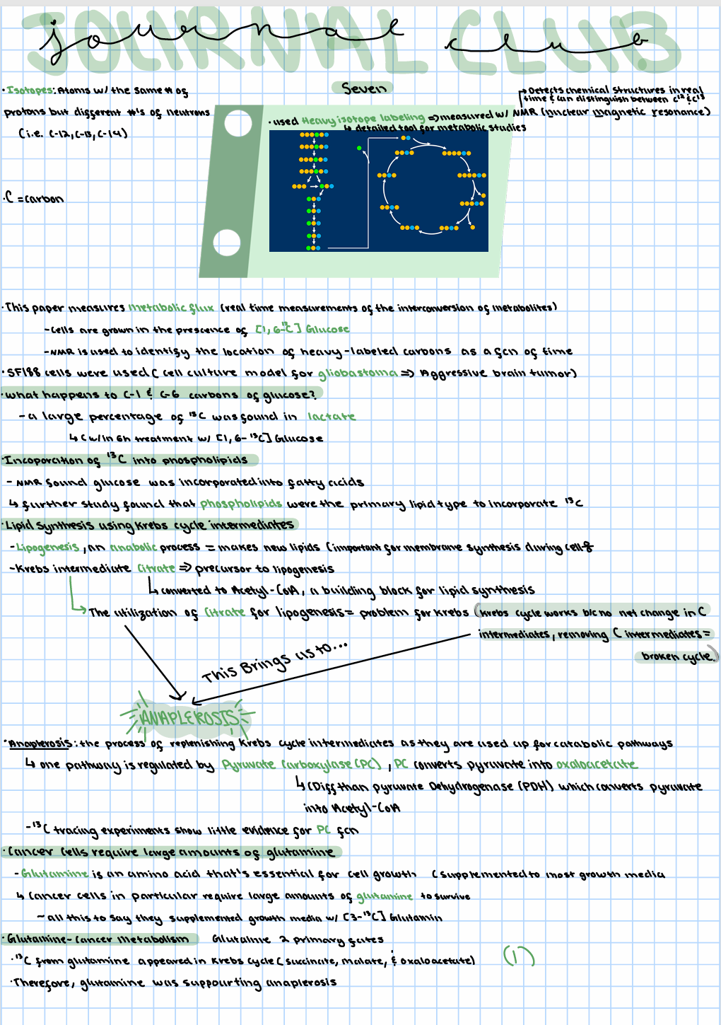
Journal Club 7:
Beyond aerobic glycolysis: Transformed cells can engage in glutamine metabolism that exceeds the requirement for protein and nucleotide synthesis
• This paper measures metabolic flux
• Real time measurements of the interconversion of metabolites
• Cells are grown in the presence of [1,6- 13C]Glucose
• NMR is used to identify the location of heavy-labeled carbons as a function of time
(Monitoring metabolic flux using heavy-labeled carbons)
Journal Club 7:
Beyond aerobic glycolysis: Transformed cells can engage in glutamine metabolism that exceeds the requirement for protein and nucleotide synthesis
• Heavy isotope labeling is powerful tool for detailed metabolic studies • This study uses [1,6- 13C]Glucose
( --Atoms with the same number of protons but different numbers of neutrons are known as isotopes)
• The carbons in the first and sixth position (normally 12C) are replaced with 13C
• Nuclear magnetic resonance (NMR) detects chemical structures in real time and can distinguish between the presence of 12C and 13C
This paper uses SF188 (Brain cancer cell line) cells, a cell culture model for glioblastoma
What happens to C-1 and C-6 carbons of glucose?
• SF188 cells grown the present of ample oxygen
• Within 6h of treatment with [1,6- 13C]Glucose, a large percentage of 13C is found in lactate
This is confirmation of the Warburg Effect^
• Further study found that phospholipids were the primary lipid type to incorporate 13C
Clear example of anabolism^
Anaplerosis using the enzyme Pyruvate Carboxylase
• Citrate is converted to Acetyl-CoA, which is used as a building block for lipid synthesis
• The utilization of citrate for lipogenesis poses a problem for the Krebs cycle
- The cycle is possible because there is no net change in carbon intermediates
- Removing carbon intermediates will ultimately break the cycle
• Anaplerosis is the process of replenishing Krebs cycle intermediates as they are used up for catabolic pathways
> The enzyme Pyruvate carboxylase (PC) regulates one pathway of anaplerosis, PC converts pyruvate into oxaloacetate
Cancer cells require large amounts of glutamine
• Glutamine is an amino acid that is essential for cell growth
• Supplemented to most growth media
• It is known that cancer cells in particular require large amounts of glutamine to survive
They found that glutamine had two primary fates:
1.)
• 13C from glutamine appeared in Krebs cycle
-Succinate, Malate, and Oxaloacetate
• Therefore, glutamine was supporting anaplerosis
2.)
• 13C from glutamine was found in secreted lactate and pyruvate
• This is related to an additional pathway in which malate is converted to pyruvate
• This pathway generates NADPH, an energetic molecule necessary for lipogenesis
Journal Club 7:
Beyond aerobic glycolysis: Transformed cells can engage in glutamine metabolism that exceeds the requirement for protein and nucleotide synthesis
Summary and Conclusions
• Heavy isotope labeling is a powerful tool for a detailed understanding of metabolic flux
• Confirmation of Warburg effect (i.e. most glucose-derived carbon is used to generate lactate in cancer cells)
• The remaining glucose-derived carbon enters the Krebs cycle, but is almost immediately diverted in the form of citrate for lipogenesis
• Depletion of Krebs cycle intermediates by anabolic pathways requires anaplerosis to maintain cycle activity
• Tumor cells consume large amounts of glutamine to supplement anaplerosis and lipogenesis
Journal Club 7:
Beyond aerobic glycolysis: Transformed cells can engage in glutamine metabolism that exceeds the requirement for protein and nucleotide synthesis
Written Notes

Acute Inflammation
Acute inflammation refers to a transient activation of inflammatory pathways that resolve the initiating challenge
Chronic Inflammation
During chronic inflammation, the initiating stress remains unresolved leading to a persistent state of inflammation
Extrinsic Stimulation
Extrinsic Stimuli (pathogen infection, physical wounds, environmental exposure)
Intrinsic Stimulation
Intrinsic Stimuli (genetic disease, oncogene activation, cell stress)
Chemokines
Chemokines: Small peptides that act as chemoattractant signals to recruit more immune cells (Examples = CCL2, CCL5, CXCL12)
Cytokines
Cytokines: Small peptides that activate pro- or antiinflammatory pathways in immune cells (Examples = TNFα, IL1β, IL-10)
DAMPs
Damage-Associated Molecular Patterns (DAMPs)
• Extracellular proteins
• Extracellular ATP
• Extracellular DNA
PAMPs
Pathogen-Associated Molecular Patterns (PAMPs)
• Bacterial cell wall components
• Viral DNA
TLR
Toll Like Receptors (TLR)
• Expressed on immune cells membranes
• Ten types of TLR in humans, each recognizes different PAMPs/DAMPs
Toll Like Receptors (TLR) are expressed by immune cells and play a role in the initial recognition of an inflammatory challenge
NF-κB
The NF-κB signaling pathways is the most well understood inflammatory signaling cascade
This pathway revolves around the activation of the NF-κB transcription factor
• Formed as homo- or hetero-dimers of various subunits
• The most important form is a heterodimer of p65 (RelA) and p50
• NF-κB (p65/p50)
p50/p65
NF-κB
• The most important form is a heterodimer of p65 (RelA) and p50
Under non-inflammatory conditions
• NF-κB (p65/p50) is sequestered in the cytoplasm by interaction with the Inhibitor of NF-κB (IκB)
• This prevents NF-κB signaling under basal conditions
Under pro-inflammatory conditions:
• Receptor activation at the cell surface (e.g. TLR receptors or cytokine receptors) activates the protein IKK (IκB Kinase)
• IKK phosphorylates IκB leading to IκB ubiquitination and degradation
• NF-κB (p65/p50) now translocates to the nucleus and activates expression of immune response genes
IκB
• NF-κB (p65/p50) is sequestered in the cytoplasm by interaction with the Inhibitor of NF-κB (IκB)
• This prevents NF-κB signaling under basal conditions
IKK
IKK (IκB Kinase)
Under pro-inflammatory conditions:
• Receptor activation at the cell surface (e.g. TLR receptors or cytokine receptors) activates the protein IKK (IκB Kinase)
• IKK phosphorylates IκB leading to IκB ubiquitination and degradation
• NF-κB (p65/p50) now translocates to the nucleus and activates expression of immune response genes
STAT
Intracellular inflammatory signaling: STAT signaling
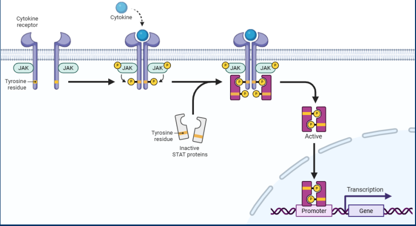
• STAT signaling pathways are also regulated by inflammatory stimuli
-> Generally activated by cytokine receptors (many different types in different cells)
• Ligand binding to cytokine receptors recruits JAK kinase enzymes to intracellular receptor domains
—-- Recruited JAK kinases phosphorylate the intracellular receptor domains
—-- Phosphorylated receptor domains recruit STAT family transcription factors
• JAK kinases phosphorylate/activate STAT transcription factors
—-- Activated STAT transcription factors dimerize and translocate to the nucleus to regulate gene expression
• Traditionally, STAT activation promotes proliferation, survival, angiogenesis, and inflammation
JAK Kinase
STAT signaling pathways are also regulated by inflammatory stimuli
• Generally activated by cytokine receptors (many different types in different cells)
- Ligand binding to cytokine receptors recruits JAK kinase enzymes to intracellular receptor domains
• Recruited JAK kinases phosphorylate the intracellular receptor domains
• Phosphorylated receptor domains recruit STATfamily transcription factors
JAK kinases phosphorylate/activate STAT transcription factors

Cancer Associated Fibroblasts
• Inflammatory activation of a fibroblast can lead to the development of cells with functions that support carcinogenesis
• This is known as a cancer-associated fibroblast (CAF)
Features of CAFs :
• Immunosuppressive function to prevent immune cell killing of cancer cells
• Secretion of pro-angiogenic factors
• Secretion of pro-growth factors
• ECM remodeling to be permissive for metastasis
Hepatitis B
The association of Hepatitis B and Hepatocellular carcinoma
• Hepatitis B infection causes a persistent state of chronic inflammation that adversely impacts the liver
• Hepatitis B carriers have a 10-25 fold greater risk of developing hepatocellular carcinoma
• Chronic inflammation in the liver leads to:
— -An oxidative environment due to ROS production by macrophages
— -A continual process of cell death followed by cell proliferation
• Evidence also suggests that hepatitis B infection may have more direct affects on carcinogenesis
— - Viral DNA has a tendency to insert into the genome near the TERT gene (Telomerase expression)
— -This disrupts normal Telomerase regulation and leads to its expression in non-stem cells
Hepatocellular Carcinoma
• Hepatitis B carriers have a 10-25 fold greater risk of developing hepatocellular carcinoma
Epithelial liver tumor
Ebstein-Barr Virus
• Epstein-Barr virus infects lymphocytes such as B-cells and is associated with increased risk of developing Burkett’s lymphoma
Lymphoma
We will consider three viral/carcinogenic relationships:
• Epstein Barr Virus/Lymphomas
Epstein-Barr virus infects lymphocytes such as B-cells and is associated with increased risk of developing Burkett’s lymphoma
• This association can be explained in part by the expression of the viral protein LMP1
— -• Integral membrane protein that, when expressed, activates NF-κB and STAT1 signaling pathways
• This leads to increased cell survival and viral replication, but may also play a role in carcinogenesis
Herpesvirus
• Herpesvirus infection is associated with Kaposi’s sarcoma, which generally occurs in vascular endothelial cells near the skin
• The viral genome contains:
— -Cell cycle regulators (e.g. v-Cyclins)
— -Anti-apoptotic proteins (e.g. v-BCL2, v-FLIP)
• The viral genome also contains proteins evolved to manipulate host cell pathways such as the protein LANA that interferes with the function of the tumor suppressor genes RB and p53
• Overall an example of a virus that specifically has learned how to activated a cancer-like phenotype
• Can also cause infected cells to release paracrine factors to nearby cells that promote cell growth, angiogenesis, etc
Kaposi’s sarcoma
• Herpesvirus infection is associated with Kaposi’s sarcoma, which generally occurs in vascular endothelial cells near the skin
Critical thinking questions
1.) What is homeostasis? How do the concepts of acute and chronic inflammation apply to the body’s ability to achieve homeostasis via inflammation?
2.) What is the difference between extrinsic and Intrinsic inflammation? How does the difference between these two stimuli relate to the difference between DAMPs and PAMPs?
1.) Homeostasis is the process the body uses to maintain a stable environment. Healthy inflammatory responses rely on the ability to re-establish homeostasis. Acute inflammation causes tissue repair/ fixes the issue and homeostasis is restored. In chronic inflammation the immune system remains activated which causes a failure to return to equilibrium or homeostasis is lost.
2.) Extrinsic inflammation is caused by foreign pathogens or substances while intrinsic is caused by internal factors like mutations. These two inflammation kinds related to the two categories of recognized stimuli because DAMPs are cell components like DNA and proteins while PAMPs are associated to foreign things like bacterial cell wall components.
or
(The primary difference between extrinsic and intrinsic inflammation lies in the origin of the stimulus that triggers the immune response, which directly correlates to whether the inflammation is caused by foreign invaders (PAMPs) or internal tissue damage (DAMPs).)
Critical thinking questions
3.)What are the three phenotypes associated with inflammation? What are the cell and molecular events that are responsible for these phenotypes?
4.) What are examples of inflammatory stimuli that play a role in the activation of NF-κB and STAT pathways? In what ways is NF-κB related to carcinogenesis?
5.) What are different ways in which viral infection can lead to carcinogenesis? What are the specific virus/cancer relationships discussed in class?
3.)
Three phenotypes often associated with inflammation:
1) Redness → b/c blood supply increase (angiogenesis)
2) Swelling → Vascular permeability facilitates the recruitment of immune cells and fluid
3) Pain/Itchiness→ Inflammatory responses require cell-cell communication, which is often done via the release of paracrine stimuli (chemokines,cytokines)
4.) PAMPs, DAMPs, Cytokines.
During carcinogenesis, NF-κB can be activated by:
• Oncogene activation
• Growth factors • Hypoxia
• The transcriptional effects of NF-κB activation are generally unique to cell types. During cancer, NF-κB can behave as a proto-oncogene and activate:
• Transcriptional expression of MDM2 (suppressing p53 expression)
• Cell division via transcriptional regulation of cyclin genes
• Suppression of pro-apoptotic genes (e.g. Bak, Bax)
5.)
Epstein Barr Virus/Lymphomas
• This association can be explained in part by the expression of the viral protein LMP1
*****• Integral membrane protein that, when expressed, activates NF-κB and STAT1 signaling pathways
• This leads to increased cell survival and viral replication, but may also play a role in carcinogenesis
Herpesvirus/Kaposi’s sarcoma
The viral genome contains:
• Cell cycle regulators (e.g. v-Cyclins)
• Anti-apoptotic proteins (e.g. v-BCL2, v-FLIP)
• The viral genome also contains proteins evolved to manipulate host cell pathways such as the protein LANA that interferes with the function of the tumor suppressor genes RB and p53
Hepatitis B/Liver Cancer
- Chronic inflammation in the liver leads to:
• An oxidative environment due to ROS production by macrophages
• A continual process of cell death followed by cell proliferation
—
• Viral DNA has a tendency to insert into the genome near the TERT gene (Telomerase expression)
• This disrupts normal Telomerase regulation and leads to its expression in non-stem cells