Physio. Ch10 Part 2 Special Senses
 Call Kai
 Learn
Learn Practice Test
 Spaced Repetition
 Match
Match Flashcards
 Knowt Play
1/51

There's no tags or description
Looks like no tags are added yet.
Name | Mastery | Learn | Test | Matching | Spaced | Call with Kai |
|---|
No analytics yet
Send a link to your students to track their progress
52 Terms
Hair cells of the ear
Mechanoreceptors
3,500 sit in the ear on a basilar membrane
Responsible for changing sound waves into nerve impulses
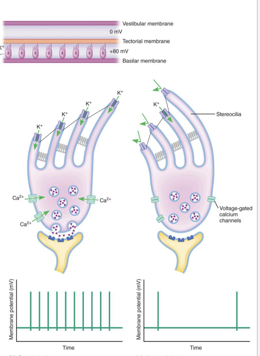
Apical surface has hair like projections called stereocillium that bend
Stereocilia hearing
Part of hair cells, located in the cochlea
Hair like projections that bend when pushed against tectorial membrane
Bend towards tallest stereocillium causes K channels to close, depolarization
- Neurotransmiter is released and action potential fires
Bend away from tallest stereocillium causes K channels to open, hyperpolarization
- Neurotransmiter stopped from releasing and no action potential fires
Sound Intensity
The further a Stereocillium bends the more intense the sound is
- Further bending causes more neurotransmitter releases
Sound Pitch/frequency
Regulated by what hair cells are stimulated
- Higher frequency stimulate stiffer hair cells at the base of the cochlea
Vestibular apparatus
Part of the inner ear
Helps to maintain balance and equilibrium
Detects head movement and acceleration to do this
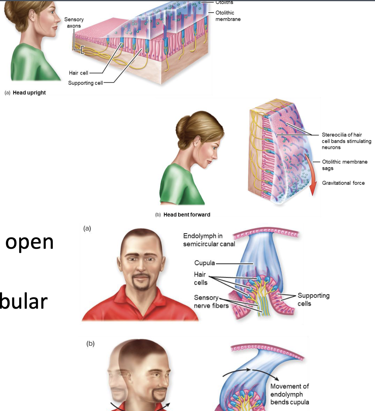
- Vestibule detect static equilibrium and linear acceleration of the head
- Semicircular ducts detect angular rotation of the head
Stereocilia equalibrium
Part of hair cells, located in the vestibule and semicircular
Bend due to changes in head/body acceleration
- Opens mechanical gated ion channels, depolarizing cells and sending action potentials
Gustatory chemoreceptors
Chemical receptors in the tastebuds
- 50, 150 per taste bud
Tastants interact with receptors to cause sensation of taste
Tastants
Chemicals that interact with Gustatory chemoreceptors to produce taste
Only 4 exist
- Salty
- Sour
- Sweet and Umami
- Bitter
Olfactory chemoreceptors
Chemical receptors
Responsible for smell
Highly sensitive receptors
Act through GPCR pathways that cause depolarization
Photoreceptors
Receptors that change light energy into electrical energy
Only 750mV to 350mV can exit these receptors (visible light spectrum)
Reflection
Light waves strike and bounce off an object
We can only see light that reflects
Refraction
The way that light waves bend when they pass through something
The density of the object makes a big difference here
- Cornea and lense do this in the eye before the retina is reached
Accommodation
The ability of the lens to focus light from an object on the retina as the distance between the object and the eye decreases
Accomplished by thickening and thinning the lens
Ciliary Muscle
The muscle the surrounds the lens and powers accommodation
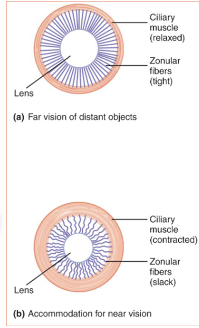
Attached to the lens by zonular fibers
- When contracts the zonular fibers are slackened
- When relaxed the zonular fibers are tightened
Zonular fibers
Fibers that connect the ciliary muscle to the lens
When tight the lens is flattened
When slackened the lens is rounder
Near objects
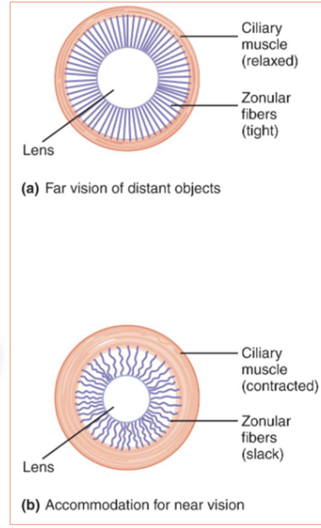
Light rays coming in are more spread out
Lens needed to be rounded to account for this
- Ciliary muscle contracts, slackening zonular fibers
Far objects
Light rays coming in are less spread out
Lens needed to flatter to account for this
- Ciliary muscle relax, tightening zonular fibers
Iris
The colored part of the eye
Contracts to protect the eye from excessive light
- Controlled by parasympathetic nervous system
Dilates to allow the eye to take in more light
- Controlled by sympathetic nervous system
Myopia
Pathophysiology corrected by a concave lens
Commonly referred to as “near sightedness”
The lens / cornea are to strong for the eyeball
- Distant images are brought to a point of focus in front of the retina, making distant objects appear blurry
Hyperopia
Pathophysiology corrected by a convex lens
Commonly referred to as “far sightedness”
The lens / cornea are to weak for the eyeball
- Near images are not brought to a point of focus before reaching the retina, making near objects appear blurry
Astigmatism
Pathophysiology corrected by an uneven lens
The lens / cornea are irregularly curved
Creates several points of focus in the retina
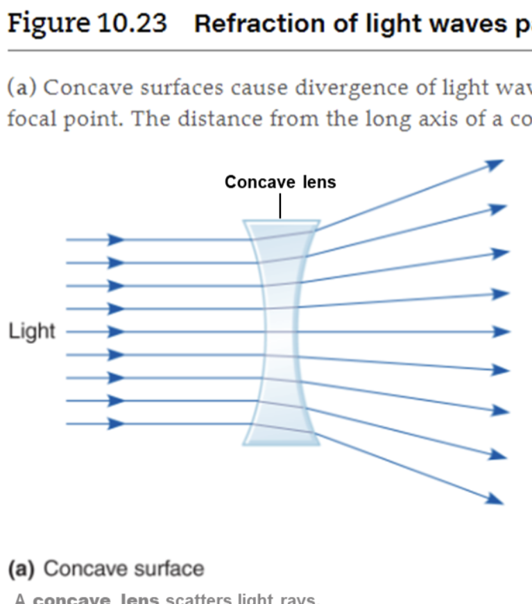
Concave lense
A lense that causes light rays to spread out when it is passed through
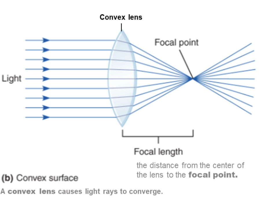
Convex lense
A lease that causes light rays to come to a point when it is passed through
Visual Acuity
Sharpness of the vision dependent on resolving power
Resolving power
The ability to tell two closely spaced objects apart
Snellen Eye Chart
The eye doctor chart
Exam to be done from 20 feet away
Retina
Located in the back of the eye
Responsible for taking in light rays and deciphering them
- Uses photoreceptors to do this
Photoreceptors
Receptors located in the retina that change visual light energy into electrical energy
Subdivided into rods and cones
Rods
Used for vision in low light situations
Sees in black and white
Communicate with bipolar cells cells
Uses Rhodopsin as its photopigments
Cones
Used for vision in high light situations
Sees in color
Communicates with bipolar cells
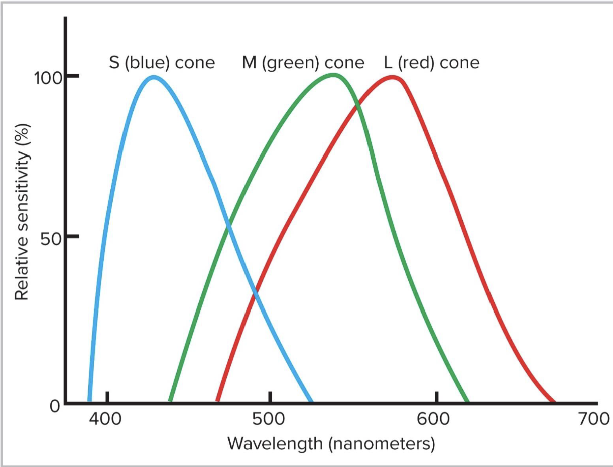
Uses L opsin, M opsin, S opsin as their photopigments
- Used in long, medium, short cones respectively
Bipolar cells
Cells that are the bridge between photoreceptors and ganglion cells
Ganglion cells
Receive information from bipolar cells and transmit it down their axons to the brain
Phototransduction
Conversion of light energy into electrical signals, carried out by photoreceptors
Photopigments
Also called “retinal”
Light absorbing portion of photoreceptor
Made from vitamin A
Four different types
- Rhodopsin
- L opsin
- M opsin
- S opsin
Membranous disks
Outer segment of photoreceptors
Contain retinal and opsin (photopigments)
- Uses G-protein transduction
Phototransduction of Light
Step 1: Rhodopsin molecule is activated by light
Step 2: Retinal and Opsin dissociate
Step 3: Opsin activates transducin
Step 4: Transducin’s alpha subunit activates phosphodiesterase (PDE)
Step 5: PDE facilitates degradation of cGMP, causing Na to stop coming in and hyperoplarization of photoreceptor
- cGMP activates Na channels
Rhodopsin molecule
Found in the retinal
Activated by light
Activation causes dissociation of retinal and opsin
Opsin
Found in photoreceptor
Disassociates from retinal when Rhodopsin is activated by light
Transducin
Found in photoreceptor
Activated by opsin
Alpha subunit activates phosphodiesterase (PDE)
Phosphodiesterase (PDE)
Activated by Transducin
Facilitates degradation of cGMP
cGMP
Degraded by Phosphodiesterase (PDE)
Opens Na channels
- Causes the release of neurotransmitter glutamate
Gutamate
Inhibitory neurotransmitter
Inversely related to levels of cGMP
Dark conditions
Na channels that are active, membrane potential -35 to -45mV
Causes depolarization that travels down the membrane and opens Ca channels
- Causes more glutamate release
Glutamate stops bipolar cells from stimulating ganglion cells
Light conditions
Na channels that are inactive, membrane potential -70mN
Causes hyperpolarization that travels down the membrane and closes Ca channels
- Causes less glutamate release
Bipolar cells stimulating ganglion cells
Color vison
Colors are perceived based on patterns of activation of the different
cones
Red-green most common
Most common form of color blindness
Affects men more because its genetically linked
- 6% of men
Ishihara charts
Charts used to detect color blindness
Visual Fields
Retina divided into nasal and temporal sides
Right visual field projected onto left side of retina
- Left visual field projected onto right side of retina
Optic chiasm
X shaped nerve pathways behind the eyes
Allows for the nasal side of each eye to cross over
- Nasal retinal nerves run to left side of brain
- Temporal retinal nerves run to right side of brain
Neural Pathways for Vision
Visual cortex processes the information coming from the retina as action potentials in ganglion cell axons
Pupillary reflex
Contraction of pupils
Parasympathetic innervation
Fovea
The sight of highest visual acuity