A&P 2: Lab Final Practical
1/230
There's no tags or description
Looks like no tags are added yet.
Name | Mastery | Learn | Test | Matching | Spaced | Call with Kai |
|---|
No analytics yet
Send a link to your students to track their progress
231 Terms
right lung lobes
3 lobes (superior, middle, inferior)
left lung lobes
2 lobes (superior and inferior)
left lung fissure
oblique fissure
right lung fissure(s)
oblique and horizontal
cardiac notch
impression formed on the left lung by the apex of the heart
division of the pharynx
nasopharynx, oropharynx, laryngopharynx
nasopharynx
stretches from the internal nares to the posterior end of the soft palate
function: solely respiratory, passageway for air
oropharynx
extends from the soft palate to the superior margin of the epiglottis
function: conveys air for respiration and food for digestion
larynogopharynx
extends from the superior margin of the epiglottis to the openings for the larynx and esophagus
function: conveys air for respiration and food for digestion
tidal volume
the volume of air either inhaled or exhaled during single breath
Residual volume
amount of air still remaining in the lungs after a maximal exhalation
Inspiratory Reserve Volume (IRV):
amount of air that is forcibly inhaled after a normal tidal volume
Expiratory Reserve Volume (ERV):
amount of air that is forcibly exhaled after a normal tidal volume.
vital capacity
maximum amount of air that can be inhaled or exhaled
trachea
identify the organ
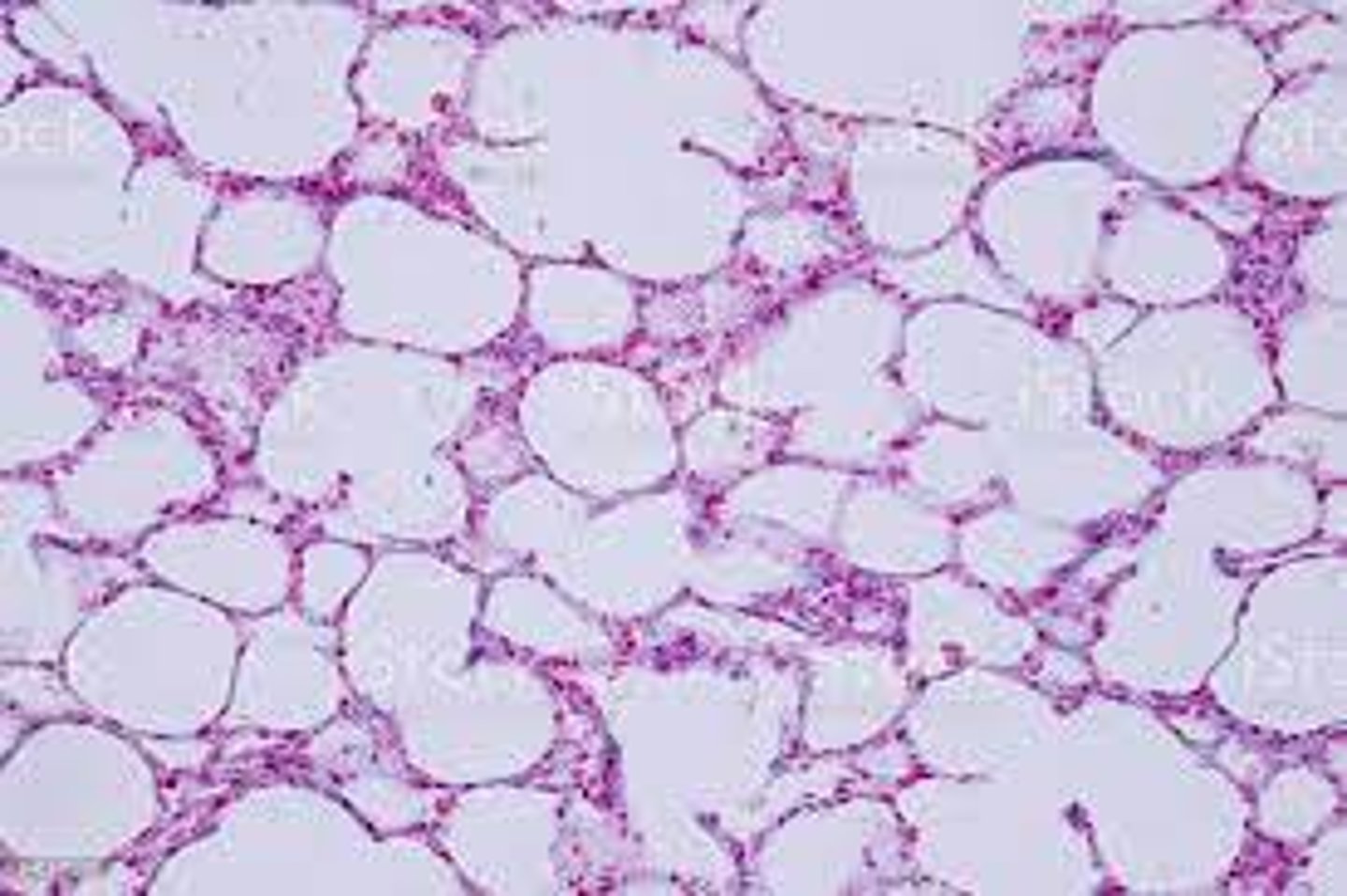
organ: lungs
cell: alveolus
identify the organ and the functional cell of the organ in the micrograph.
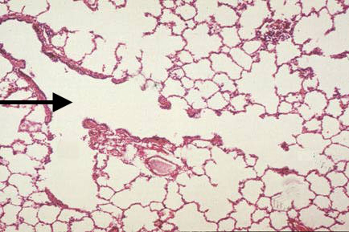
aveolar duct
Identify the organ indicated by the arrow.
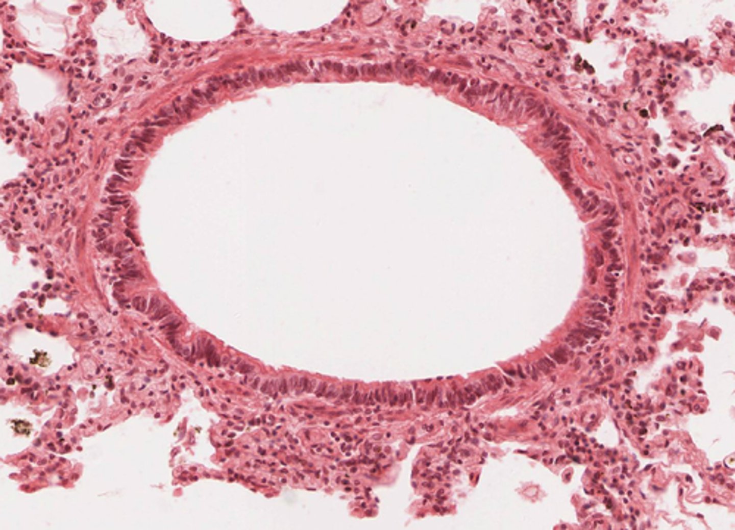
bronchioles
Identify the organ in the micrograph below.
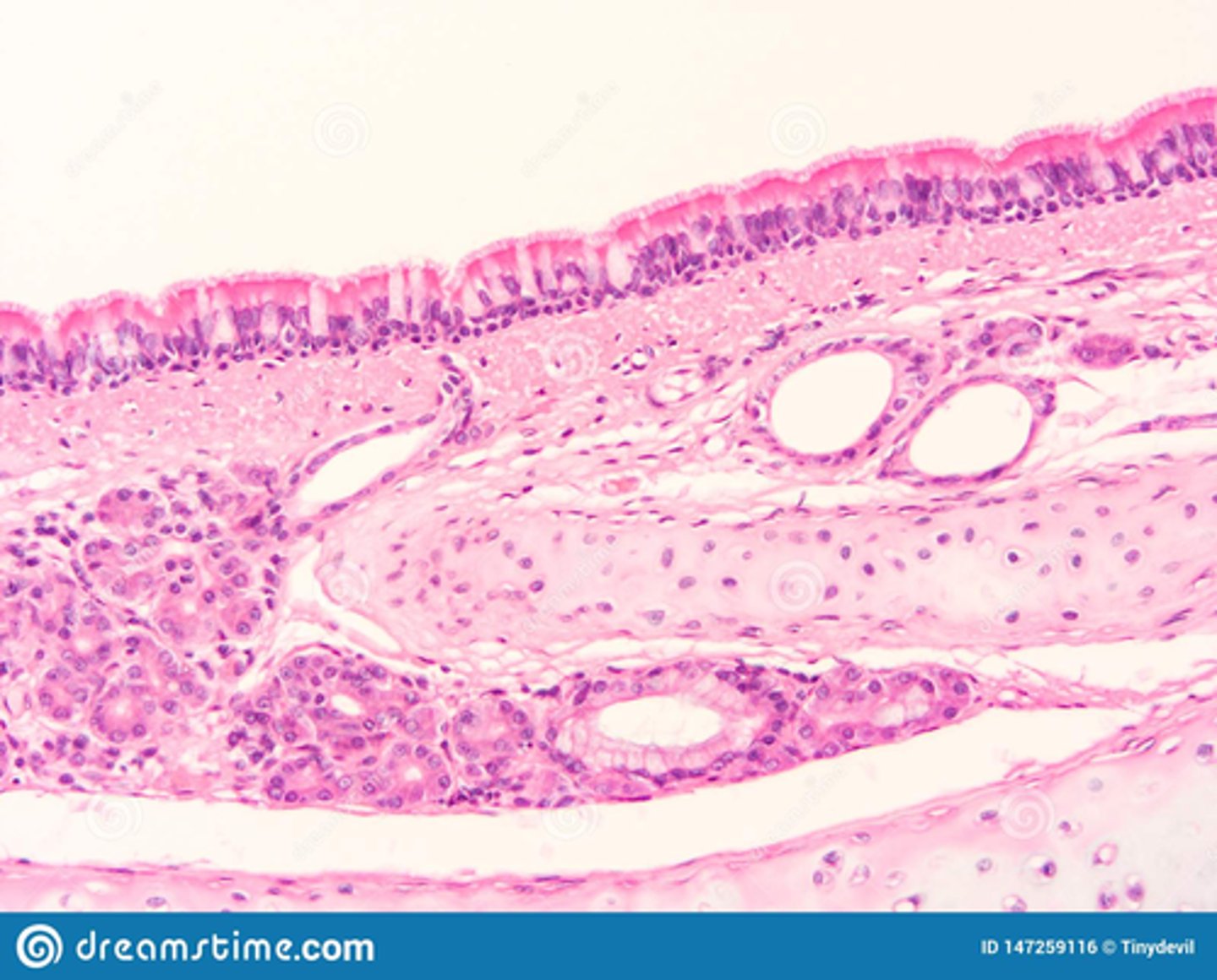
What is the epithelial tissue that makes up the mucosa of the trachea?
Pseudostratified columnar
What is the cellular extension in the respiratory epithelium that allow the filtering of foreign particles?
cilia
What is the function of the numerous goblet cells of the nasal cavity?
The goblet cells secrete mucous to trap foreign microbes.
The area that separates the trachea into the left and right primary bronchi is the __________.
carina
There is a gradual decline in the amount of cartilage until it reaches the bronchioles, where there is the presence of _________ _________ tissue.
smooth muscle
Place the layers of the trachea in order from deep to superficial.
mucosa, lamina propia, submucosa, adventitia
The serous membrane that surround and protect the lungs is the _____________.
pleural cavity
Which respiratory cartilage is elastic cartilage?
epiglottis
primary bronchi
airways enter the lungs at the hilum
terminal bronchioles
represent the end of the conducting airways
tertiary bronchi
airways deliver air to the bronchopulmonary segments
Alveoli
the exchange of carbon dioxide and oxygen occur in these airways
secondary bronchi
transport air to the lung lobes
bronchioles
small airways that deliver air to the lung lobules
The structure that provides an entry and exit site for bronchi, vessels, nerves, and lymphatics of the lungs is the ____________.
hilum
nasopharynx
A

nasal cavity
B
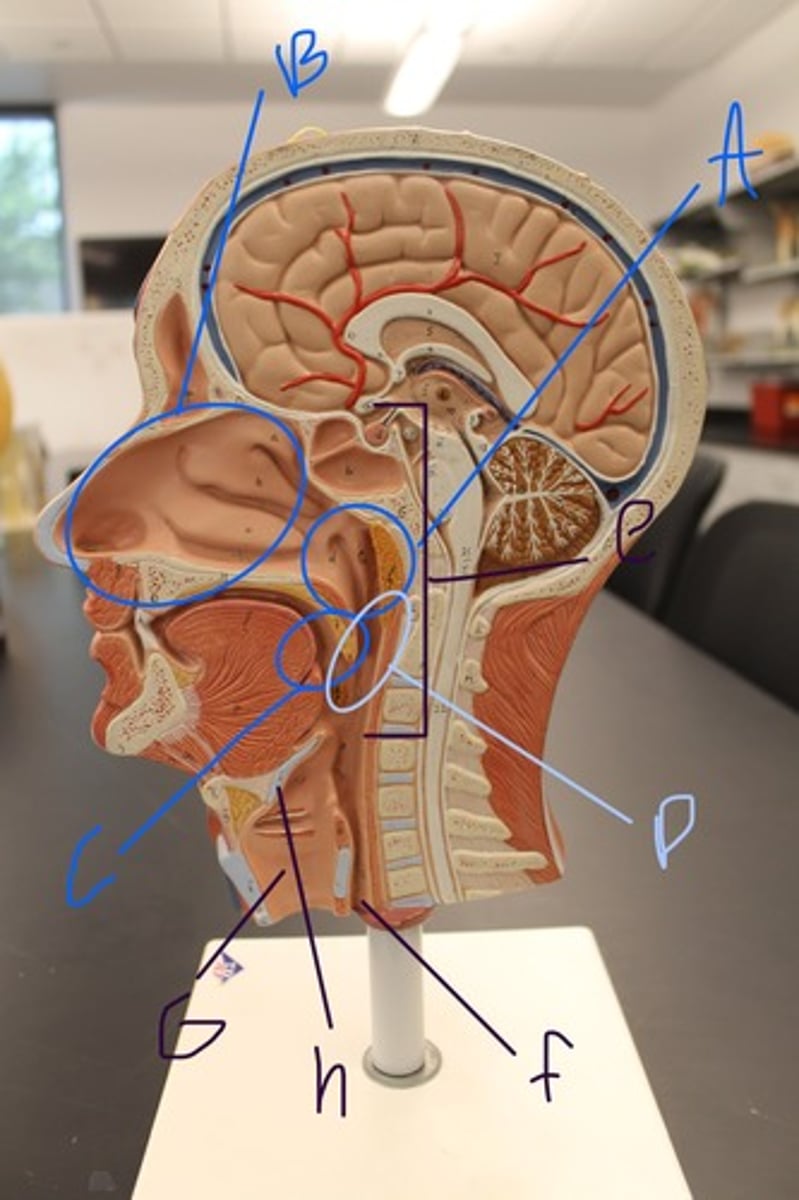
ulva
C

oropharynx
D

Pharynx
E

Esophagus
F

trachea
G

Epiglottis
H

carina
A

primary bronchi
B

Secondary bronchi
C

tertiary bronchi
D

trachea
E
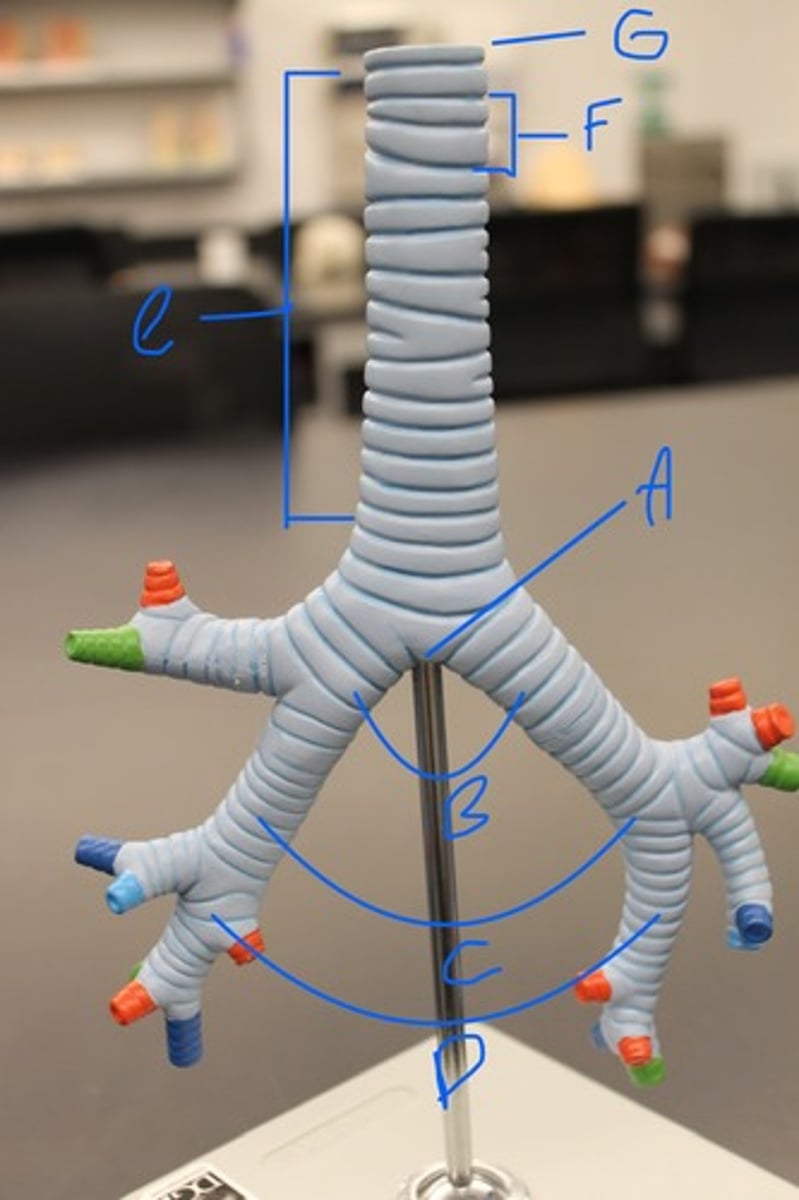
trachial cartilage
F

hilum
G

left oblique fissure
A

Cardiac Notch
B
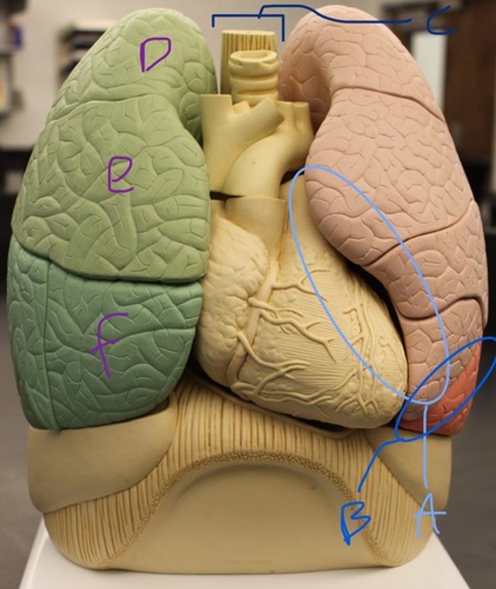
hilum
C

superior lobe
D

middle lobe
E

inferior lobe
F

Left lung
A

Right lung
B
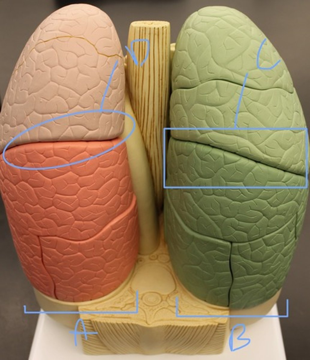
Horizontal fissure
C

left oblique fissure
D

Superior concha
A
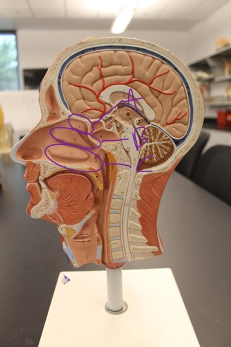
Middle concha
B

Inferior concha
C

infundibulum
7

fimbriae
8

ovary
9

uterus
10
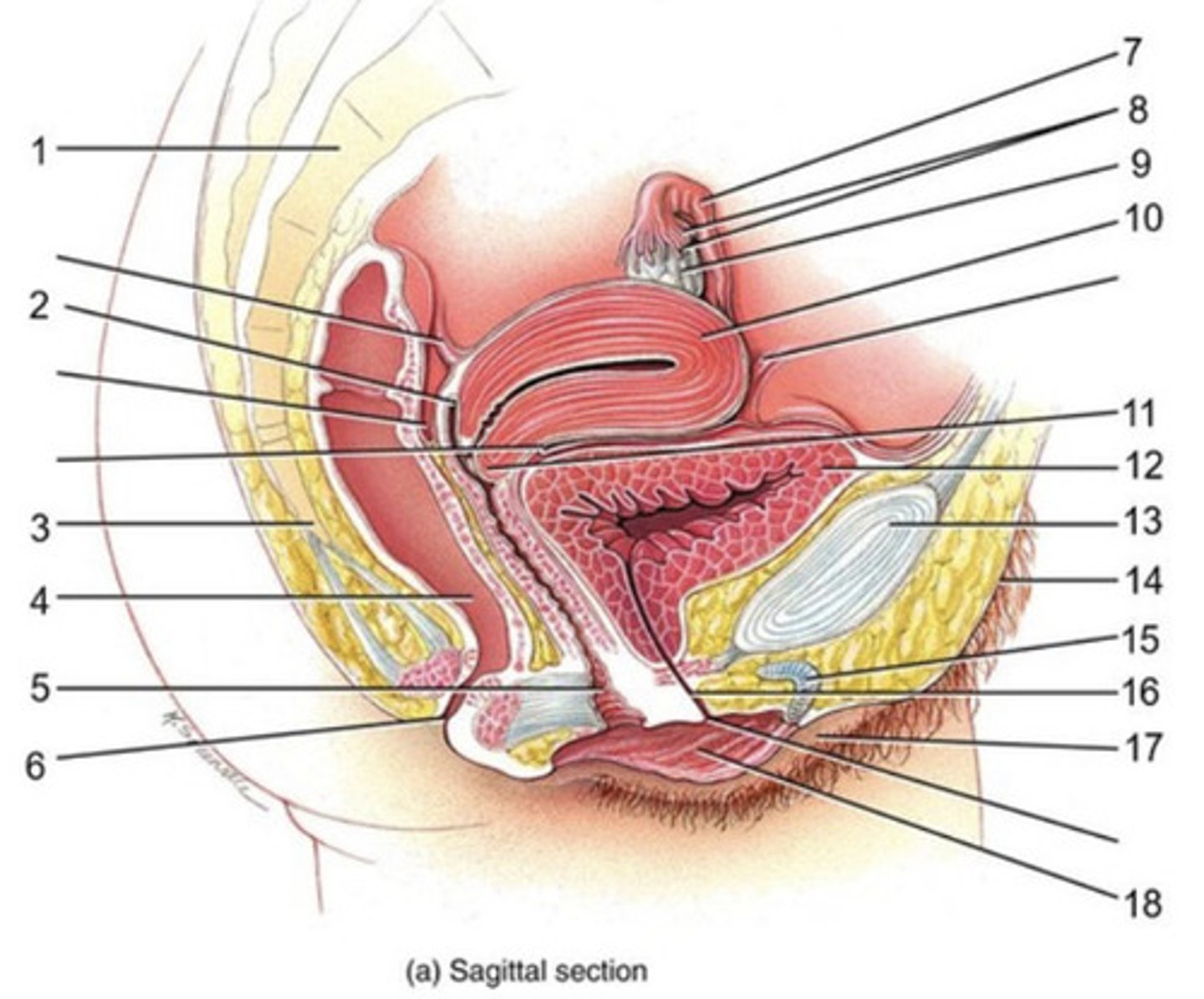
cervix
11

urinary bladder
12

pubic symphasis
13

mons pubis
14

clitoris
15

urethra
16

labium majus
17

labium minus
18

uterus
A

Cervix
B

Fallopian Tube
C

Ovary
D
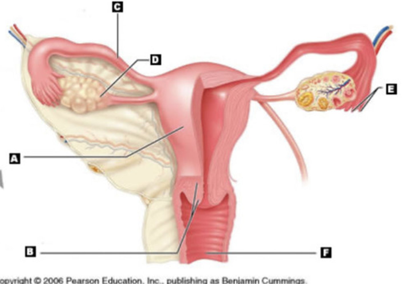
Fimbriae
E

Vaginal Canal
F

urinary bladder
7

bulbourethral gland
8
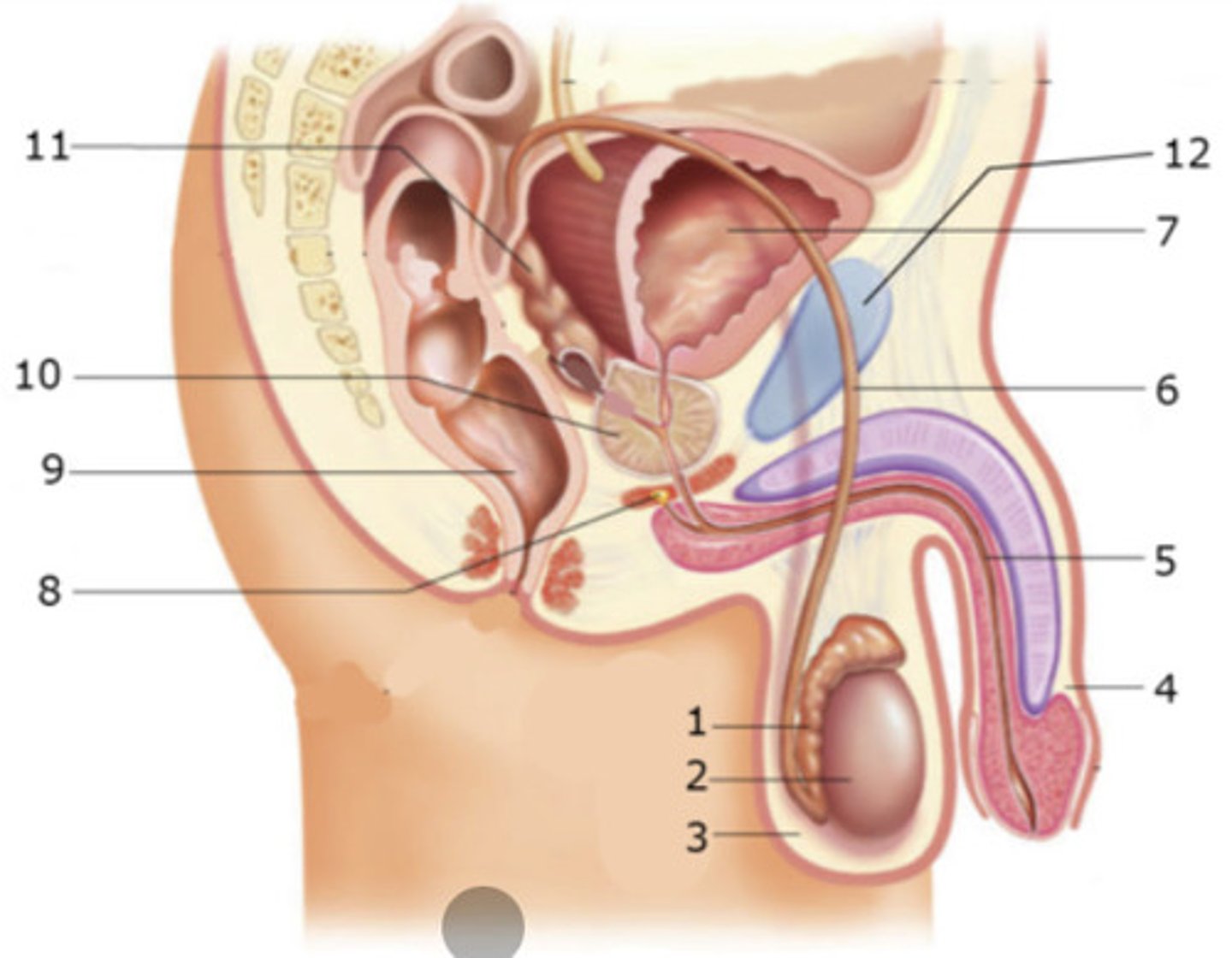
rectum
9

prostate glands
10

seminal gland
11

mons pubis
12

epididymis
1

testes
2

scrotum
3

penis
4

spongy urethra
5

vas deferens
6

Mons pubis
A

Urinary bladder
B

Prostate gland
C

bulbourethral gland
D

Rectum
E
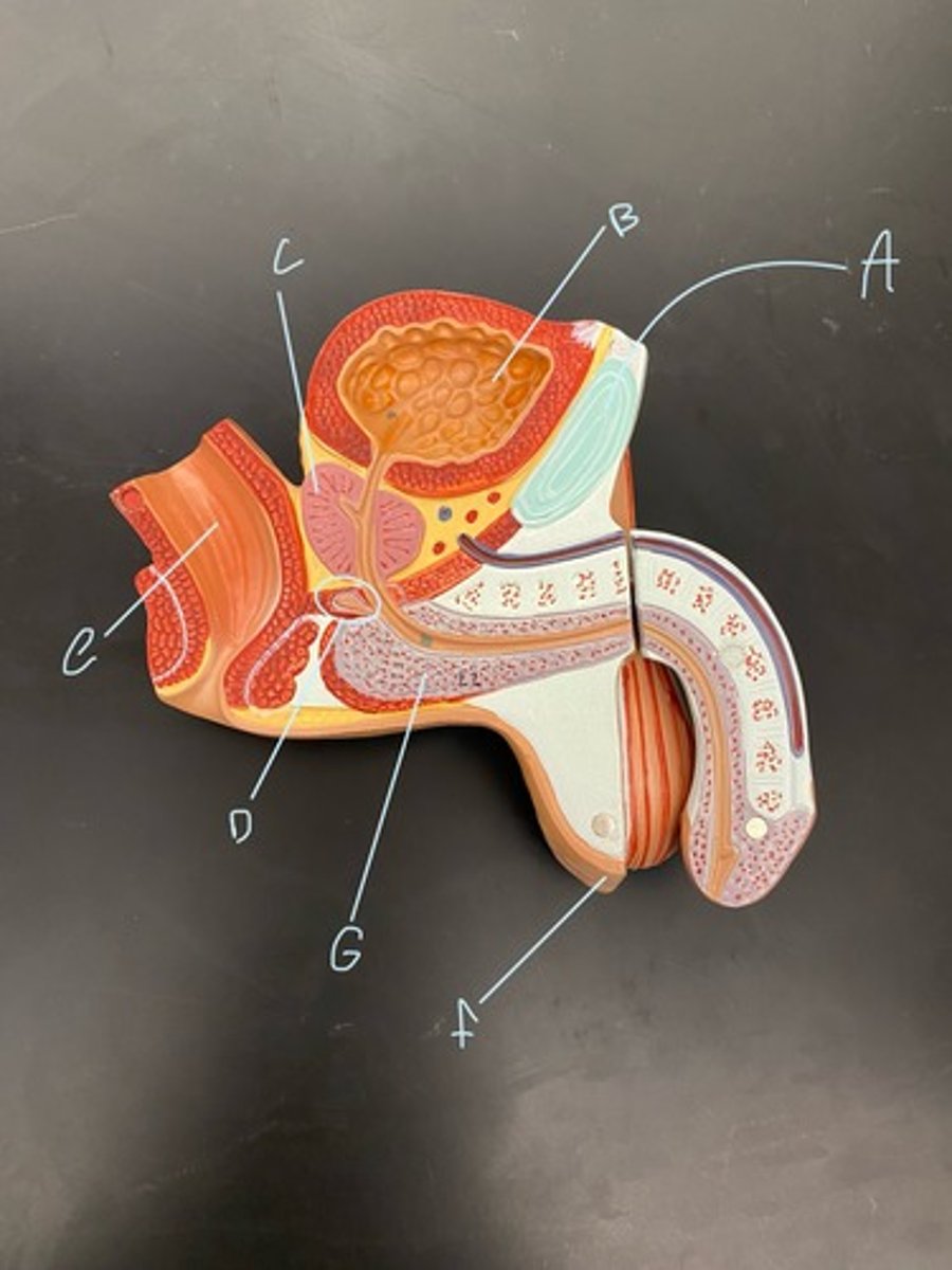
Scrotum
F

spongy urethra
G

Vas Deferens
A
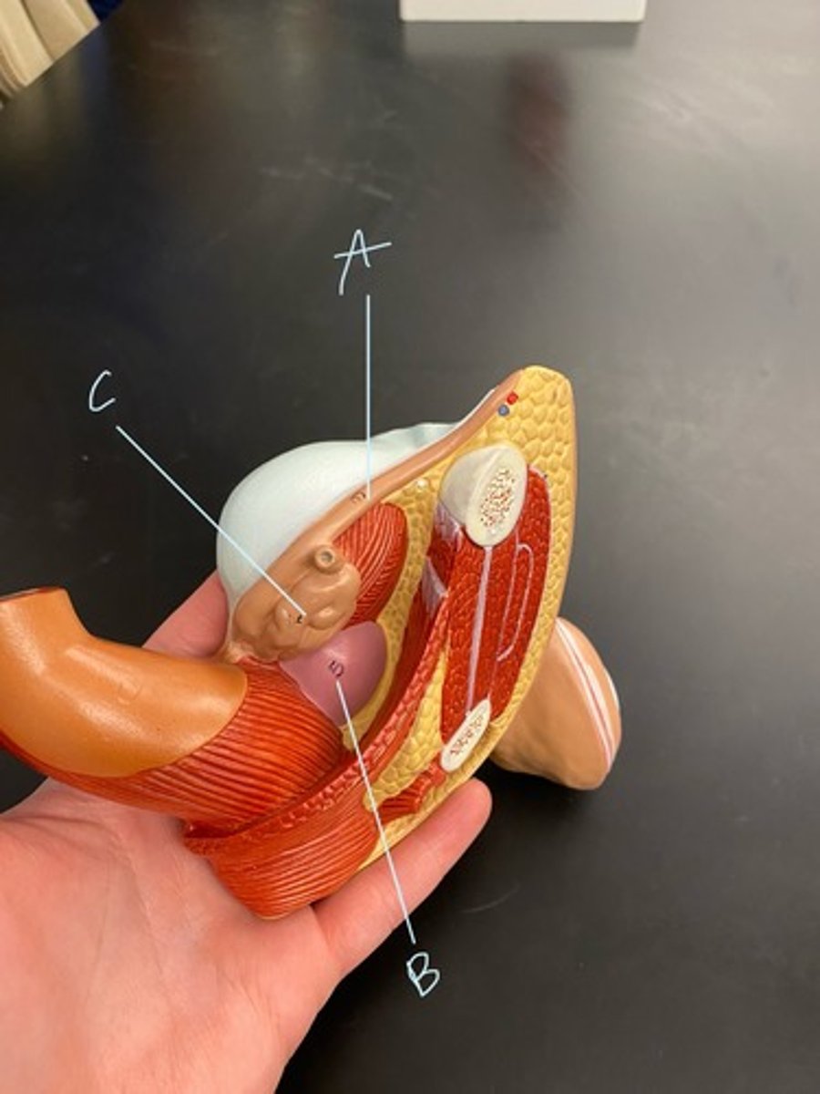
Urinary bladder
B
