Musculoskeletal Imaging, Breast & superficial structure
1/15
There's no tags or description
Looks like no tags are added yet.
Name | Mastery | Learn | Test | Matching | Spaced | Call with Kai |
|---|
No analytics yet
Send a link to your students to track their progress
16 Terms

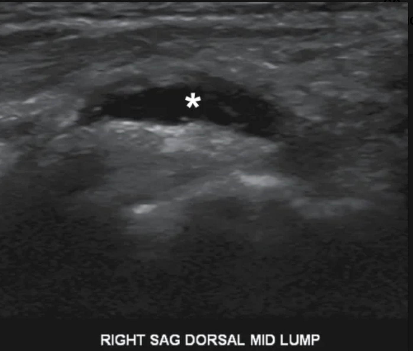
The masses in image were discovered in lactating patient who were suffering from focal breast erythema, swelling, fever, & rain. What is the most likely diagnosis
breast abscess
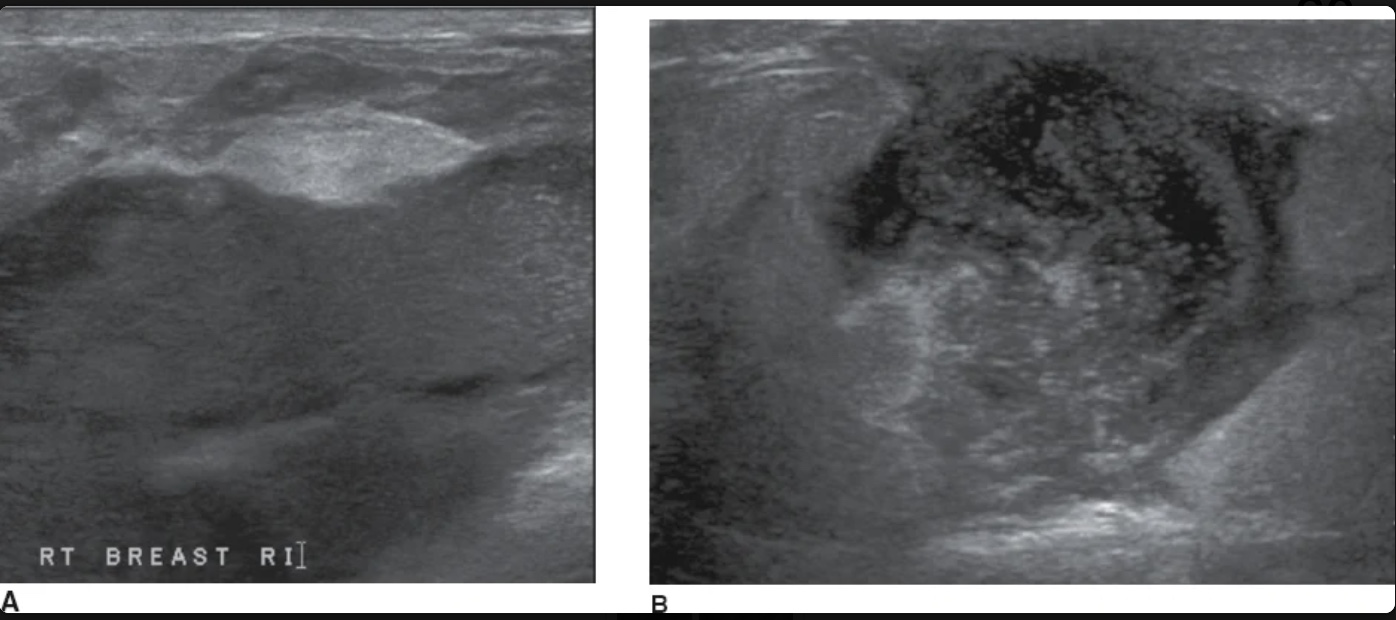
What lab would most likely be elevated in the patient with this image
White blood cell count
Which of the following would be most likely noted in a patient following the surgical removal of a malignant breast lesion?
Seroma
What is the painless abnormality noted in the wrist of patient in image
Ganglion cyst
Which of the following would be most likely noted in a patient following the surgical removal of malignant breast lesion
Seroma

If the abnormality in the image was noted in the region of the radial artery, it would so referred to as
Volvar
A sonographer is asked to analyze a palpable, painful mass w/ in the lateral shoulder muscle following an injection. What is the name of this muscle.
Deltoids
What is the medical term for the wrist bones?
Carpals
Which of the following is NOT typically a cause of gynecomastia?
Renal adenocarcinoma
Which of the following is a clinical test for developmental hip dysplasia that is used to evaluate the hip for the reduction or relocation of a dislocated hip?
Ortolani
Which of the following best describes the most common sonographic appearance of gynecomastia
Hypoechoic, retroareolar mass
A patient presents to the sonography department w/ a hx of cellulitis on his abdomen. The patient has a fever, edema, & complains of local tenderness in a specific region affected by cellulitis. Sonographically, you identify a localized complex collection of fluid. What is the most likely diagnosis?
Superficial abscess
A complicated bake- cyst may contain a thin flap of tissue referred as
pannus
Sonographically - normal muscle appears as
hypoechoic tissue that contain, linear, ehogenic strands
Hyperemic flow w/ in or around a structure is often indicative of
inflammation
Superficial lipomas may appear as all of the following except
anechoic to the surrounding issue