A2.2 cell structure
1/45
There's no tags or description
Looks like no tags are added yet.
Name | Mastery | Learn | Test | Matching | Spaced | Call with Kai |
|---|
No analytics yet
Send a link to your students to track their progress
46 Terms
what is the equation for magnification?
I = AM, M = I/A
how big is a bacteria?
1µm
how big is a cell?
up to 100µm
how big is a virus?
100nm
who discovered cells?
Robert Hooke
when were cells discovered?
1655
How were cells discovered?
looking at a cork with a microscope
why do electron microscopes have a higher resolution?
because beams of electrons have a much shorter wavelength
what is fluorescence?
when a substance absorbs light then re-emits it at a longer wavelength.
what feature do fluorescence microscopes have?
intense light sources, such as high-power LEDs or single wavelength lasers, and give particularly bright images.
how does immunofluorescence work?
Antibodies that bind to specific antigens in the cell are produced. Fluorescent markers of different colours are then linked to them. A multicoloured fluorescent image can then be produced showing the location of the antigens.
what is freeze fracture electron microscopy used for?
producing images of surfaces within cells or the internal structure of membranes
how does freeze fracture electron microscopy work?
a sample is plunged into liquified propane (-190oC) and rapidly freezes to stop movement.
Frozen cell is fractured with a knife; the membrane splits between the phospholipid layers.
A thin layer of metal (usually platinum) is evaporated onto the fractured surface.
Organic material is dissolved, leaving a metal replica of the membrane surface.
Replica is viewed under the electron microscope.
Why do membranes fracture in freeze fracture microscopy?
the membrane is made of two layers of phospholipids, the ice crystals usually cause it to fracture between the two layers.
what is cryogenic electron microscopy used for?
to research protein molecular structure.
how does cryogenic electron microscopy work?
a thin layer of pure protein solution is applied to a grid
solution is flash frozen to create smooth vitreous ice and prevent water crystal formation - usual coolant is liquid ethane (-182oC).
many patterns of protein molecules are produced
computers combine the patterns to produce a 3d image of the molecules

what is prokaryotic cell wall made of?
peptidoglycan
what size are prokaryotic ribosomes?
70S
draw a prokaryote.
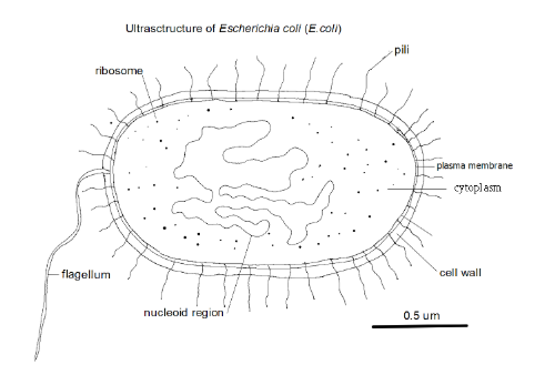
what area of a prokaryotic cell contains DNA?
nucleoid
what does compartmentalised mean?
divided up by partitions into compartments - by internal membranes
what size are eukaryotic ribosomes?
80S
what are plastids found in and what are they?
plants - membrane bound organelles e.g chloroplasts
what are centrioles in plants and fungi?
only present in plant and fungi cells with swimming male gametes which have centriole at base of flagellum
what are centrioles in animals?
used to construct spindle, that moves chromosomes in mitosis, and the microtubules in cilia and flagella.
what are undulipodia?
cilia and flagella
what does atypical mean?
not usual
how are red blood cells atypical?
red blood cells have no nucleus
how are phloem sieve tube cells atypical?
during development the nucleus and most other cell contents are broken down, leaving only the plasma membrane
how are skeletal muscle cells atypical?
some large multinuclear structures form when cells fuse - syncytium. this is how muscle fibres develop
how are Aseptate fungal hyphae atypical?
in some cells the nucleus repeatedly divides without cell division resulting in an unusually large multinucleate structure
draw and label a eukaryotic cell
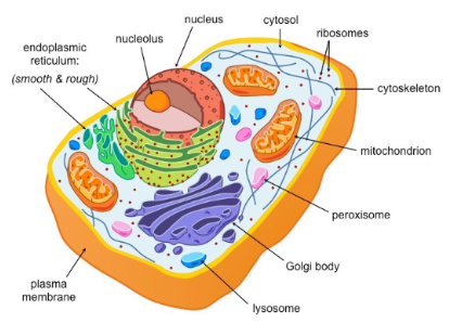
what is the rough endoplasmic reticulum?
consists of flattened membrane sacs (cisternae). ribosomes are attached to the outside. its main function is to synthesise proteins. the proteins are carried by vesicles which bud off and are moved to golgi body.
what is the smooth endoplasmic reticulum?
used to synthesise lipids and package into vesicles - no ribosomes attached
what are lysosomes?
vesicles that contain digestive enzymes
what is the golgi apparatus?
consists of cisternae. it processes proteins brought in vesicles by RER
what are mitochondria?
double membranes, have their own DNA and 70S ribosomes. they produce ATP by aerobic respiration
what are free ribosomes? what size are they?
sites of translations for proteins that will typically remain in the cell. 80S
what are vacuoles and vesicles?
membrane bound structures used for transport and storage.
what are microtubules?
protein tubes that have many functions including the movement of organelles within the cell. e.g spindle fibres and protein motors for cilia and flagella
what are chloroplasts?
double membraned, have their own DNA and 70S ribosomes. contain enzymes and structures for photosynthesis such as chlorophyll.
what is the cytoskeleton?
a system of structures made of tubulin including microtubules that maintain the shape of the cell
what evidence is there for the endosymbiosis theory?
mitochondria and chloroplasts are double membrane bound. they contain their own DNA and ribosomes and have their own cytoplasm and enzymes/.
what is the endosymbiosis theory?
that mitochondria and chloroplasts were once free living prokaryotic organisms that were taken in by endocytosis by larger prokaryotes - both benefiting from this - resulting in eukaryotic cells
what are the advantages of multicellularity?
organisms are larger
allows for complexity