ANAT 225 IU Block 3
1/201
There's no tags or description
Looks like no tags are added yet.
Name | Mastery | Learn | Test | Matching | Spaced | Call with Kai |
|---|
No analytics yet
Send a link to your students to track their progress
202 Terms
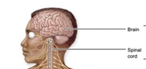
Central Nervous System
brain and spinal cord (control center)
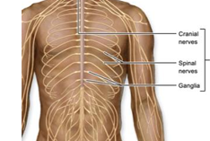
Peripheral Nervous System
cranial nerves, spinal nerves, and ganglia
Sensory division - afferent
receives information FROM body and transmits it TO the CNS for processing, CNS and PNS
Somatic Sensory
receives sensory information from skin, joints, muscle, special senses - vision, hearing, balance, smell, taste
Visceral Sensory
receives sensory info from blood vessels and viscera, not consciously perceived, but still happening
Motor Division - efferent
transmits info FROM CNS TO muscles and glands, CNS and PNS
Somatic motor
innervates skeletal muscle
Autonomic motor
innervates smooth muscle, cardiac muscle, and glands of viscera
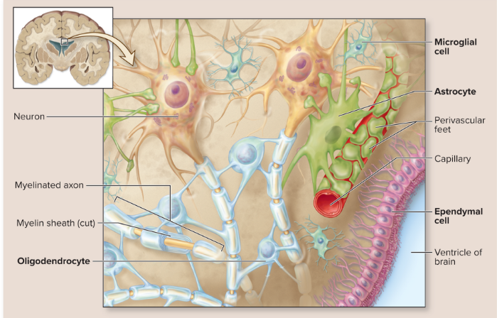
Neuron
respond to stimuli and conduct nerve impulses; amitotic
Glial Cells
support and protect neurons, many kinds in the CNS
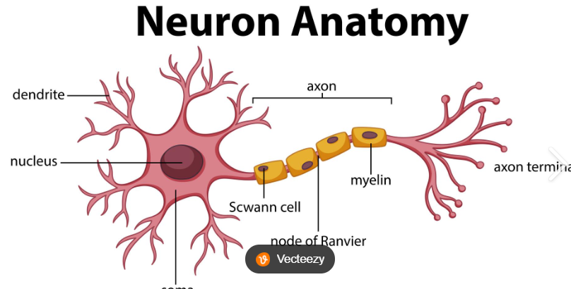
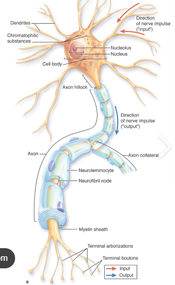
Cell body
neuron’s control center or head

Nucleus
contains nucleolus in the center of nucleus
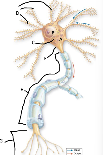
chromatophilic substances
rough ER, protein synthesis, looks like darkened clumps within the cytoplasm, none in axon hillock

Dendrites
nerve input, neurons can have one or many dendrites, short nerve cell processes, each long branch coming out of cell body

Axon
primary transmission line for electrical signals, transfer input, 1 axon per neuron

Axon hillock
portion of cell body from where axon originates, lacks chromatophilic substance

Synapse
site of neuronal communication, termination of axons, axons can communicate with other neurons, muscle cells or gland cells

Multipolar Neuron
one axon and many dendrites; most common, ex: motor nuerons, interneurons
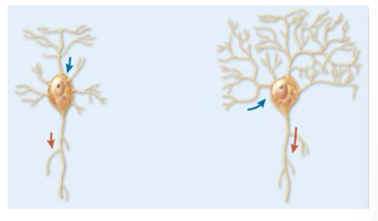
Bipolar Neuron
two processes with one axon and one dendrite; limited/rare, ex: in retina of eye, olfactory neurons in nose
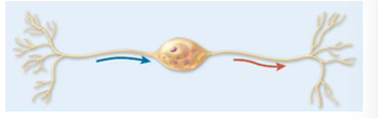
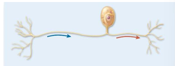
Unipolar Neuron
single process from cell body and divides into two branches; common, ex: most (not all) sensory neurons
Sensory Neuron - afferent
Brings information TO the CNS, unipolar or bipolar structure, input

Motor Neuron - efferent
takes information FROM CNS to other parts of the body, multipolar structure, output
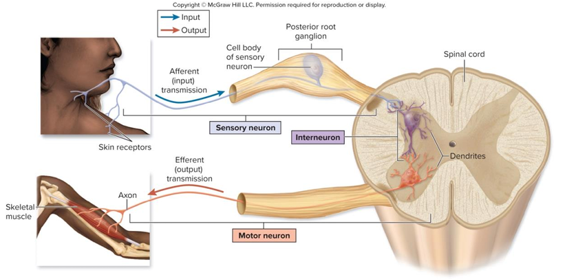
Interneuron
helps coordinate and integrate info between sensory and motor neurons, multipolar structure, mediator, only in the CNS

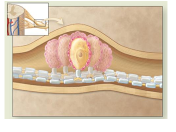
Satellite Cells
surround neuron cell bodies in spinal ganglia, pink around cell body

ganglion
group of neuron cell bodies located outside CNS, in PNS, protect cell bodies, regulate nutrient exchange and waste removal
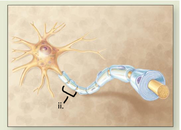
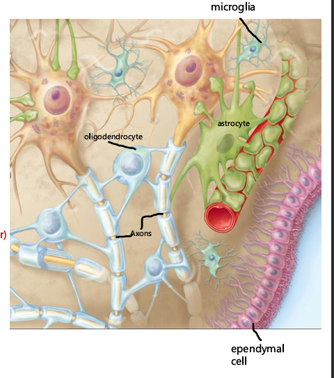
Neurolemmocytes
myelinate axons in PNS, protective covering around axon, insulates axon to produce faster nerve impulses, cna help regenerate damaged PNS axons
Astrocytes
regulate transfer of materials from blood to the brain - help the workings of “blood - brain barrier”, makes vessels less leaky
blood brain barrier
keeps harmful substances away from brain, some needed substances can’t pass the barrier like chemotherapy drugs, no chemo for brain cancer, associated with Parkinson’s disease
Oligodendrocytes
myelinate axons in CNS

Microglia
phagocytize (remove and eat up) damaged neurons, they replicate when there is CNS damage and they need to clean up an area

Ependymal Cells
line central canal and ventricles - help circulate cerebrospinal fluid (CSF)

Myelination
process of wrapping an axon with myelin, insulates axon and produces a faster nerve impulse

Myelination procedure in PNS
neurolemmocyte wraps around a 1 mm portion of axon, cytoplasm and nucleus of neurolemmocyte is “squeezed” to the outside, inner successive layers of cell membrane make up the myelin sheath
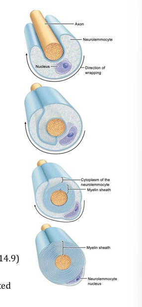
neurofibril nodes
separates neurolemmocytes, gap between 2 adjacent neurolemmocytes where nerve impulse generated, unmyelinated
Myelination procedure in CNS
one oligodendrocyte myelinates 1 mm portions of many axons
Epidural space
space superficial to the dura mater, filled with fats and blood vessels

Dura mater
Dense connective tissue, most superficial and thick, first line after epidural space

arachnoid mater
thin, spiderweb like membrane, second line after epidural space
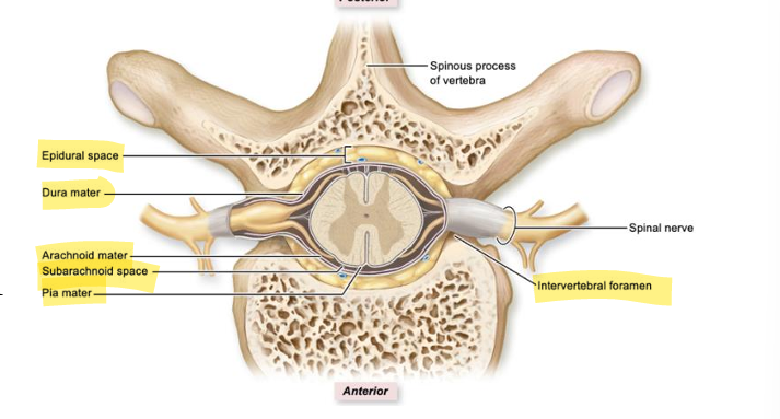
subarachnoid space
cerebrospinal fluid filled space, between the arachnoid mater and pia mater

Pia mater
thin layer of connective tissue, adheres (sticks) to the spinal cord

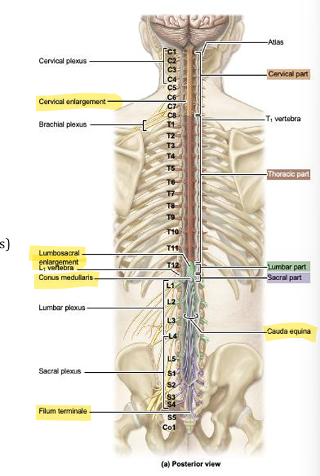
Cervical enlargement
contains neurons that innervate upper limb
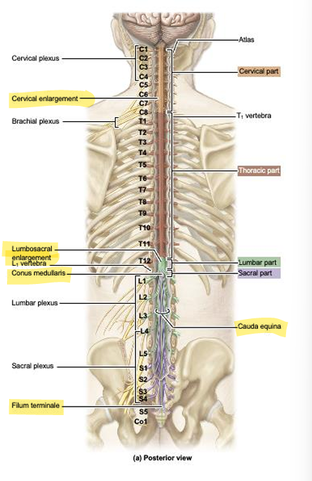
lumbosacral enlargement
contains neurons that innervate lower limb

conus medullaris
pointed end of the spinal cord

cauda equina
collection of nerve roots coming off conus medullaris
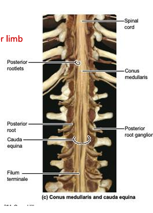
filum terminale
thin strand of pia mater that anchors the conus medullaris to the coccyx

spinal nerves
31 pairs, 8 cervical, 12 thoracic, 5 lumbar, 5 sacral, 1 coccygeal, exit through intervertebral foramen adjacent to the vertebrae with the same name, C1 exits above the first cervical (altas), and C8 exits below the 7th cervical vertebrae
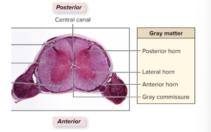
gray matter
inner, contains nerve cell bodies, unmyelinated axons, glial cells and interneurons
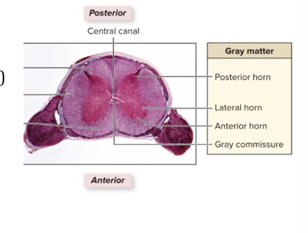
central canal
lined with ependymal cells, contains CSF

anterior horns
cell bodies of somatic motor neurons, innervate skeletal muscle

lateral horns
cell bodies of autonomic (sympathetic) motor neurons (T1 - L2) innervation of cardiac muscles, smooth muscles and glands, involuntary
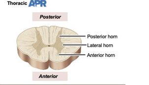
posterior horns
interneurons and axons of sensory neurons

grey commissure
unmyelinated axons crossing from one side to the other, contains central canal
white matter
bundles of myelinated axons, anterior, lateral, posterior funiculi, contains ascending and descending spinal cord tracts
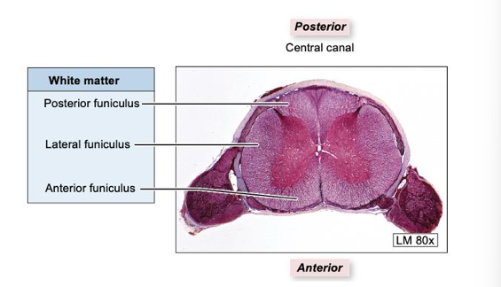
Spinal cord tract
bundles of axons that run in the white matter of the spinal cord
ascending tracts
sensory axons

descending tracts
motor axons

spinal nerve
formed from the unification of anterior and posterior roots

anterior root
axons of motor neurons

posterior root
axons of sensory neurons, organized by function (sensory/motor)
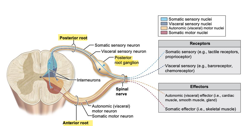
posterior root ganglion
contains cell bodies of sensory neurons, organized by function (sensory/motor)

posterior ramus
innervates deep back muscles and skin of back, smaller, organized by location (anterior/posterior)
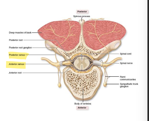
anterior ramus
innervates everything else, larger, organized by location (anterior/posterior)

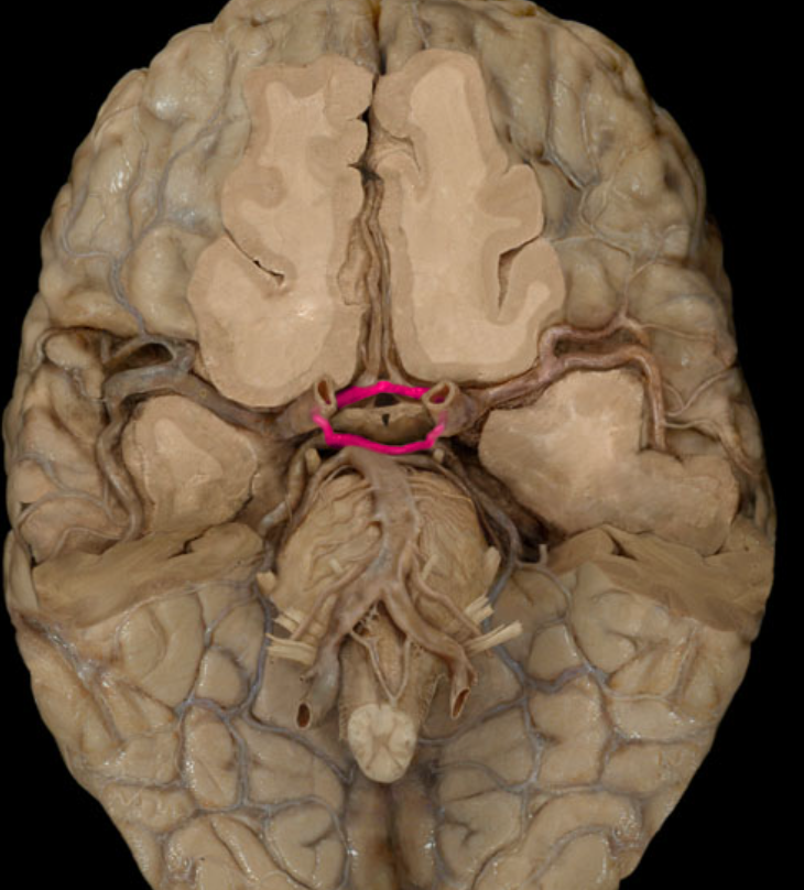
vertebral arteries
supply blood to the brain, passes through the transverse foramen of the cervical vertebrae, enter the cranial cavity through the foramen magnum, and then join to form a single vessel
basilar artery
the single vessel formed after the vertebral arteries connect, sends off various branches to the brain, some of which join with branches from the internal carotid arteries
cerebral arterial circle
provides alternative vascular pathways if one of the major vessels is blocked
dural venous sinuses
drainage of blood through these, within dura mater, drain into the internal jugular veins

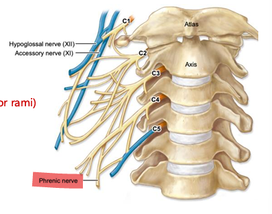
cervical plexus
anterior rami of C1-C4 spinal nerves, sensory innervation of skin of neck ear and shoulders, motor innervation of anterior neck muscles

phrenic nerve
part of cervical plexus, C3, C4, C5, supplies the diaphragm, sensory component, some diaphragm and thorax. FOR DONOR: on neck or over lung, lateral to vagus nerve
Brachial plexus
anterior rami of C5-T1 spinal nerves, primarily innervates upper limb, each nerve has both sensory and motor components - MARMU
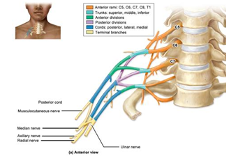
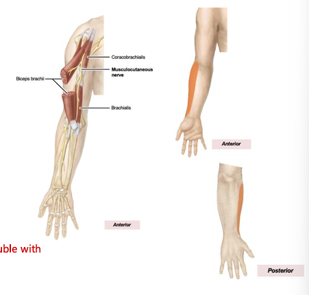
Musculocutaneous Nerve
sensory: lateral forearm, motor: most muscles of the anterior compartment of the arm: coracobrachialis, biceps brachii, brachialis. FOR DONOR: shortest one on the M
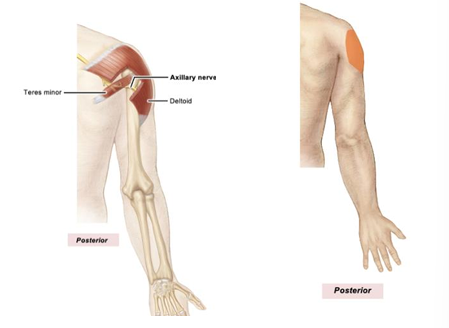
axillary nerve
sensory: lateral shoulder, motor: deltoid, teres minor
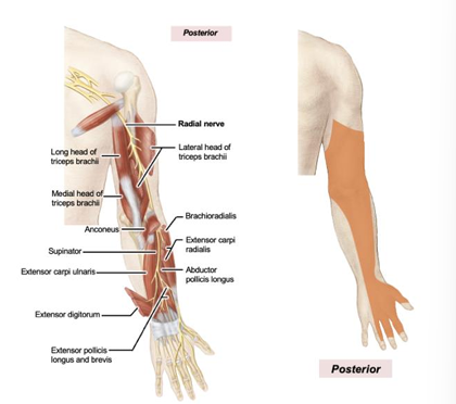
radial nerve
sensory: posterior arm, forearm, hand, motor: muscles of the posterior arm, posterior forearm. FOR DONOR: thicker, underneath the M
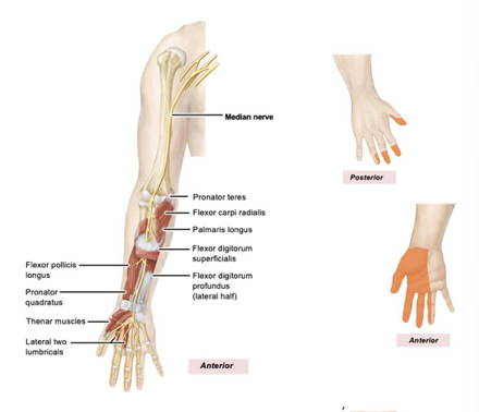
median nerve
sensory: lateral palm, motor: most anterior forearm muscles, hand: thenar muscles, lumbricals going to fingers 2 and 3. FOR DONOR: middle of the M
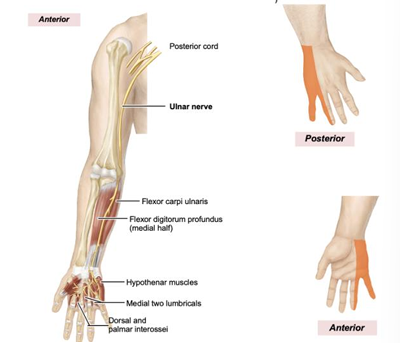
ulnar nerve
sensory: medial hand, motor: anterior forearm - just medial half of flexor digitorum profundus and flexor carpi ulnaris, hand: hypothenar muscles, lumbricals to fingers 4 and 5, interossei. FOR DONOR: most medial on the M
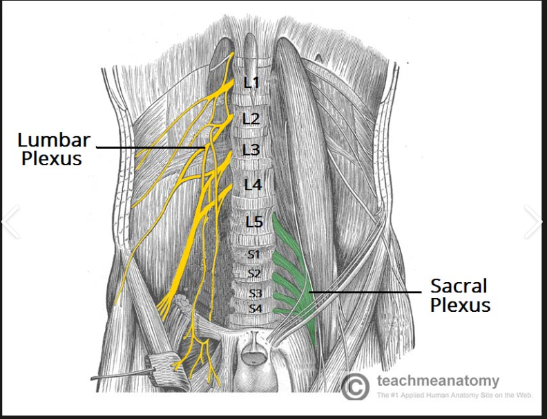
lumbar plexus
anterior rami of L1-L4 spinal nerves, innervates inferior abdominal wall and part of lower limb, each nerve has sensory and motor components
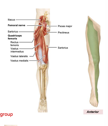
femoral nerve
sensory: anterior and inferomedial thigh, medial leg, medial foot, motor: anterior thigh muscles
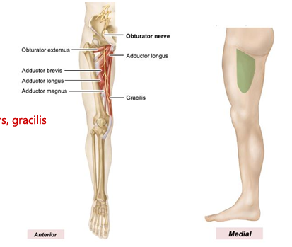
obturator nerve
sensory: medial thigh, motor: medial thigh muscles
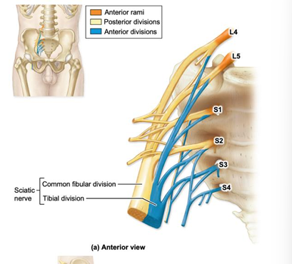
sacral plexus
anterior rami of L4-S4 spinal nerves, innervates the buttocks, pelvic structures, and majority of lower limb, each nerve has sensory and motor components

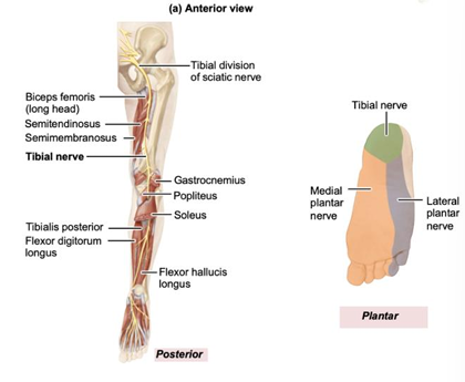
sciatic nerve
splits into tibial nerve and common fibular nerve. FOR DONOR: very large down to middle posterior thigh until split
tibial nerve
sensory: posterior leg/sole of foot, motor: most posterior thigh muscles, posterior leg muscles, muscles on sole of foot. FOR DONOR: medial, posterior only
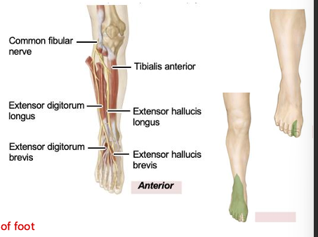
common fibular nerve
sensory: anterior and lateral leg and dorsum of foot, motor: anterior leg muscles, lateral leg muscles, muscles on dorsum of foot. FOR DONOR: lateral, runs to anterior
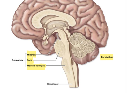
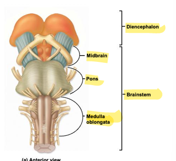
brainstem
consists of: midbrain, pons, medulla oblongata. Relay center for sensory input and motor output, also responsible for many basic reflex actions
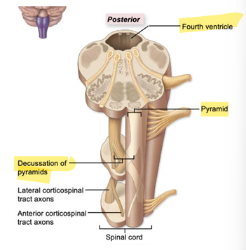
medulla oblongata
most inferior part of brainstem, contains pyramids, autonomic nervous system centers and inferior part of the fourth ventricle
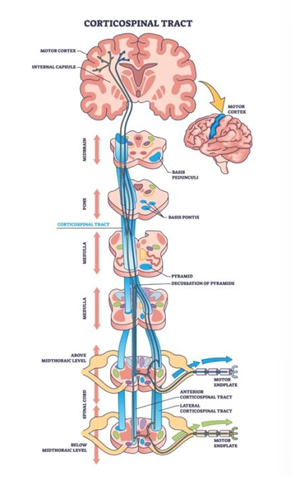
pyramids
bilateral ridges on the anterior side, motor axons, most of the axons decussate (cross over to opposite side)
cardiac center
in the medulla oblongata, regulates heart rate and strength of contraction
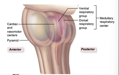
vasomotor center
in the medulla oblongata, constricts (high bp) and dilates (low bp) arterioles

respiratory center
in the medulla oblongata, regulates breathing rate (works with pontine respiratory center in the pons)

fourth ventricle
in the medulla oblongata, inferior part, continuous with the central canal of the spinal cord, communicates with the third ventricle via the cerebral aqueduct
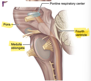
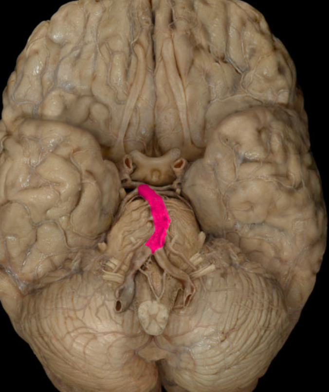
Pons
chiefly composed of groups of axons, below midbrain, above medulla oblongata

pontine respiratory center
in the pons, helps control rate and depth of breathing
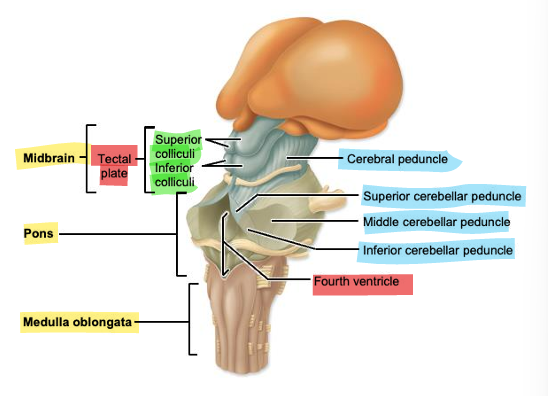
midbrain
above the pons

tectal plate
located on the posterior side of the midbrain, 4 bumps

superior colliculi
2, visual reflex centers, coordinate head and eye movement to sudden image

inferior colliculi
2, auditory reflex centers, coordinate head and eye movements to sudden sound

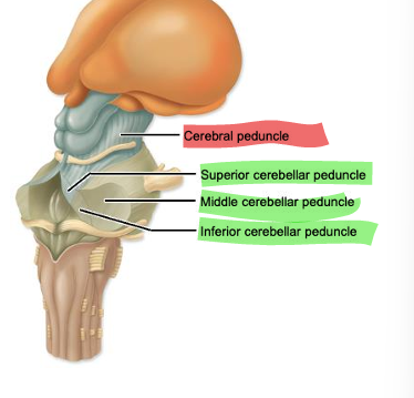
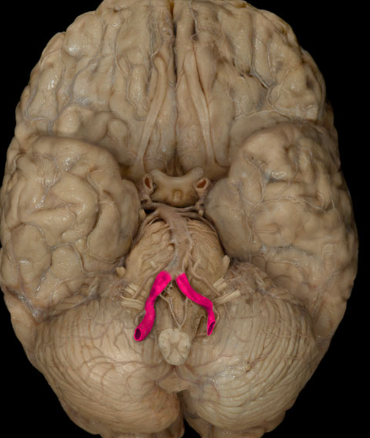
cerebral peduncles
groups of axons (nerve fiber tracts) on anterior side of midbrain, conduct nerve impulses between the cerebrum and brainstem
cerebral aqueduct
connects 4th ventricle to 3rd ventricle, the “chute”
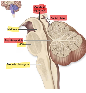
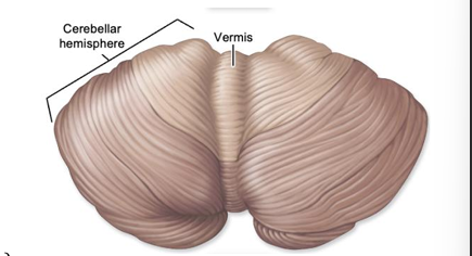
cerebellum
right and left cerebellar hemispheres, several functions such as coordinating and fine tuning movements but doesn’t initiate them, maintains balance and posture in response to info from proprioceptors, assists cerebrum with regulation of behavioral expression, some cognitive skills, some language retrieval
superior cerebellar peduncles
connect the midbrain to the cerebellum

middle cerebellar peduncles
connect the pons to the cerebellum