Chapter 12.6 & 13 Neural Integration and Spinal Cord
1/116
There's no tags or description
Looks like no tags are added yet.
Name | Mastery | Learn | Test | Matching | Spaced | Call with Kai |
|---|
No analytics yet
Send a link to your students to track their progress
117 Terms
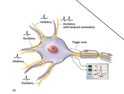
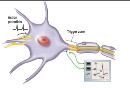
Postsynaptic potentials - most important determinants of neural activity are?
EPSP/IPSP interactions
EPSP
(excitatory postsynaptic potential) = depolarization
IPSP
(inhibitory postsynaptic potential) = hyperpolarization
EPSPs and IPSPs can combine through?
Temporal summation
spatial summation
Facilitation/inhibition
Mixed summation
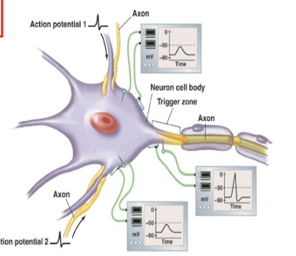
Spatial Summation
Temporal summation
Inhibition
GABA release at axoaxonal synapse inhibits opening calcium channels in synaptic knob
Reduces amount of neurotransmitter released when action potential arrives
Facilitation
Activity at axoaxonal synapses increases amount of neurotransmitter released when action potential arrives
Enhances and prolongs the effect of the neurotransmitter
Determination of the strength of a stimulus can be coded through?
Recruitment (more neurons fire) or by the rate of generation of action potentials are often used to interpret the signal
Neuronal pools
Functional group of interconnected neurons
Neural circuit patterns
Divergence
convergence
Reverberation
serial processing
parallel processing
Localized enlargements provide innervation to limbs
cervical enlargement
lumbar enlargement
Conus medularis
Tapered conical portion of the spinal cord below lumbar enlargement
Filum terminale
fibrous tissue that supports the spinal cord below the conus medularis
Dura mater
Longitudinal collagen fibers
Arachnoid
elastin and collagen fibers
lined with simple squamous epithelium
Subarachnoid space contains CSF
Pia Mater
meshwork of elastin and collagen fibers
Denticulate ligaments extend from pis mater to dura mater
White matter
Is myelinated and unmyelinated axons; exterior portion of the spinal cord
Gray matter
is cell bodies, unmyelinated axons and neuroglia; interior portion of the spinal cors
Nuclei
Are cluster of neuron cell bodies in the gray matter of the brain or spinal cord
Ganglia
are cluster of neuron cell bodies outside the CNS
Posterior gray horns
contains somatic and visceral sensory nuclei
Anterior gray horns
contains somatic motor neurons
Lateral gray horns
contain visceral motor neurons
Gray commissures
contain axons that cross froom one side to the other
Ascending tracts
relay information from the spinal cord to the brain
sensory tracts
Descending tracts
carry information from the brain to the spinal cord
motor tracts
Tracts
Are groups of nerve fibers carrying similar information to similar destinations. Anatomically, tracts are formed into columns, or fasciculi in the spinal cord
Ascending tracts
Carry sensory information toward the brain
have three neurons in series. May have collaterals or connect to interneurons that go to other regions in divergent pathways
Descending tracts
Carry motor information from the brain
have two neurons in series. May have collaterals or connect to interneurons that go to other regions in divergent pathways
first order neurons
Sensory neurons that deliver information to the CNS
Cell bodies are in the dorsal root ganglia
Second order neurons
Interneurons in the CNS that synapse with axons from first order neurons
they are in nuclei within the spinal cord or lower brain regions
send axons to the thalamus
Third order neurons
Found in the thalamus
if the sensation will reach conscious awareness (perceived) they send axons to the primary sensory area of the cerebral cortex (on the same side of the body)
Collaterals go to other regions (sensory association areas, limbic system, other interpretation areas)
Somatic Sensory Pathways three major pathways carry sensory information, that are further subdivided
posterior (dorsal) column pathways
Fasciculus gracilis
fasciculus cuneatus
Anterolateral pathways (spinothalamic and others)
Spinocerebellar pathways
Fasciculus gracilis
Carry information from inferior portion of the body
Fasciculus cuneatus
Carry information from superior portion of the body, upper limbs and neck
Second order neurons are in the nucleus gracilis or nucleus cuneatus within the medulla
They decussate and then relay information to the thalamus via a tract called the medial lemniscus
Anterolateral Pathway
Carries poorly localization sensations of crude (light) touch, pressure, pain, itchm and temperature
Second order neurons
are in the posterior gray horn of the spinal cord. Axons decussate in the spinal cord and ascend within the anterior and lateral spinothalamic tracts
Spinocerebellar pathway
Carries sensation to the cerebellum concerning proprioception
Proprioception
position of muscles, tendons and joints
Posterior spinocerebellar tracts
uncrossed axons (ascend on the same side)
Enter cerebellum via interior peduncle
Anterior spinocerebellar tracts
mostly crossed but some uncrossed axons
most crossed axons re-cross in the pons or cerebellum
Upper motor neuron
Cell body lies in the CNS processing center
Lower motor neuron
Cell body located in the motor nucleus of the brain (for motor cranial nerves) or spinal cord and send axons to skeletal muscles
Each lower motor neuron innervates a single motor unit and cause contraction of a muscle
Somatic Motor Pathways (3)
Corticospinal & cortical bulbar pathway
medial pathways
lateral pathway
Basal nuclei and cerebellum monitor and adjust activity within these pathways
Facilitate or inhibit neurons
The corticospinal pathway (pyramidal system)
Starts with pyramidal cells of the primary motor cortex
Provides voluntary skeletal muscle control
Corticobulbar tracts
Terminate at cranial nerve nuclei
control jaw, eye, face, some neck muscles
Corticospinal tracts
Synapse on motor neurons in the anterior gray horns of the spinal cord
Visible along medulla as pyramids
Gracile Fasciculus/Cuneate fasciculus
posterior
in medulla
Sensation of limb and trunk position and movment, deep touch, visceral pain, and vibration, below level T6
Spinothalamic
lateral and anterior
in spinal cord
sensation of light touch, tickle, itch, temperature, pain, and pressue
Spinoreticular
lateral and anterior
spinal cord (some fibers)
Sensation of pain from tissue injury
Posterior/Anterior(spinal cord) spinocerebellar
lateral
None of one of them and in spinal cord for the other
Feedback from muscles (proprioception)
Lateral corticospinal
lateral
in medulla
frin control of limbs
Anterior corticospinal
anterior
in spinal cord
fine control of limbs
Tectospinal
Anterior
in midbrain
Reflexive head turning in response to visual and auditory stimuli
Lateral reticulospinal
lateral
none
Balance and posture; regulation of awareness of pain
Medial reticulospinal
anterior
none
same as lateral reticulospinal
Lateral vestibulospinal
anterior
none
balance and posture
Medial vestibulospinal
anterior
in medulla (some fibers)
Control of head position
Dorsal roots contain
SENSORY AXONS
Ventral roots contain
MOTOR NEURONS
Nerve plexus
Complex interwoven network of nerves from different
spinal nerves made of mixed nerves (contain both
sensory and motor neurons)
Four large plexuses
Cervical plexus
Brachial plexus
lumbar plexus
Cervical plexus
Innervates neck, thoracic cavity, diaphragm, phrenic nerve
Brachial plexus
Innervates pectoral girdle and upper limbs
Lumbar plexus and sacral plexus
innervate the pelvic girdle and lower limbs, sciatic nerve
Reflex Arc
• Arrival of stimulus and activation of receptor
• Activation of sensory neuron (afferent pathway)
• Integration / Information processing (interneurons)
• Activation of motor neuron (efferent pathway)
• Response by effector (muscle or a gland)
Innate reflexes
Result from connections that form
between neurons during development (e.g. chewing, sucking,
tracking).
Acquired reflexes
Learned, and typically more
complex (e.g. driving skills, bell ringing and leave class, typing)
Cranial reflexes
Reflexes processed in the brain (startle reflex)
Spinal reflexes
Interconnections and processing events
occur in the spinal cord (e.g. knee jerk reflex)
Somatic Reflexes
• Control skeletal muscle
• They are imprecise and crude (e.g. the knee jerk reflex)
• Provide a rapid response (e.g. pull away from a hot
surface)
• Often modified by higher centers
Visceral reflexes
Control activities of other systems (e.g. blood pressure,
urination, defecation)
Monosynaptic reflex
Sensory neuron synapses directly on a motor neuron
(there is no interneuron)
Polysynaptic reflex
• At least one interneuron between sensory afferent and
motor efferent
• Because of synaptic delay, the more interneurons
there are the slower the reflex i.e. the longer delay
between stimulus and response
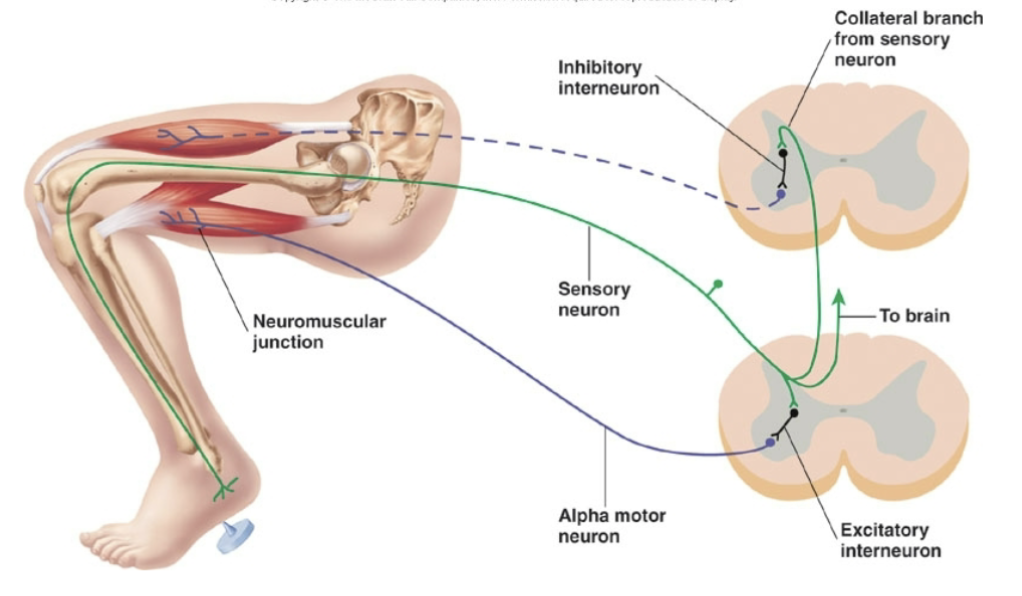
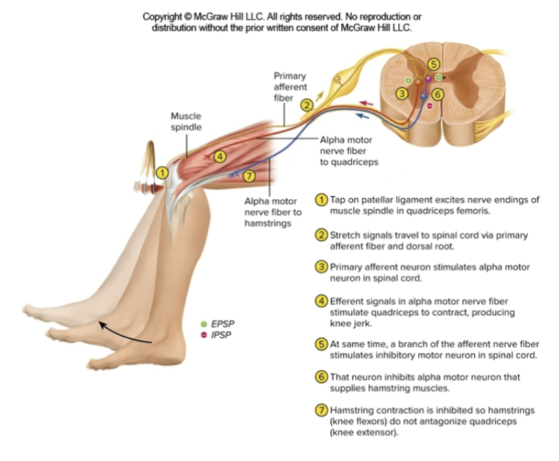
Stretch reflex
automatically monitors skeletal muscle
length and tone
• Patellar (knee jerk) reflex
sensory receptors are?
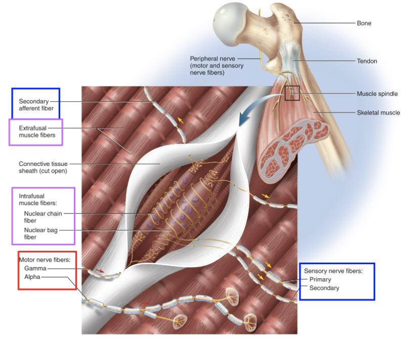
Muscle spindles
Postural reflexes
Maintains upright position
Muscle spindles
Specialized muscle regions used as sensory stretch receptors
Extrafusal muscle fibers
alpha (a) motor neurons
Intrafusal muscle fibers
gamma (y) motor neurons
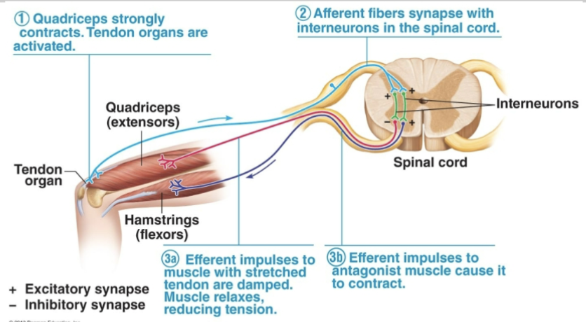
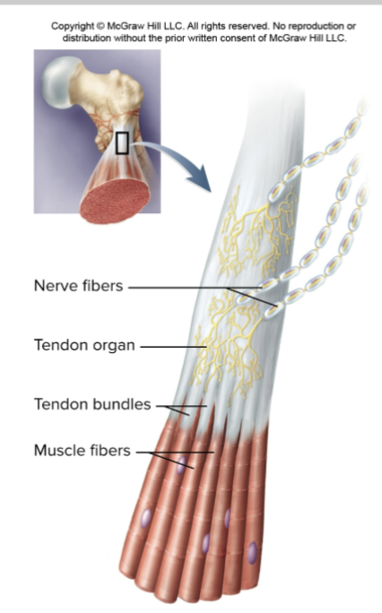
Golgi tendon reflex
prevents contracting muscles fromapplying excessive tension to tendons
Produces sudden relaxation of the contracting muscle and activation of the antagonistic muscles
Reinforcement and inhibition
brain can facilitate motor patterns based in spinal cord
complex movements such as walking can work by having the brain initiate reflex movements
Reinforcement
Reinforcement
Facilitation that enhances spinal reflexes
Spinal relexes can also be inhibited an example is?
Babinski relex replaced by the planter reflex
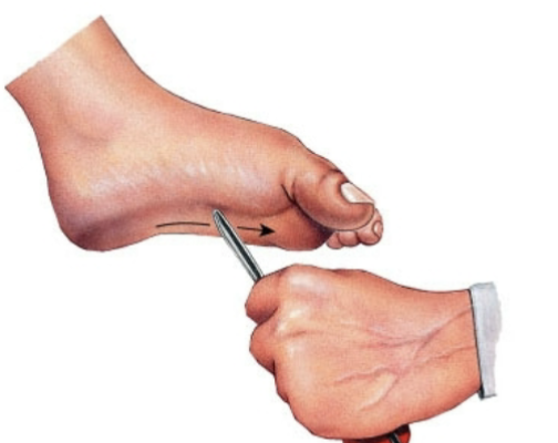
Planter reflex
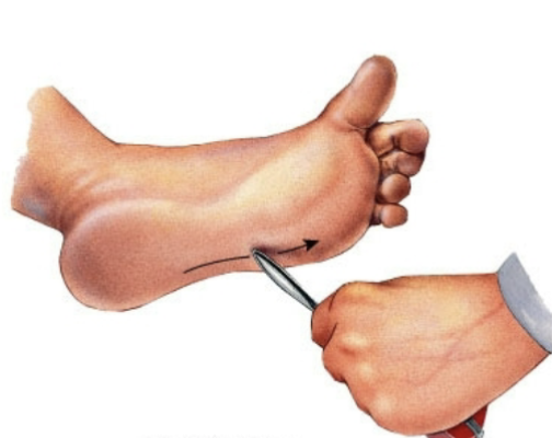
Babinski sign
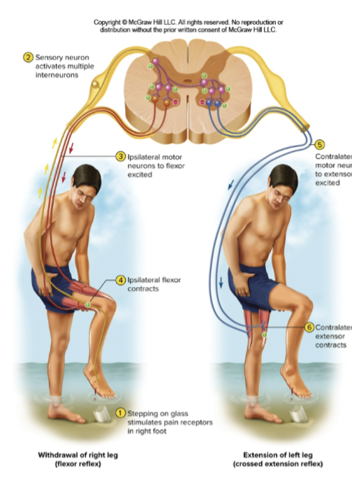
Withdraw and crossed extensor reflexes
ipsilateral
Contralateral
Flexor and Inhibitory Reflexes
Golgi Tendon organ
Muscle spindle relfec arches
Stretch Reflex (e.g. patellar reflex)

1
Phrenic nerve

2
Sciatic nerve

3
Cervical plexus

4
Brachial
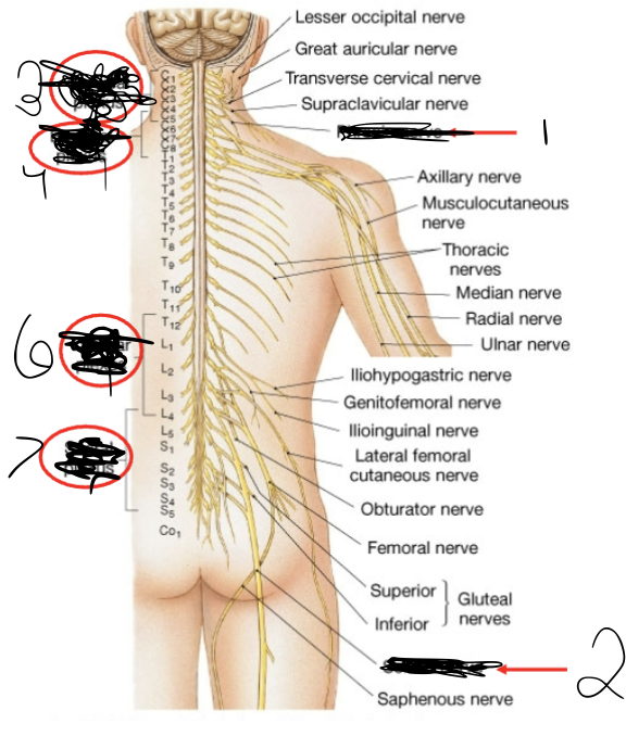
5
Lumbar plexus