digestive tract
1/66
There's no tags or description
Looks like no tags are added yet.
Name | Mastery | Learn | Test | Matching | Spaced | Call with Kai |
|---|
No analytics yet
Send a link to your students to track their progress
67 Terms
Mucosal epithelium, lamina proprio, and muscularis mucosae are found in what layer
The mucosa
What cells secrets pepsinogen
Chief. Cells
Which hormones stimulates gallbladder contraction
Cholecytostokinin
What layer contains the myeneric plexus
Muscularis external
What increases surface aéra in the small intestine
Villi and pelican circulares
What absorbs large lipid complexe
Lacteals
Hepatopancreatic. Sphincter controls the release of what
Pancreatic juice and bile
What absorbs the most nutrients
Jejuneum of small intestine
What type of movement mixes chyme
Segmentation
What organ stores bile
Gallbladder
Stimulation of parietal and chief cells increases acid and enzyme secretion is the function of what
Gastrin
What nerve increases gastric activity
Vagus nerve
Branch of hepatic artery and branch of hepatic portal vein and bile duct form what
The portal triad
What artery supplies the stomach from the celiac trunk
Left gastric artery
Partially digested food and gastric secretions are
Chyme
Water reabsorbtion from waste happens when
Compaction
I’m going contractions that churn intestinal contents is the action of what
Segmentation
Water réabsorptions and feces formation is the main function of what
Large intestine
Parietal cell produces what
Acid
Intrinsic factor functions in what
Vb12 absorption
This carries blood in liver lobule
Sinusoids
Mixing contractions is known as
Segmentation
Wave like propulsion forward movement is know as
Peristalsis
what is the main function of the pancreas
produce digestive enzymes
what type of cells release hydrochloric acid and what’s its function
parietal cells gets rids or bacteria and viruses also denatures proteins
what cells produces gastrin that increase stomach activity
G cells
what sphincter goes to small intestines
pyloric sphincter
what are the ridges in the duodenum of the small intestine called
plicae circulares
What is the duct from the gallbladder and liver come together called
common bile duct
what is the duct called in the pancreas
main pancreatic duct
what is the combination of the pancreatic duct and common bile duct as it enters the duodenum of the small intestine
hepatopancreatic ampulla
what surrounds the duodenal ampulla
duodenal papilla
what the sphincter going from the ileum of the small intestine to the cecum of the large intestine
ileocecal sphincter
what are the two top curvatures of the large intestine called
right colic/hepatic flexure and left colic/splenic flexure
what are the individual pouches of the large intestine called
haustra
what is known as the brush border of the small intestine
microvilli
paneth cells secrets what
defensins and lysozymes
function of the hepatic portalcirculation
venous delivery of newly absorbed nutrients being carried to the liver
change in the epithelium of the lower esophagus from stratified squamous to columnar epithelium is know as what
barrettes esophagus
what are mixed together at the hepatopancreatic ampulla
bile and pancreatic enzymes
what is the main molecule digested in the stomach
proteins
what is the only function that is essential to life is
secretion of intrinsic factor
the myenteric nerve plexus is located in which tunic of the alimentary canal
muscularis externa
pepsin enzymatically digest what
protein

this layer is the most external tunic and contains connective tissue and peritoneum represented by letter B
Serosa
what does letter a represent
mesentery
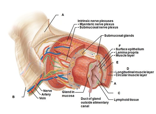
what layer the second most external and contains the circular muscles and longitudinal muscles and the oblique in the stomach and represented by letter D
muscularis externa

this is the second most inner layer represented by letter E contains blood vessels lymph ductless and submucosal plexus
Submucosa

this is the most internal layer of the tunics and contain mucous epithelium lamina propria and muscularis mucosae represented by letter F
mucosa

what does letter C represent
lumen
deep folds of the mucosae and submucosa that extend completely of partially around the circumference of the small intestine
circular folds
conduit for both air and food
pharynx
folds of gastric mucosa
rugae
pocket like sacs of the large intestine
haustra
valve at the junction of the small and large intestine
ileocecal valve
membrane securing the tounge to the floor of the mouth
frenulum
valve controlling food movement from the stomach into the duodenum
pyloric sphinter
where are peyers patches located
ILleum
where is blood from the digestive tract drains first into what
Hepatic portal vein
what is the functional unit of the liver
liver lobule
the sphincter ’ controlling bile entry into duodenum is
oddi
the tineae coli create
haustra
brunner’s glands are located where
duodenum
the hormone that increases acid secreations
gastrin
the nerve plexus in the musclaris externa is the what plexus
myenteric
the large intestine muslce bands are called what
tenaie coli
the accumulation of fluid in the peritoneal cavity is called what
ascites