Unit 5 - Nervous System
1/138
There's no tags or description
Looks like no tags are added yet.
Name | Mastery | Learn | Test | Matching | Spaced | Call with Kai |
|---|
No analytics yet
Send a link to your students to track their progress
139 Terms
Abducens nerve function
abducts the eyeball by innervating the lateral rectus muscle
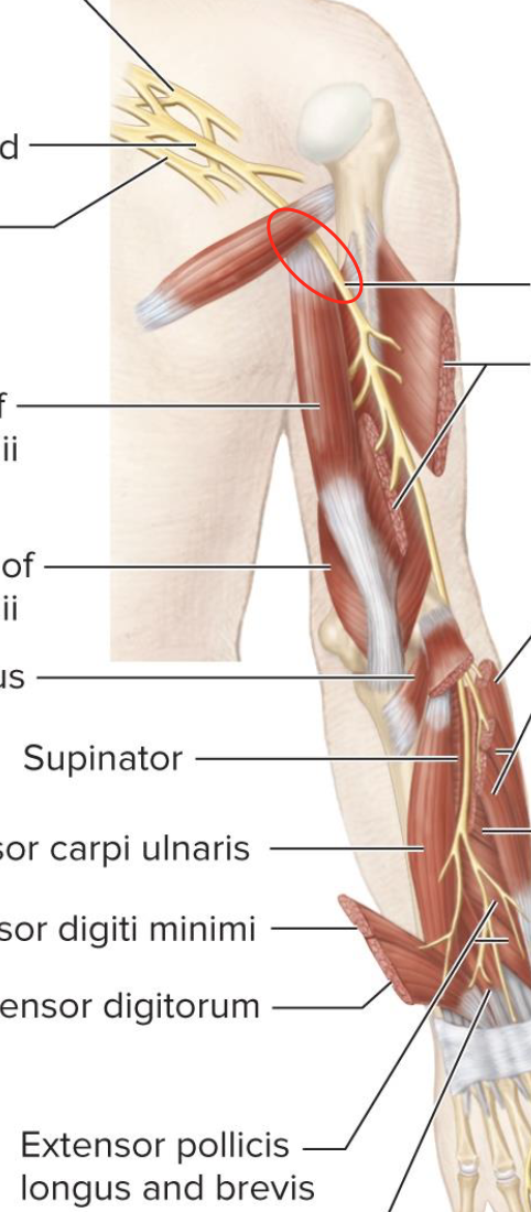
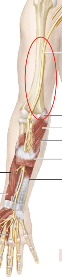
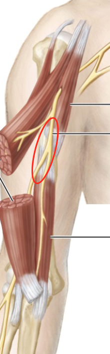
Radial nerve
innervates the triceps, supinator, and brachioradialis
Olfactory
Optic
Oculomotor
Trochlear
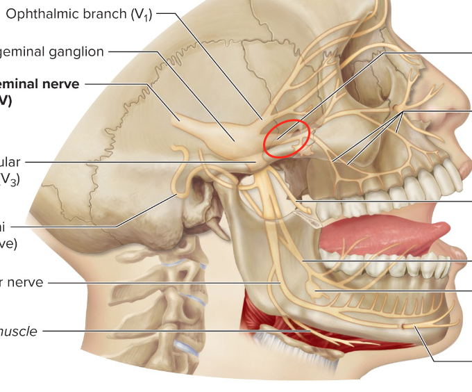
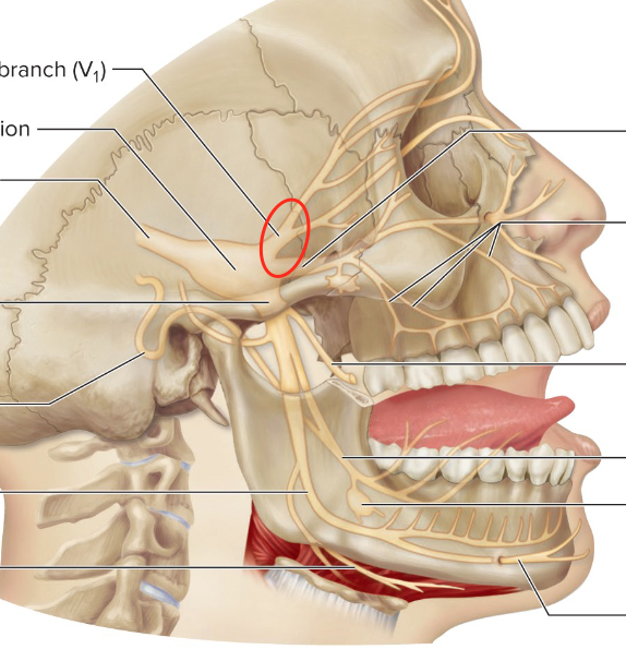
Trigeminal
Abducens
Facial
Vestibulocochlear
Glossopharyngeal
Vagus
Accessory
Hypoglossal
Accessory Nerve
innervates the trapezius muscle, formed from ventral rootlets of spinal cord
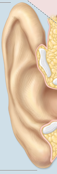
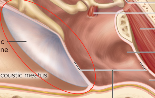
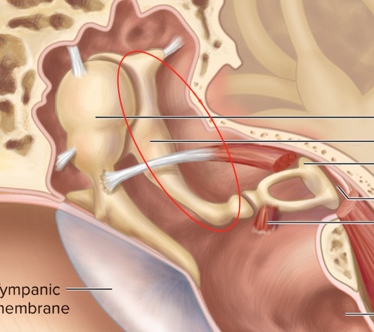
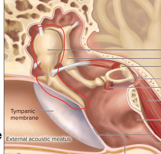
External acoustic meatus
ear canal
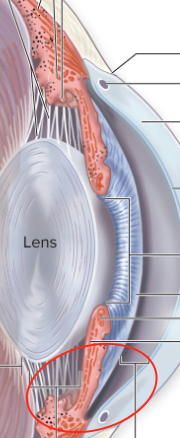
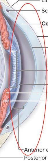
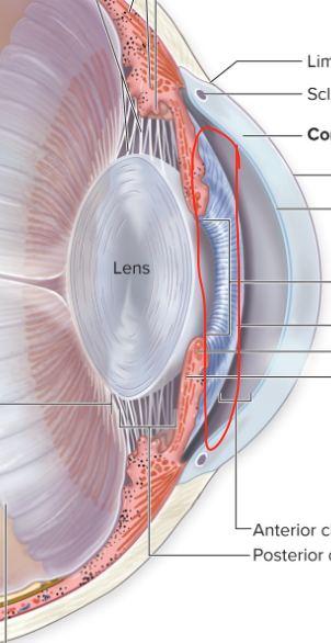
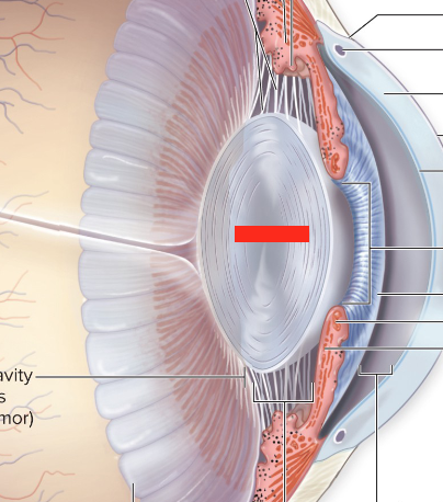
Anterior cavity
Anterior and posterior chambers
Arachnoid mater function
contains major blood vessels, has web-like threads
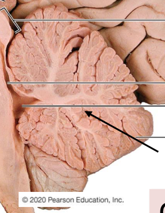
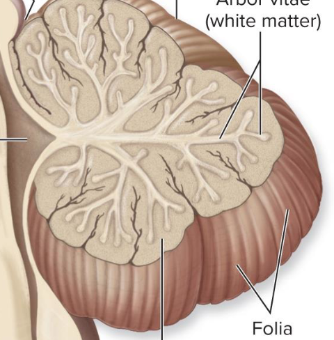
Arbor vitae
Auricale/pinna
Autonomic motor
involuntary innervation of smooth muscle, cardiac muscles and glands
Axillary nerve
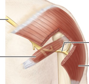
Axillary nerve function
innervated the deltoid and teres minor; stems from the posterior cord
5 Rami, 3 Trunks, 6 Divisions, 3 Cords, 5 Nerves
Brachial plexus hierarchy
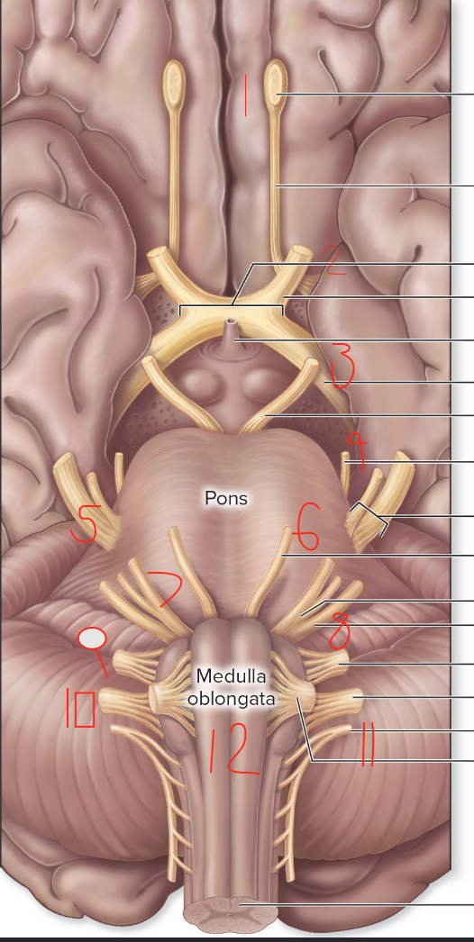
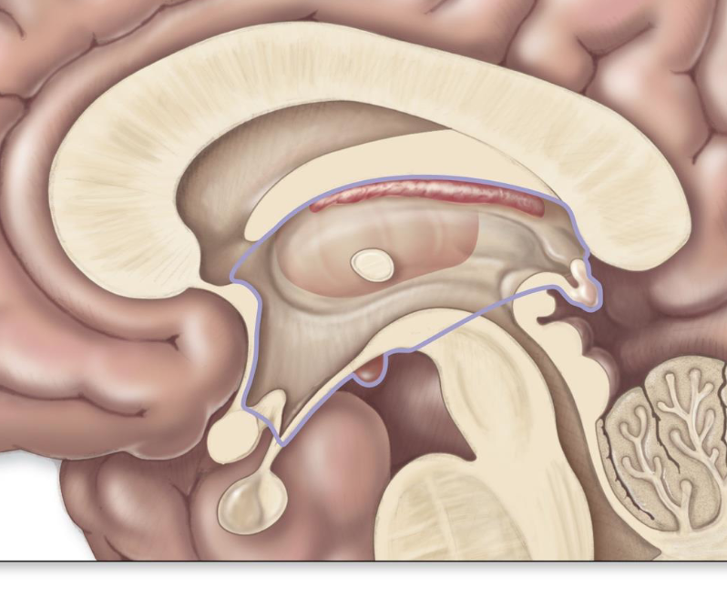
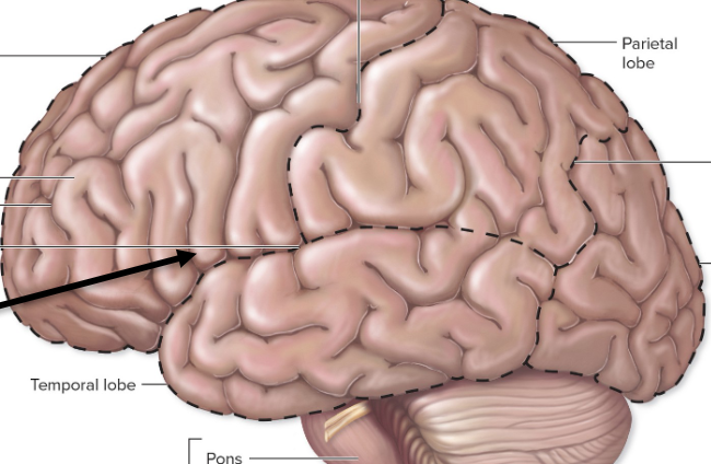
Brain stem
made up of the midbrain, pons, and medulla oblongata
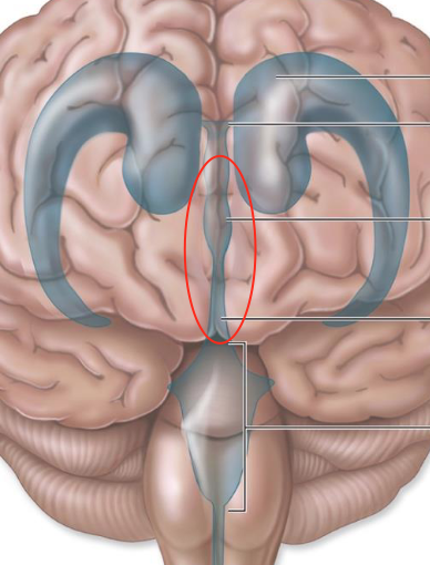
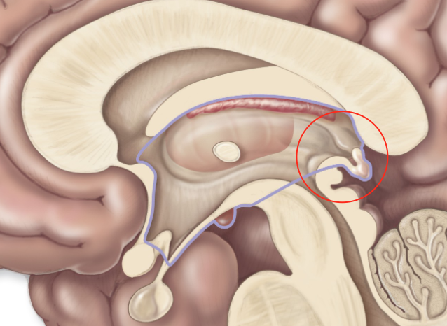
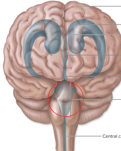
Brain ventricles
expansions of the brain’s central cavity filled with CSF; continuous with eachother and the central canal
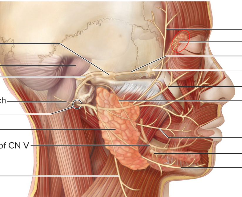
Buccal branch
Central canal
Cerebral hemispheres
symmetrical halves on cerebrum
Cerebellar nuclei
deeply situated in the gray matter of the cerebellum
Cerebellum
Cerebrospinal fluid
fills the hollow cavities of the brain and spinal cord to provide a liquid cushion
Cerebral aqueduct
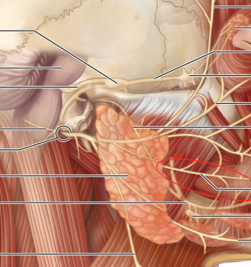
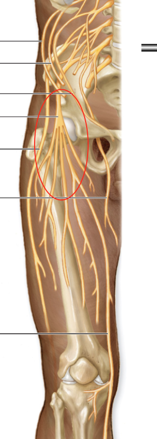
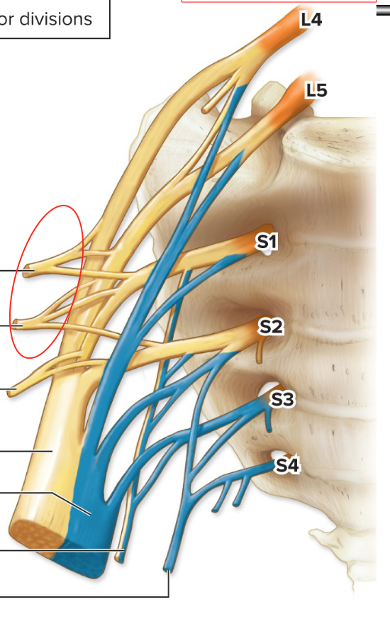
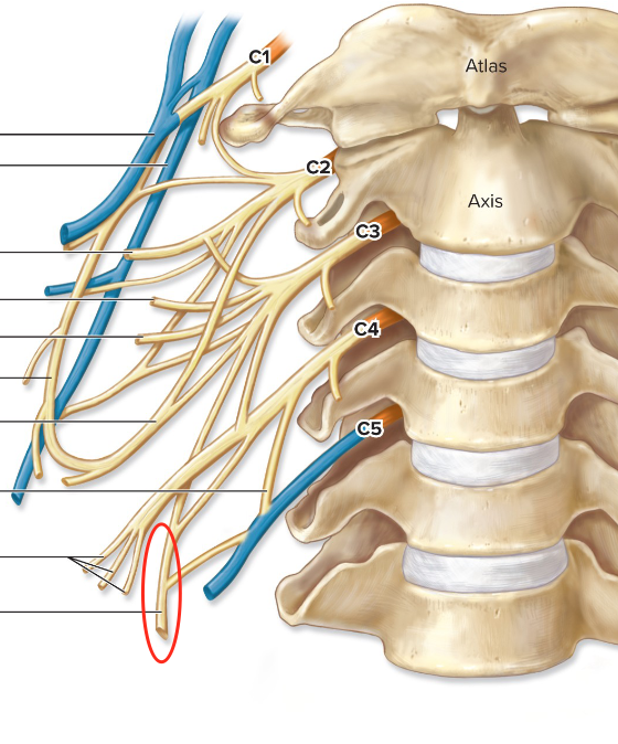
Cervical plexus
formed by ventral rami of C1-C4; under the sternocleidomastoid muscle
Choroid function
posterior portion that prevents light scatter due to brown colour; has vasculature, connective tissue and melanocytes
Choroid
Ciliary body
anterior portion that controls the shape of the lens; acts as an anchor and filled with aqueous fluid
CNS
brain and spinal cord
Cochlea
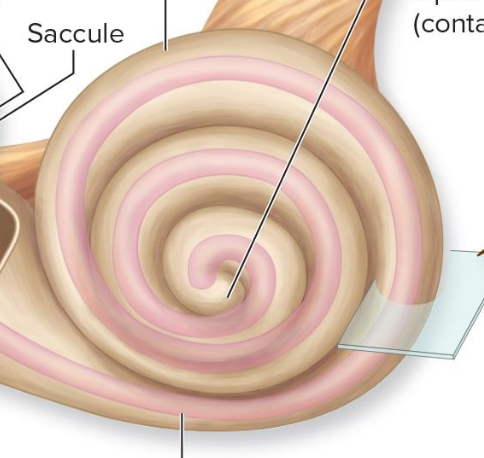
Cochlea function
spiraling chamber that contains the organ of Corti
Cornea
made up of a fibrous tunic
Cortex
grey matter of cerebellum
Diencephalon
forms the centre core of the forebrain
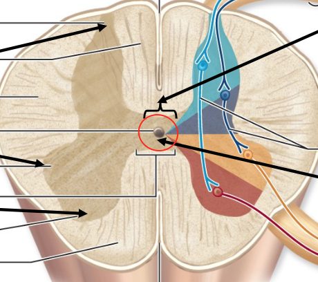
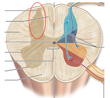
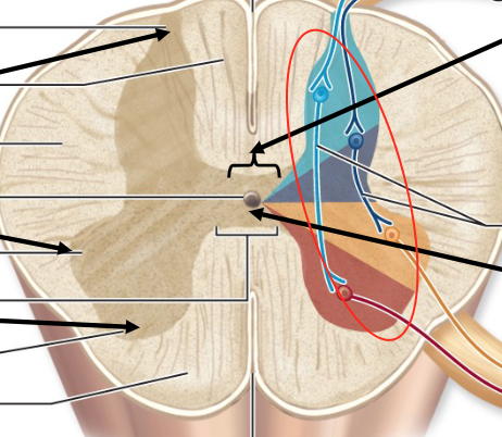
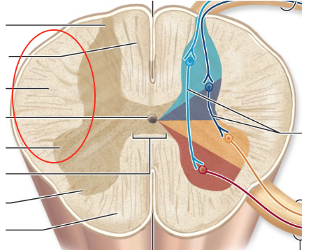
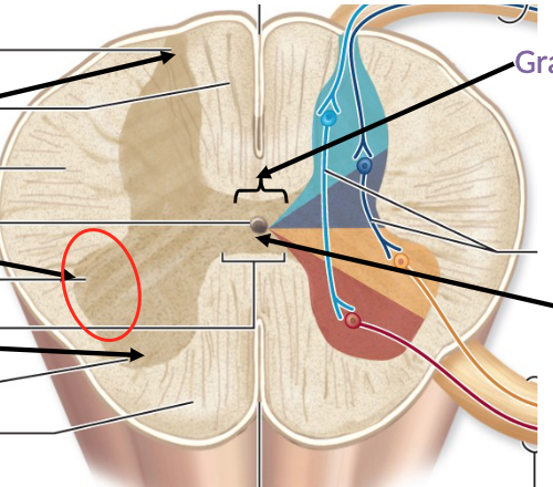
Dorsal funiculus
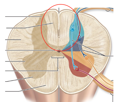
Dorsal median sulcus
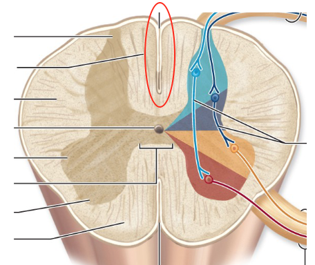
Dorsal horn
consist of interneurons
Dorsal ramus function
supply forsum of the neck and the posterior trunk
Dorsal ramus
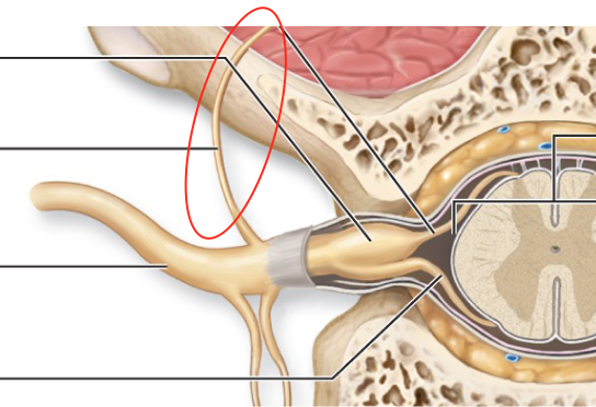
Dorsal root function
contains sensory fibres; cell bodies found in the DRG
Dorsal root
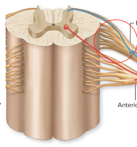
Dura matter function
strong meningeal layer that consists of two layers: periosteal and meningeal
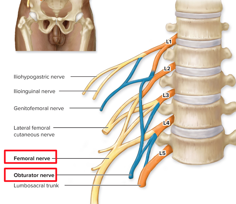
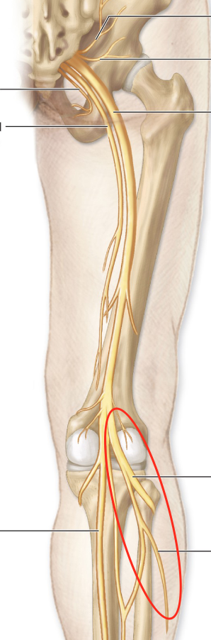
Obturator nerve
Obturator nerve function
innervates adductor muscles: adductor brevis, longus and magnus
Eardrum (tympanic membrane)
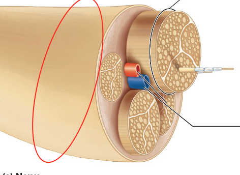
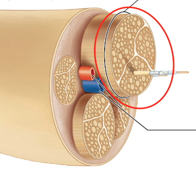
Endoneurium
wraps around axon
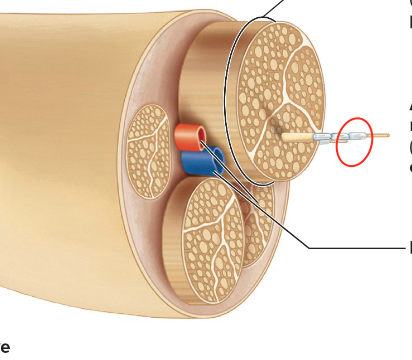
Epineurium
wraps around nerve
Epithalamus
forms part of the roof of the third ventricle
Exteroceptors
sensitive to stimuli arising from outside the body
Facial nerve
innervates muscles of facial expression; taste to anterior portion of the tongue; motor control to the lacrimal glands, mucus glands, and saliva; and sensory to part of the face and mouth
Femoral nerve function
innervates anterior thigh muscles: sartorius, rectus femoris, vastus lateralis, vastus medialis, and vastus intermedius
femoral nerve
Ascending Fibers
go up to the brain
Descending fibres
go down to the body
commissural fibers
cross the midline
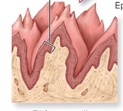
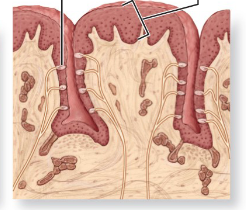
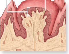
Filiform function
most numerous papillae but do not contain taste buds
Filiform papillae
Foliate function
papillae found on each side of the tongue
Foliate papillae
Foramen magnus
hole where the spinal cord begins
Fourth ventricle
Fungiform function
taste buds found all over the tongue
Fungiform papillae
Glossopharyngeal nerve
innervates structures of the tongue (posterior third for sense of taste) and pharynx
Gluteal nerves function
part of the sacral plexus that innervates the gluteal muscles
Gluteal nerves
Gray commissure
contains the central canal
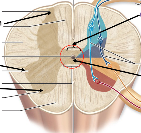
Gray matter
neuronal cell bodies, dendrites, short unmyelinated neurons
Hypoglossal nerve
runs inferior to tongue and innervates the tongue muscles for movement
Incus
Insula
deep sulcus within the cerebral cortex
Functions of the nervous system
i) sensory receptors monitor changes inside and outside the body
ii) processes and interprets sensory input
iii) dictats a response by activating effector organs
Interneurons
relay signals onto other neurons
Iris
pigmented anterior portion with muscle fibres that surround the pupil
Lacrimal glands
produce tears
Lateral funiculus
Lens
divides the posterior and anterior cavities
Lateral horn
Lateral ventricles
location of CSF production
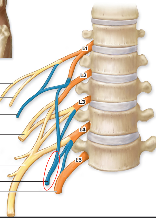
Lumbar plexus
Malleus
Mandibular branch
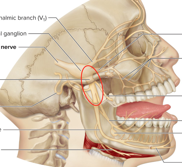
Maxillary branch
Median Nerve
Innervates anterior forearm muscles and lateral palm: pronator quadratus and pronator teres; helps with sensation in 3.5 fingers
Meningeal branch
branch of spinal nerves that goes back into the vertebral canal to innervate spinal structures
Musculocutaneous nerve
innervates biceps brachii and brachialis
Nerve plexus
a network of nerves that originate from the ventral rami to serve the limbs
Oculomotor nerve
innervates muscles that open the eyelid, constrict the pupil, and change lens shape when accommodating for near vision (adduction)
Olfactory nerve
sensory nerves of smell that innervate the olfactory mucosa
Cribiform plate of ethnoid bone
Which bone do olfactory nerves cross?
Optic nerve
sensory nerves of vision
Optic chiasm
crossover of the visual nerves
Opthalamic branch
Palprebrae
eyelids
Perineurium
sheath that covers fassicle
Peroneal (common fibular) nerve
Pharyngotympanic tube
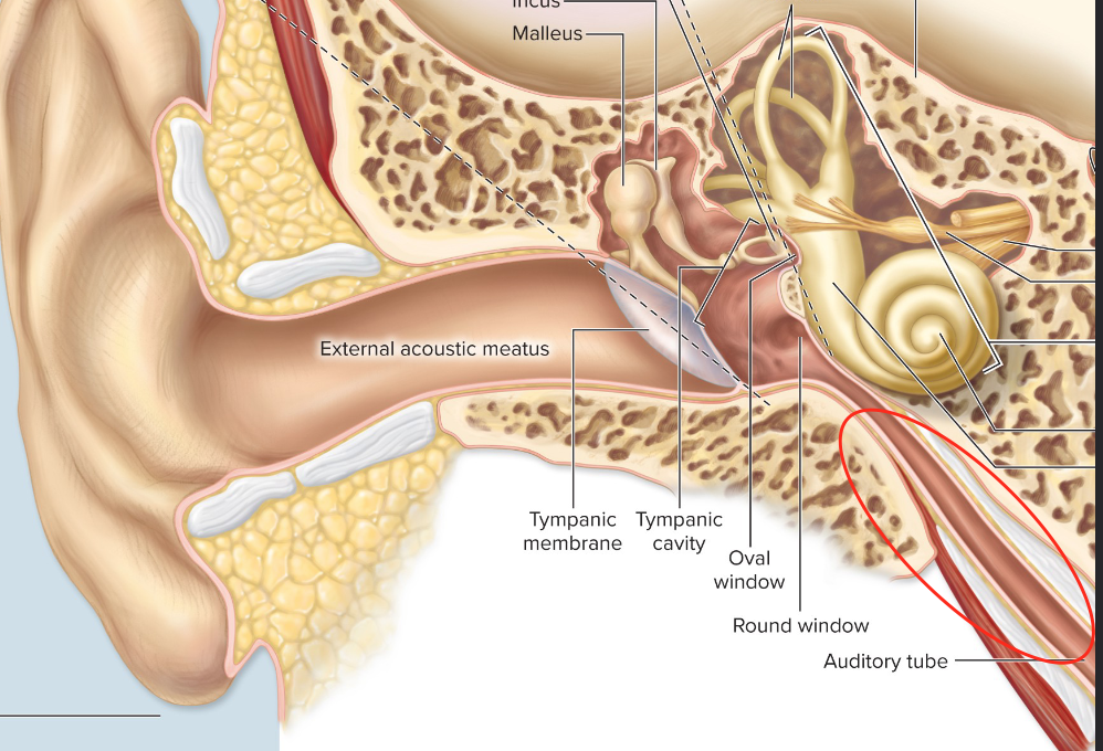
Pharyngotympanic tube function
links middle ear to pharynx; is usually closed off but can help relieve pressure
Phrenic nerve
goes to the thoracic cavity; innervates diaphragm
PNS
all peripheral nerves outside of the CNS; neuronal cell bodies = ganglia
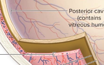
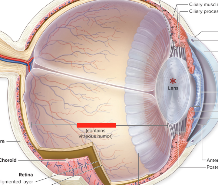
Posterior cavity
filled with vitreous humor
Proprioceptors
located in skeletal muscles, tendons, joints, and ligaments to maintain posture