A&P LAB Urinary and Reproductive System
1/168
There's no tags or description
Looks like no tags are added yet.
Name | Mastery | Learn | Test | Matching | Spaced | Call with Kai |
|---|
No analytics yet
Send a link to your students to track their progress
169 Terms
what is pharyngotympanic tube
a tube that opens into the lateral walls of the nasopharynx and connects the nasopharynx to the middle ear
what is the function of the pharyngotympanic tube
allows the middle ear pressure to equalize with atmospheric pressure
what are the paranasal sinuses
air-filled cavities surrounding the nasal cavity, named for the bones in which they are located
what are the functions of the paranasal sinuses
act as resonance chambers for speech, warm and moisten incoming air

what is #1
nasopharynx

what is #2
opening of pharyngotympanic tube
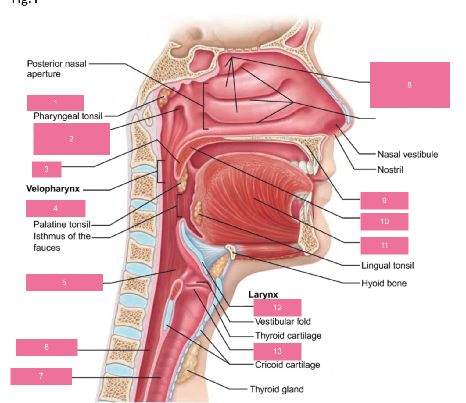
what is #3
uvula

what is #4
oropharynx

what is #5
laryngopharynx

what is #6
esophagus

what is #7
trachea

what is #8
nasal cavity (nasal conchae superior, medial and inferior)

what is #9
hard palate

what is #10
soft palate

what is #11
tongue

what is #12
epiglottis

what is #13
vocal fold
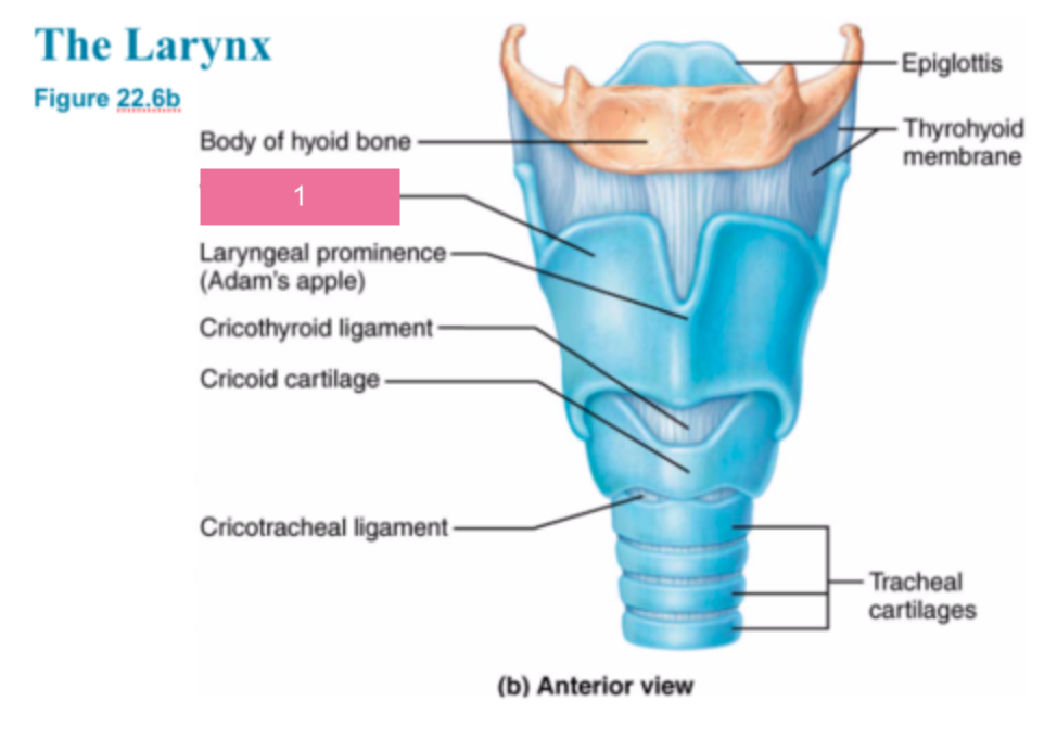
what is #1
thyroid cartilage
what type of cartilage makes up the thyroid cartilage
hyaline cartilage
what is the laryngeal prominence of the thyroid cartilage commonly called
adam’s apple
what is the function of the thyroid cartilage
form the framework of the larynx
what type of epithelium lines the trachea
ciliated, mucus-secreting, pseudostratified columnar epithelium
what is the function of the tracheal epithelium
trap debris in mucus and move it upward with cilia to keep the airway clean

what is #1
trachea

what is #2
alveolar duct
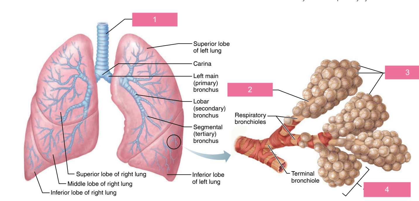
what is #3
alveoli

what is #4
alveolar sac

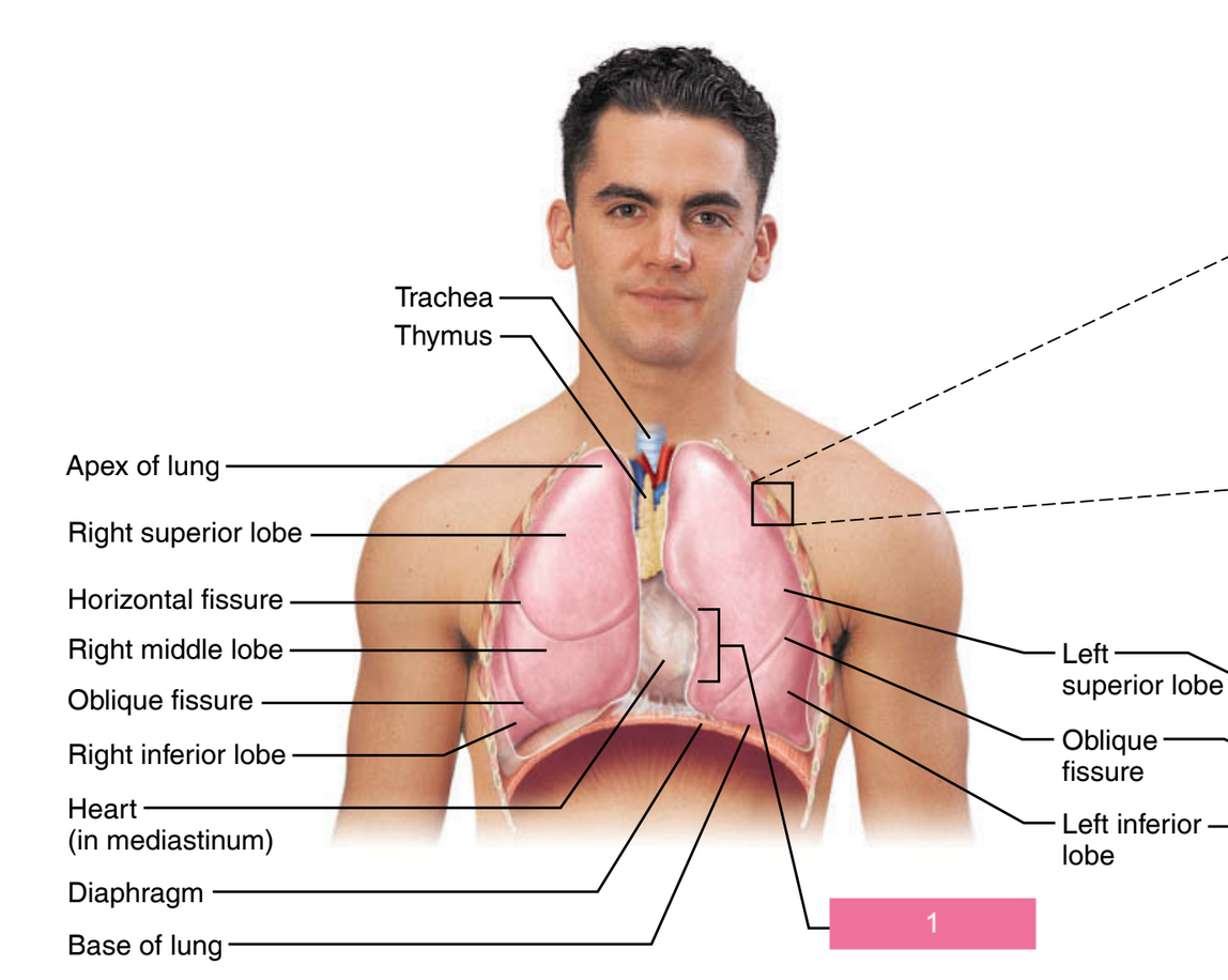
what is #1
hilum of lung
what is the medial indentation where the structures of the lung root enter or leave the lung
the hilum
what is the cardiac notch, what lung is it on?
a concavity on the medial surface of the left lung that accommodates the heart where it extends left from the body’s midline.
what is #1
cardiac notch
what serous membrane encloses each lung in a double-layered sac
the pleura

what is #1
posterior

what is #2
esophagus

what is #3
trachea

what is #4
esophagus

what is #5
trachealis muscle
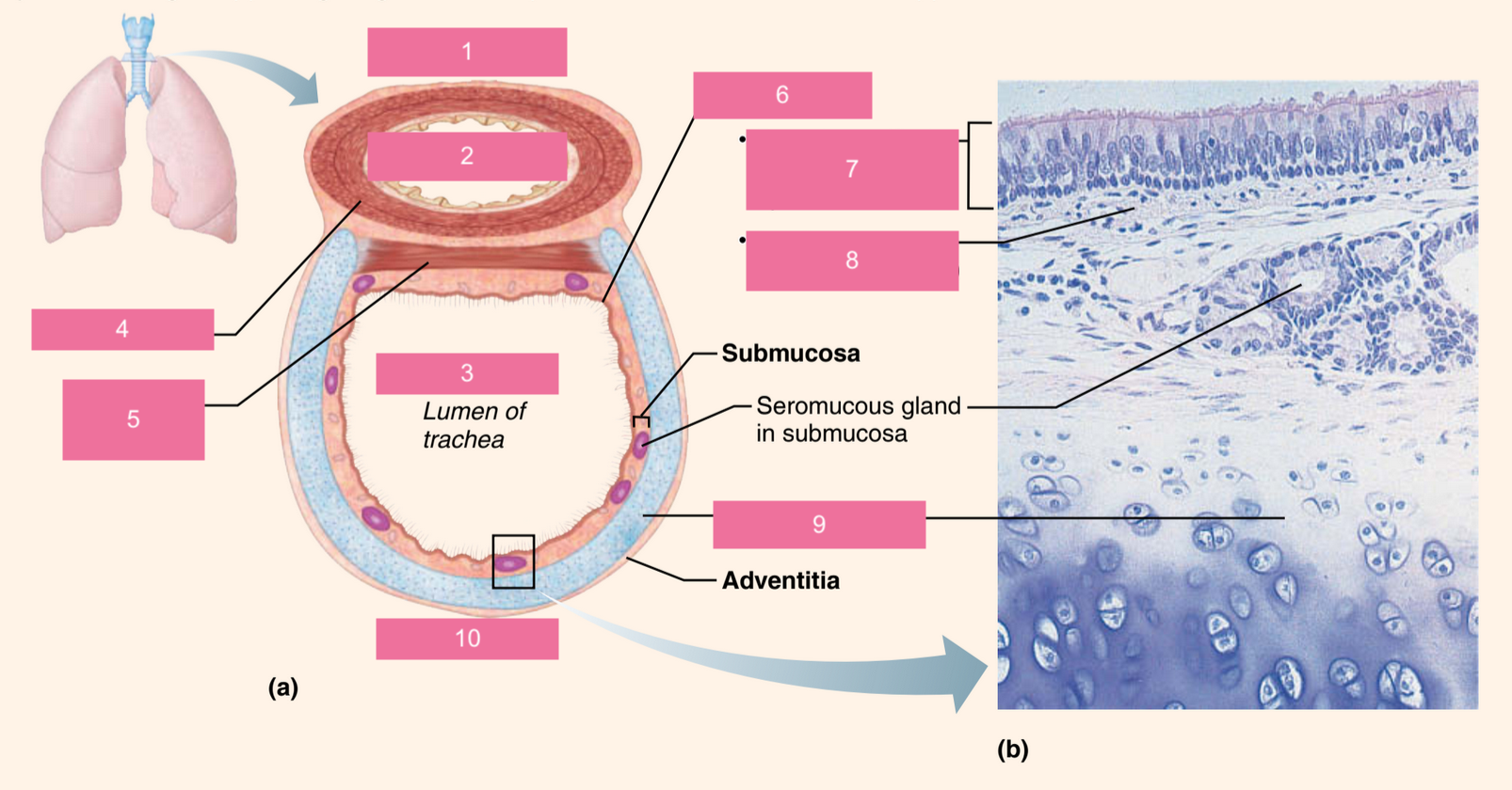
what is #6
mucosa

what is #7
pseudostratified ciliated columnar epithelium

what is #9
hyaline cartilage

what is #10
anterior
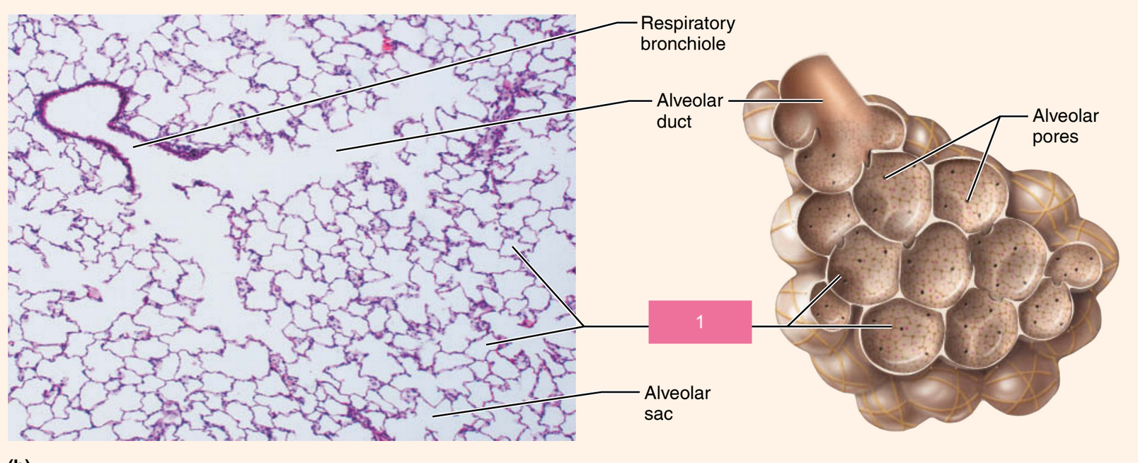
what is #1
alveoli
what is inspiration
when air is taken into the lungs
what is expiration
when air passes out of lung
how are bronchial sounds produced
air rushing through the large respiratory passageways
how are vesicular breathing sounds produced
apparently results from air filling the alveolar sacs and resembles the sound of rustling leaves
epiglottitis is a condition in which the epiglottis is inflamed. it is most often caused by a bacterial infection. explain why this type of inflammation is life-threatening.
this inflammation can quickly obstruct the airway, preventing oxygen from entering the lungs and causing asphyxiation or respiratory failure.
pneumonia is an infectious disease in which fluid accumulates in the alveoli. patients who are diagnosed with pneumonia are monitored for their oxygen saturation levels. describe how pneumonia could affect the amount of oxygen in the blood.
pneumonia fills the alveoli with fluid, which reduces the surface area available for gas exchange. because oxygen cannot diffuse effectively through fluid-filled alveoli, less oxygen enters the bloodstream. this leads to lower oxygen saturation levels.

what is #1
tongue

what is #2
esophagus

what is #3
liver

what is #4
gallbladder

what is #5
small intestine

what is #6
large intestine
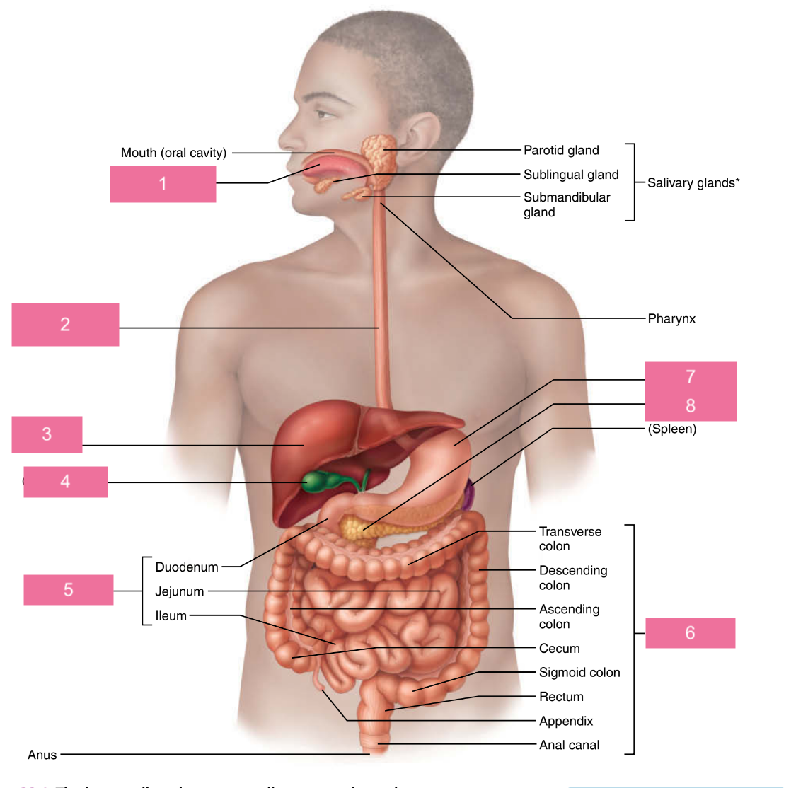
what is #7
stomach

what is #8
pancreas

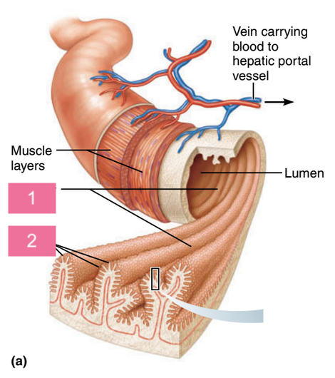
what is #1
esophagus

what is #2
muscularis externa

what is #2a
longitudinal layer
circular layer
oblique layer

what is #3
duodenum

what is #4
pyloric sphincter at pylorus
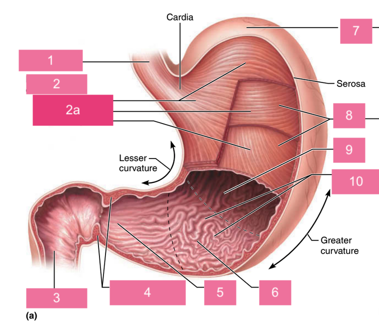
what is #5
pyloric canal

what is #6
pyloric antrum

what is #7
fundus

what is #8
body

what is #9
lumen

what is #10
rugae of mucosa

what is #1
gastric pit
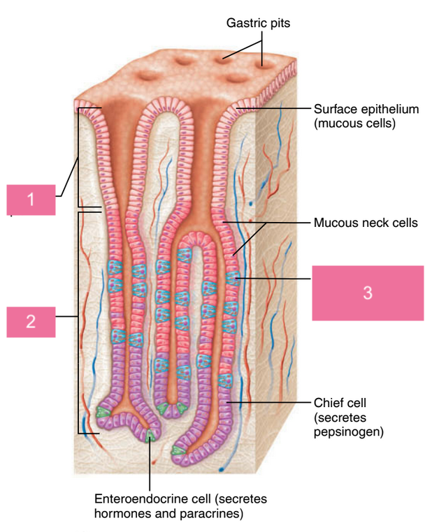
what is #2
gastric gland

what is #3
parietal cell (secretes HCl and intrinsic factor)
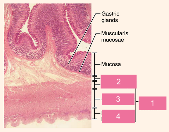
what is #1
muscularis externa

what is #2
submucosa oblique layer

what is #3
circular layer

what is #4
longitudinal layer
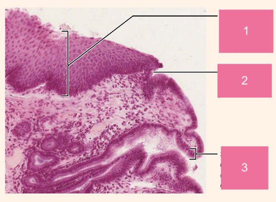
what is #1
stratified squamous epithelium of esophagus

what is #2
esophagus-stomach junction

what is #3
simple columnar epithelium of stomach
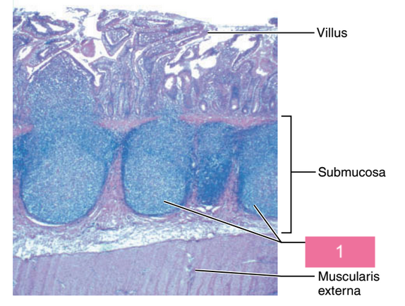
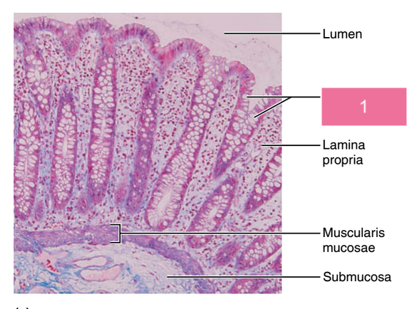
what is #1
circular folds

what is #2
villi
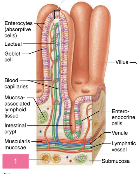
what is #1
duodenal glands
what is #1
peyer’s patches
what is #1
goblet cell in epithelium
what is the crown of a tooth
the superior portion of the tooth visible above the gingiva (gum), which surrounds the tooth
what covers the surface of the tooth crown
enamel
what is enamel made of
heavily mineralized, 95–97% inorganic calcium salts

what is #1
neck
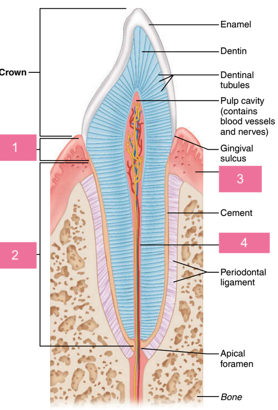
what is #2
root

what is #3
gingiva (gum)

what is #4
root canal
what happens when the common hepatic duct or bile duct is blocked (e.g., by gallstones)
bile cannot enter the small intestine, so it accumulates and backs up into the liver
how does blocked bile flow affect liver cells
exerts pressure on liver cells and begins to leak into the bloodstream
why does jaundice occur when bile enters the bloodstream
as bile pigments circulate through the body, they deposit in tissues, causing them to appear yellow

what is #1
liver

what is #2
cystic duct

what is #3
common hepatic duct

what is #4
bile duct and sphincter

what is #5
tail of pancreas
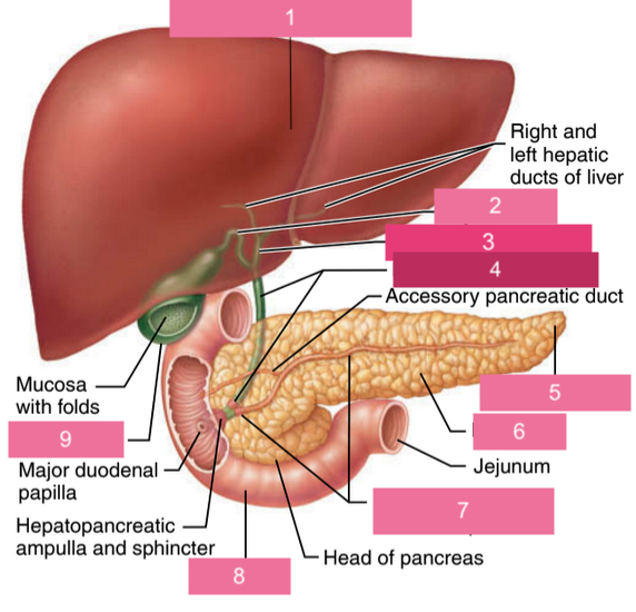
what is #6
pancreas

what is #7
main pancreatic duct and sphincter

what is #8
duodenum