Musculoskeletal system (WIP)
1/44
There's no tags or description
Looks like no tags are added yet.
Name | Mastery | Learn | Test | Matching | Spaced | Call with Kai |
|---|
No analytics yet
Send a link to your students to track their progress
45 Terms
What is cartilage, where is it found and what is it for?
strong, flexible and semi-rigid connective tissue. It can withstand compressive forces but can bend.
Found in the ear, trachea, ribs and nose
support, articulations surfaces for bones, act as a template for growth and development of bone, and is a lubricating surface to reduce friction
How is cartiledge made/ what is their structure
chondroblasts are cells in the outer layer of cartilage and secrete extracellular matrix, then become trapped in it and mature into cells called chondrocytes.
Most cartilege is then covered by a layer of dense, irregular, connective tissue called perichondrium
characteristics of cartilage
nourished by diffusion from capillaries in the perichondrium so it must be thin
slow healing
no blood vessels or nerves
Hyaline cartilage
most common cartilage and a precursor to bones.
Bluish white colour found in ribs, nose, larynx, trachea and ends of bones
translucent with a high proportion of collagen
second most flexible
White Fibrous catilage
glistening white colour in invertabral discs and join capsuole ligaments
the collagen is organised in dense fibres which makes it strongest but also the lest flexible
the fibres are oriented in multiple directions of functional stresses
no perichondrium
Yellow elastic cartilage
yellow colour found in external ear, epiglottus, laryut
numerous chondrocytes in a threadlike network of elastic fibres made of the elastin proteins
most flexible and most chonrocytes
What is bone
a stong, flexible and semi rigid support tissue resistant to bending, compression and stretch
what are the functions of bone
-support- the structural framework for the body, providing attachment sites for muscles
-Protection- protecting internal organs
-assisting movement
-mineral homeostasis for calcium and phosphorus
-blood cell production in bone marrow
What is the extracellular matrix of bone made of
-30% organic- mainly collagen providing bone’s flexibility
-70% inorganic- main component being hydroxyapatite which contains calcium and phosphate
how are bones made/ what stages do they go through
before the extracellular matrix is calcified the tissue is called osteoid tissue. When the concentrations of calcium and phosphate rise high enough they are deposited into the extracellular matrix and the bone calcifies
Features of compound bone
-surrounds most bones
-hard and gives bones the smooth white appearance
-strong and rigid
Features of spongy bone
-found at ends of bones and in vertebrae
-contains marrow where blood cells are made
osteoprogenitor cells
‘stem’ cells of bone and the source of new osteoblasts
osteoblasts
line the surface f bones, secrete collagen and the organic matrix of bone (osteoid). As they become trapped in organic matrix they become osteocytes
osteocytes
maintain bone tissue. Fine processes from these cells pass through bone and form gap junctions with other osteocytes
How do osteoclasts break down bone
They are large, multinucleated, ruffles border, secretory cells with a prominent golgi body and vesicles. They secrete enzymes lik carbonic anhydrase which acidifies the matrix and causes it to decalcify. Then they break down the matrix
How are osteoclasts made
monocytes/macrophages are derived from hematopoietic cells in the bone marrow. Monocytes travel in the bloodstream and collect at sites of bone destruction where they fuse to form osteoclasts.
How does bone remodelling occur
relies on the correct balance between bone resorption and bone deposition by osteoblasts
1-mechanical stresses on the skeleton cause release of calcium, that stimulate bone-remodelling
2- Bone breakdown is stimulated by parathyroid hormone and inhibited by calcitonin
structure of haversian systems/ osteons
-1 alignation
-2 osteocytes
-3 lacunae
they are aligned in the same direction along lines of stress which change with the stresses. Osteocytes sit in the calcified matrix in small space called lacunae
Long processes from the osteocytes lie in small channels called canaliculi which transport nutrients and waste
lacunae are arranged in concentric sings of bone amtrix called lamellae around a central haversian canal and their processes run in interconnecting canaliculi
the Haversian canal and horizontal canals contain blood vessels and nerves from the periosteum
Where is cartilage found
found in limb, vertebrae and ribs. Form as hyaline cartilage in embryos
Ossification
cartilage cells flatten and calcium salts are deposited around them. Osteoblasts secrete layers of matrix and osteoclasts break catilage down. In limbs ossification begins at caps at the ends and in the middle
Periosteum
is a dense fibrous membrane that surrounds bone. it consists of an outer fibrous layer and an inner cellular layer (cambium)
The outer layer is mostly collagen with nerve fibres that cause pain when the tissue is damaged. it contains blood vessels with branches that penetrate the bone to supply theo osteocytes
develops from perichondrium
Axial bones
-skull, sternum, ribcage, vertebral column, pelvic girdle
Appendicular bones
scapula and clavicle, pectoral girdle, hind and forelimbs
How to identify the atlas and axis
***
How to identify the cervical bones in spine
-smallest
-triangular foramen with foramen in processes
-no articular facets
-smallest body and small transverse processes
Movement in cervical bones
-flexion and extension and rotation. Region with greatest movement
How to identify thoracic bones in spine
-circular foramen
-articular facets
-larger body
-thinner intervertebral discs
-large transverse processes
Movement in thoracic bones in spine
rotation and lateral flexion. prevents extension
how to identify lumbar bones in spine
Largest with the largest body (must carry the most weight)
-no articular facets and thickest intervertebral discs
-triangular foramen
-largest and bluntest transverse processes (wing shaped kinda)
Movement allowed by the lumbar bones
lest flexion and tension. rotation prevented
How to identify sacrum
bones all fused together
How to identify coccyx
***
Tendon
-inelastic so it remains the same length when muscles contract
-attach muscle to bone
-fibres continuous with muscle
-energy of contraction efficiently transmitted to movement of the joint
-mostly collagen
high tensile strength
Ligaments
-holds bone is position
-collagen
-allow for some elasticity and provide strength
Types of synovial joints
-hinge joins> movement in one plane
-ball and socket joints> motion in multiple planes
-gliding joint> movement in two planes
hinge joints
one articulating surface convex and the other is concave. Strong collateral ligaments restrict movements to one plane
ball and socket joints
bone surfaces reciprocally convex and concave. rotation in all three planes
gliding joints
articulating bone surfaces nearly flat and bones glide across eachother in single plane
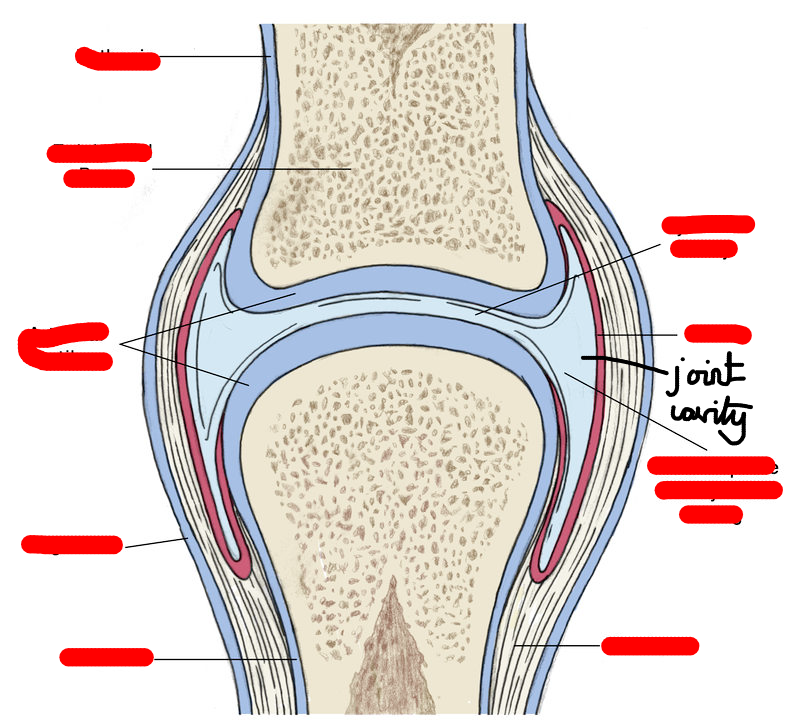
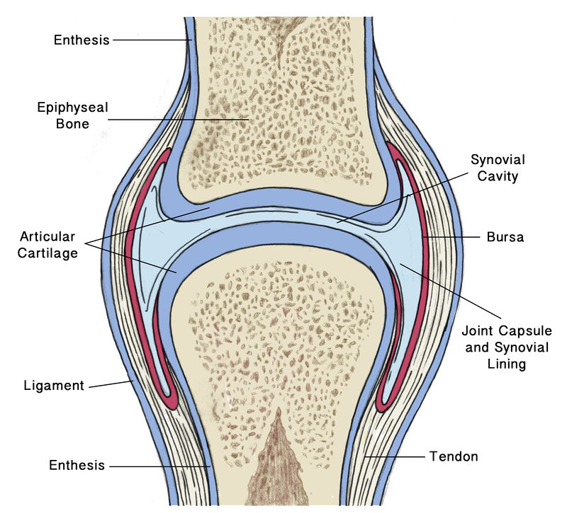
label
first class levers
-the fulcrum (axis) is between the load and the effort
-eg. joint between the head and first vertebrae
second class levers
-the load is between the axis and the effort
-eg standing on toes
why do second class levers have high mechanical advantages
-when a levers effort arms is longer than its load arm. High mechanical advantage means it can move large loads with a relatively small amount of force
third class levers
-most common
-effort is between the load and axis
How to calculate the force
F1 x d1 = F2 x d2