Lecture 8
1/15
There's no tags or description
Looks like no tags are added yet.
Name | Mastery | Learn | Test | Matching | Spaced | Call with Kai |
|---|
No analytics yet
Send a link to your students to track their progress
16 Terms
Antibody
Refers to the protein that binds to an antigen
E.g. Anti-cholera toxin antibody
Immunoglobulin
Refers to the antibody as a protein
Regardless of what antigen it binds to
Antigen
Substance that causes the body to produce specific antibodies or sensitized T cells
IgG
Main Function:
Long-term immunity, opsonization
Location:
Blood & tissues
Characteristics:
20% of all plasma proteins
Only antibody that crosses placenta
Also found in milk and colostrum
IgA
Main Function:
Protects mucosal surfaces
Location:
Saliva, tears, breast milk, mucus
Characteristics:
Large amounts in body secretions
• Has 2 forms: Blood and secretory form
IgM
Main Function:
First antibody produced in infection
Location:
Blood - as a pentamer
B cell surface – as a monomer
Characteristics:
1st antibody secreted into blood during early stages of infection – primary response.
Is synthesized by the fetus.
Indicates recent/acute infection
IgE
Main Function:
Allergic reactions, parasite defense
Location:
Body fluids: bound to basophils
Skin/tissues: bound to mast cells
Characteristics:
The fab fragments are free to bind ag to which humans cand develop allergies
Responsible for allergies & anaphylaxis
IgD
Main Function:
B-cell receptor function
Location:
Surface of B cells
Characteristics:
Main role is B-cell activation
Co-expressed with IgM
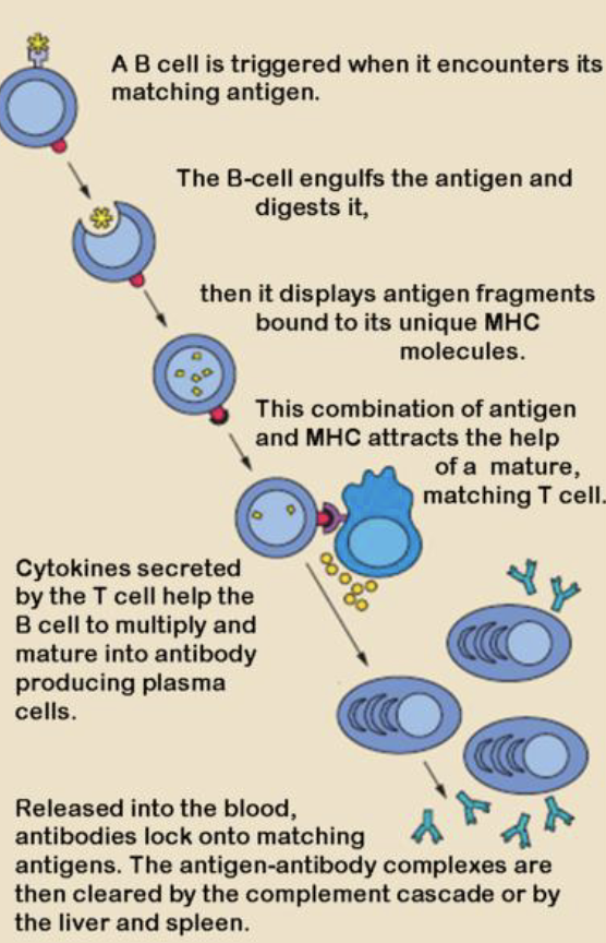
Main Function of B cells
1. Antibody production
When they come across an antigen they differentiate into plasma cells
which release large amounts of antibodies to fight infection
2. Memory formation
Some B cells differentiate into memory B cells
3. Antigen presentation
B cells can act as antigen-presenting cells (APCs)
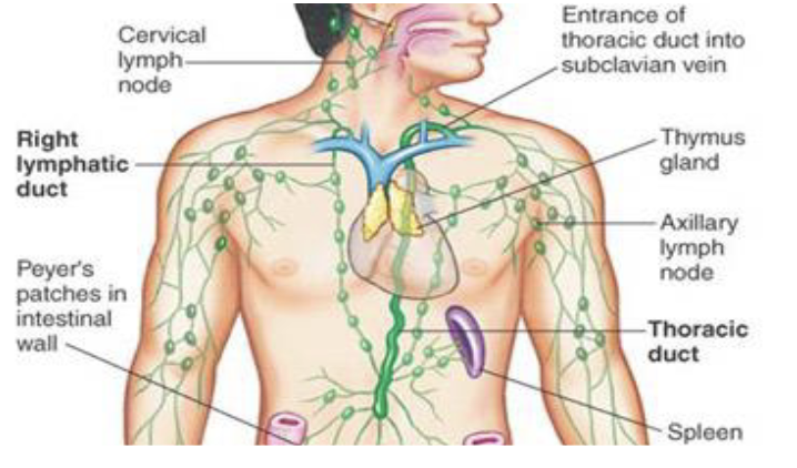
Where do we have B cells?
Blood lymphocytes
Lymph nodes
• Filter the lymphSpleen
• Filters the blood
B cell Activation
Antigen Binds to B cell Surface Ig (BCR)
B cell becomes activated
B cell Proliferates
B cell differentiates into a Plasma Cell
Plasma Cell produces antibodies
How do we activate B cells?
Antigen binding to B cell surface Ig
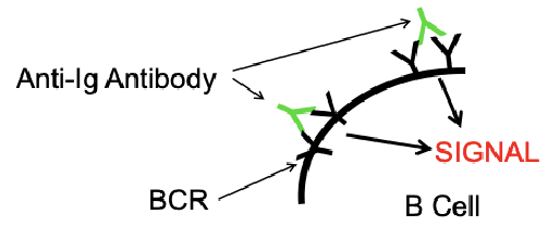
• Only activates few cells with antibody specific for that antigenAdding Antibodies to the B cell Igs
• Adding anti-mouse IgG to mouse B cells
• Two binding sites on added Ab bind B cell BCR to each other
• Activates most or all B cellsPolyclonal Activators
• Agents that can activate whole groups of cells, regardless of what antigen they are specific for.
a. Lipopolysaccharide - Activates B cells
b. Concanavalin A & Phytohemagglutinin - Activate T cells
c. Pokeweed Mitogen - Activates B and T cells
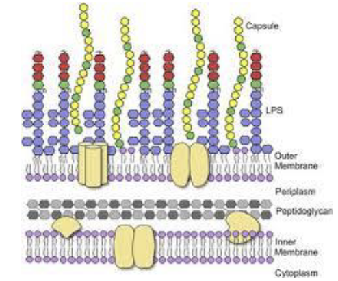
Lipopolysaccharide
Found on the Outer membrane of Gram-Negative Bacteria
• E. coli, Salmonella, Shigella
• Made of lipid and sugars bound togetherNonspecifically activates B cells
• Binds to the Toll-Like Receptors (TLRs)Only activates IgM B cells
• Results in IgM secretion
• Very little effect on IgG
Why do we need to activate B cells?
Most B cells are Resting (G0 phase)
To study how B cells are activated to produce antibodies
Study how T cells govern B cell activation and antibody production
• T cells produce cytokines (IL-4, IL-6, etc.)
• T cells and B cells interact through cell surface proteinsTo study how added agents can alter B cell activation and antibody production
• How T cell factors enhance B cell proliferation
• How T cell factors enhance antibody production
• Effect of drugs
Experiment 8
Harvest mouse spleen
Tissue Dissociation
Cell Collection
Centrifugation and Red Blood Cell lysis
Final Processing:
Count the cells using trypan blue – as in previous labs
Prepare cell suspension of mouse spleen cells - Dilute the cells in RPMI TCM to 5 x 10⁶ cells/ml in a total of 2.5 mL
Culture cells with LPS
Perform differential leukocyte counts on mouse Spleen
Dilute the cells to 8 x 10⁵ cells/mL in 2 ml of Wash Medium (2% BCS PBS). Add 1 ml to a 5 ml round bottom centrifuge tube to give 8 x 10⁵ cells/tube
Follow the procedure in the Lab manual to prepare the cells
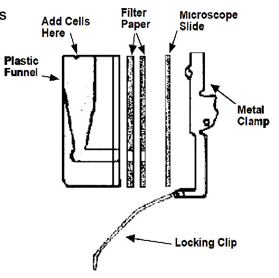
While waiting, clean and Label a microscope slide with "Spleen" and your group name.
Set up 1 Cytospin cytocentrifuge holder with 2 filters
To each slide holder, add 0.25 ml of the spleen cells you prepared in #2 to give 2 x 10⁵ cells/slide.
Centrifuge the cells at 800 rpm for 10 minutes.
Stain the cells with the Leukostat Stain as in the the previous experiment.
Counts cells.