Ch.3 part2
1/57
Earn XP
Description and Tags
This set of vocabulary flashcards identifies the key components, landmarks, and clinical considerations of the cranial bones as described in the lecture based on the 6th edition of Illustrated Anatomy of the Head and Neck.
Name | Mastery | Learn | Test | Matching | Spaced | Call with Kai |
|---|
No analytics yet
Send a link to your students to track their progress
58 Terms
Cranial Bones
The eight bones that form the cranium, including the single occipital, frontal, sphenoid, and ethmoid bones, and the paired parietal and temporal bones.
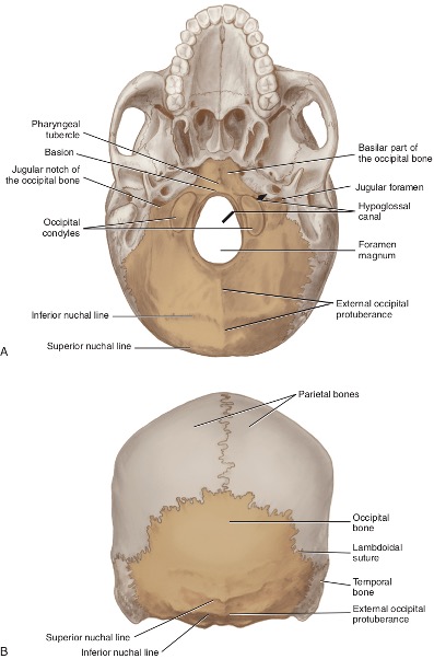
Occipital Bone #3
A single cranial bone that forms the posterior part of the skull and the base of the cranium, articulating with the parietal, temporal, sphenoid, and the first cervical vertebra (atlas).
Foramen Magnum #18
A large opening on the external surface of the occipital bone that is completely formed by the occipital bone.
Occipital Condyles #19
Paired, curved, and smooth projections on the lateral part of the occipital bone that have a movable articulation with the atlas.
Pharyngeal Tubercle
A midline projection on the basilar part of the occipital bone, anterior to the foramen magnum. Basion also called point Ba
Basion (Point Ba)
A cephalometric landmark located on the midpoint of the anterior border of the foramen magnum.
Hypoglossal Canals
Paired openings in the occipital bone that transmit the twelfth cranial or hypoglossal nerve.
Superior and Inferior Nuchal Lines
Curved ridges on the external surface of the occipital bone that serve as sites for muscle attachments such as the sternocleidomastoid, trapezius, and occipitalis muscles.
External Occipital Protuberance (Inion)
A midline projection on the posterior surface of the occipital bone and a cephalic landmark.
Frontal Bone #1
A single cranial bone forming the anterior part of the skull superior to the eyes, the majority of the forehead, and the roof of each orbit.
Superior temporal line & inferior temporal line
Curved ridges on the lateral surface of the skull that serve as attachment points for the temporalis muscle.
Frontal Sinuses
Air-filled cavities within the frontal bone that are lined with mucous membranes and help to reduce the weight of the skull.
Frontonasal Duct
A constricted canal through which each frontal sinus communicates with and drains into the middle nasal meatus of the nasal cavity.
Glabella
The smooth elevated area between the eyebrows and the supraorbital ridges.
Supraorbital Notch (Foramen) #30
An opening on the medial part of the supraorbital ridge where the supraorbital artery and nerve travel to the forehead.
Lacrimal Fossa #34
An area located internal to the lateral part of the supraorbital rim containing the lacrimal gland, which produces tears.
Parietal Bones #2
Relatively square, paired cranial bones forming the greater part of the right and left lateral walls and the roof of the skull.
Sagittal suture #15
The fibrous joint that connects the two parietal bones along the midline of the skull.
Lambdoidal suture #16
The fibrous joint connecting the occipital bone to the parietal bones at the posterior of the skull.
coronal suture #14
The fibrous joint connecting the frontal bone to the two parietal bones at the anterior of the skull.
squamosal sutures
The fibrous joints connecting the temporal bones to the parietal bones on each side of the skull.
sphenoid bone #6
A complex bone located in the middle of the skull that helps form the base of the cranium, the sides of the skull, and parts of the eye socket.
Parietal bones #2
The two large bones that form the superior and lateral aspects of the skull, situated on either side of the frontal bone.
Temporal Bones #4
Paired cranial bones forming the lateral walls in the temporal region and part of the base of the skull in the auricular region.

Squamous Part of the Temporal Bone
The large fan-shaped flat part on each of the temporal bones
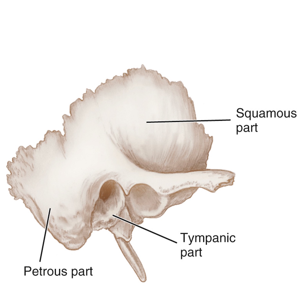
Tympanic Part of the Temporal Bone
A small, irregularly shaped part associated with the ear canal that forms most of the external acoustic meatus.

Petrous Part of the Temporal Bone
The inferiorly located part of the temporal bone that helps form the cranial floor and contains the mastoid process.
Zygomatic process of the temporal bone #28
A protrusion from the temporal bone that articulates with the zygomatic bone, contributing to the formation of the zygomatic arch.
Articular Fossa
On the inferior surface of the zygomatic process of the temporal bone. Anterior to the articular fossa is the articular eminence, and posterior to it is the postglenoid process
External acoustic meatus #25
a short canal leading to the tympanic cavity, located posterior to the articular fossa
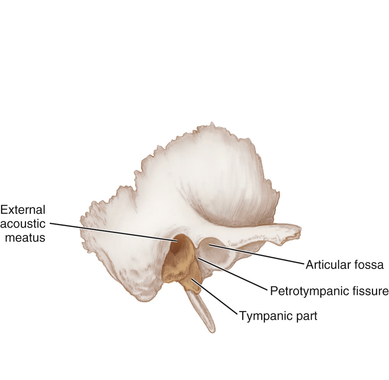
Petrotympanic fissue
Posterior to the articular fossa, the tympanic part is separated from the petrous part by a fissure
Mastoid Process #23
A large roughened projection of the petrous part composed of mastoid air cells; On the inferior aspect of the petrous part of the temporal bone and posterior to the external acoustic meatus is a large roughened projection
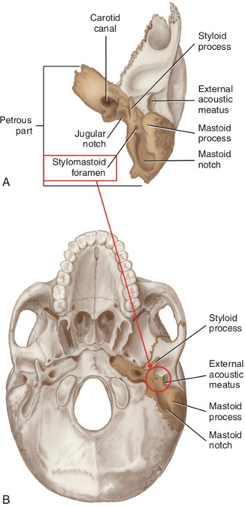
Mastoid notch
Medial to the mastoid process is the deep groove of the
Styloid process #22
Inferior and medial to the external acoustic meatus is a long, pointed bony projection

Stylomastoid Foramen #24
A foramen between the styloid and mastoid processes that carries the seventh cranial or facial nerve.

#21 Carotid canal
noted is the large circular aperture of the

Jugular Foramen #20
the jugular notch of the temporal bone is visible
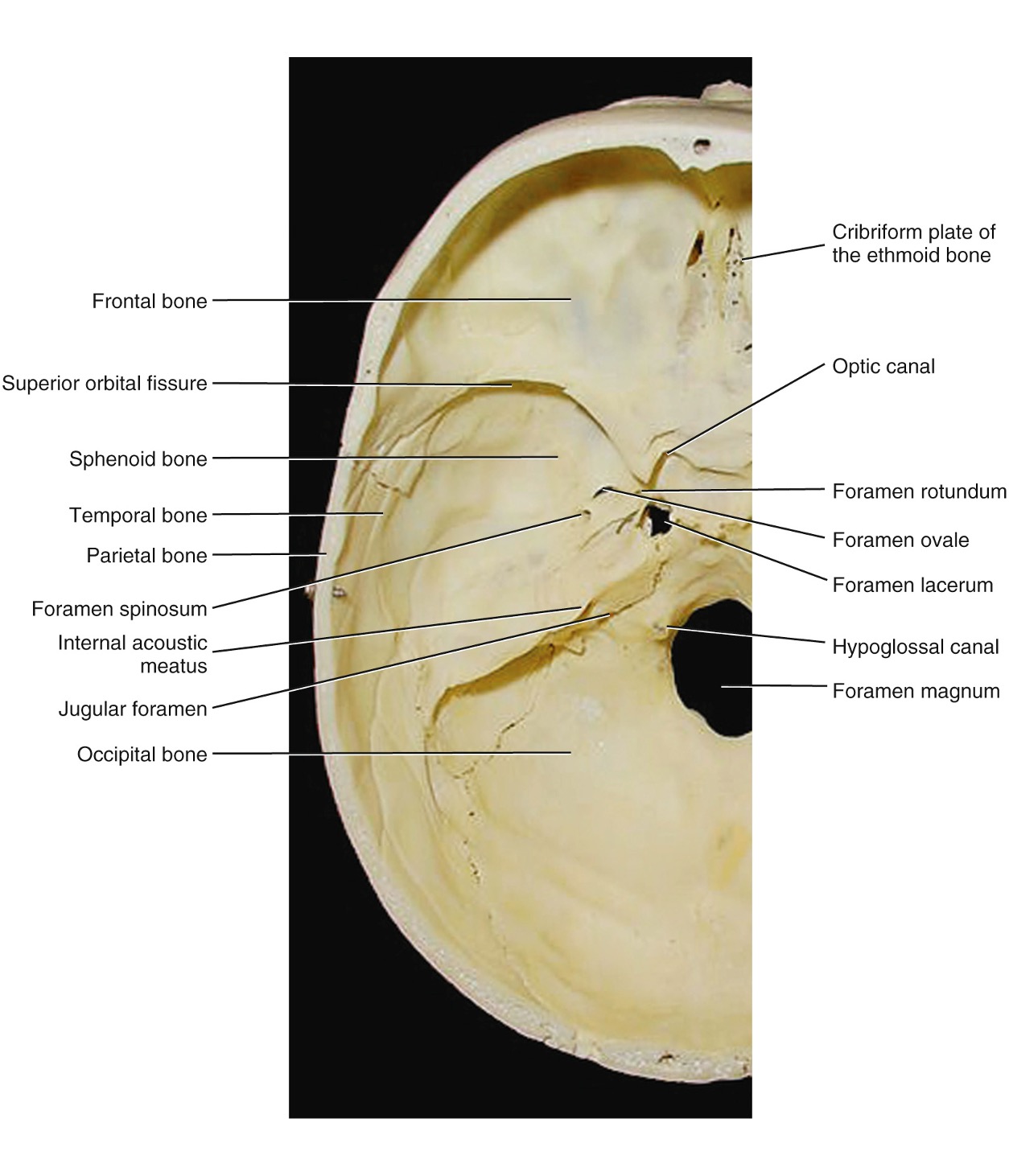
Internal Acoustic Meatus (IAM) #49
An opening on the intracranial surface of the petrous part carrying the seventh cranial (facial) and eighth cranial (vestilocochlear) nerves.
Mastoiditis
An infection within the mastoid antrum and mastoid cells, usually secondary to middle ear infection (otitis media).
Sphenoid Bone #6
A single midline cranial bone called the 'keystone' of the cranial floor because it is in contact with most other cranial bones.
ethmoid bone #5
Is a single, complex cranial bone located between the nasal cavity and the orbital cavities, playing a key role in the structure of the skull and the face. With Crista galli #40
sphenoidal sinuses
are air-filled spaces located within the wings of the sphenoid bone, involved in the drainage of mucus and regulation of air pressure in the nasal cavity. Separated by a septum.
Sella Turcica (Pituitary Fossa)
A saddle-shaped depression on the superior surface of the sphenoid body; its deepest part (hypophyseal fossa) contains the pituitary gland.
Hypophyseal fossa #43
The deepest part of the sella turcica, housing the pituitary gland within the sphenoid bone.
Lesser Wing of the Sphenoid Bone #41
An anterolateral process that forms the orbital apex, the deepest part of the orbit. is an anterolateral process that forms the orbital apex
Greater Wing of the Sphenoid Bone
A posterolateral process divided by the infratemporal crest; its orbital surface creates the posterior part of the lateral wall of the orbit.
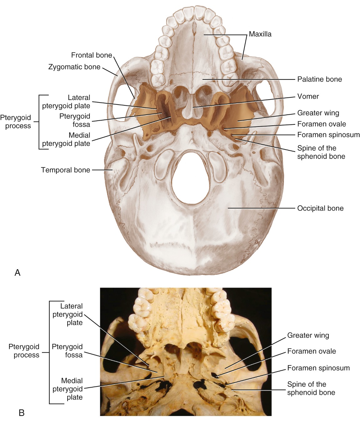
Pterygoid Process
A sphenoid process consisting of two plates ( Ptergoid plate lateral and medial) with a pterygoid fossa between them.
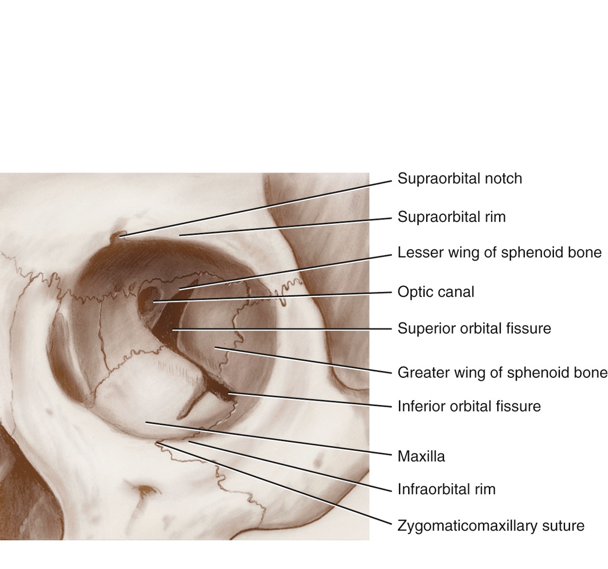
Superior Orbital Fissure #31
The most anterior curved and slit-like opening, the superior orbital fissure. A slit-like opening in the sphenoid bone that transmits the ophthalmic nerve (first division of the trigeminal nerve).
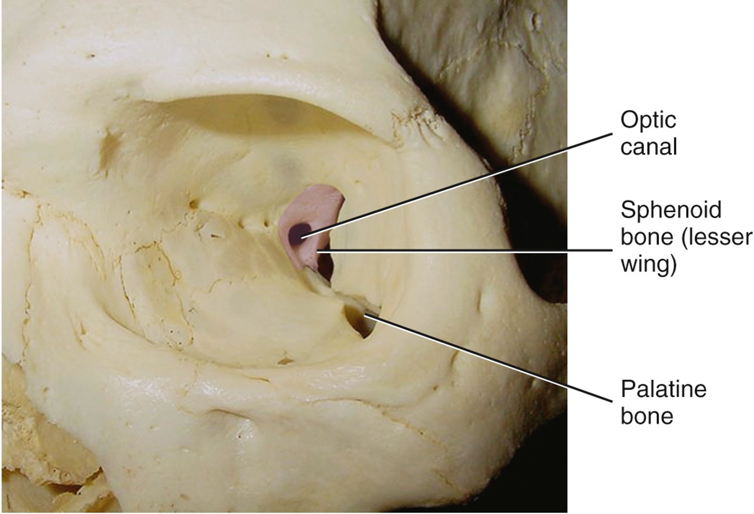
Optic canal #33
The round opening in the orbital apex, which lies between the two roots of the lesser wing.
Foramen Rotundum #44
A round opening in the sphenoid bone that transmits the maxillary nerve (second division of the trigeminal nerve).
Foramen spinosum #46
with the middle meningeal artery, middle meningeal vein, and the meningeal branch of the mandibular nerve
Foramen laceram #47
Foramen Ovale #45
A large oval opening in the sphenoid bone that transmits the mandibular nerve (third division of the trigeminal nerve).
Optic Canal
A round opening in the orbital apex of the sphenoid bone through which the optic nerve and ophthalmic artery pass.
Ethmoid Bone
A single midline cranial bone located anterior to the sphenoid bone, consisting of the perpendicular plate and the cribriform plate.
Cribriform Plate
The horizontal plate of the ethmoid bone perforated by olfactory foramina for the passage of olfactory nerves.
Crista Galli #40
A vertical midline wedge-shaped continuation of the perpendicular plate that serves as an attachment for layers covering the brain. next to the ethmoid
Ethmoidal Sinuses (Ethmoid Air Cells)
A variable number of small cavities in the lateral masses of the ethmoid bone located between the orbital plate and the nasal conchae.