Image Analysis Quiz 3
1/154
There's no tags or description
Looks like no tags are added yet.
Name | Mastery | Learn | Test | Matching | Spaced | Call with Kai |
|---|
No analytics yet
Send a link to your students to track their progress
155 Terms
What injury is being evaluated in the PA oblique projection of the shoulder?
Shoulder dislocation
How much is the patient obliqued and which way for the PA oblique projection of the shoulder?
45-60° toward IR
Which border of the scapula is thicker and is demonstrated on the image as a double cortical line?
Lateral border
Which border of the scapula is thinner and is demonstrated as a single line?
Medial border
CR location for a PA oblique projection of the shoulder:
Scapulohumeral joint
Upon analysis of your image, you notice that the thicker border of the scapula is demonstrated laterally and the thinner border of the scapula is superimposed over the ribs. What is your positioning error?
Under rotated
Upon analysis of your image, you notice no superimposition of the scapular borders and the medial border appears more laterally and the lateral border is medial. What is your positioning error?
Over rotated
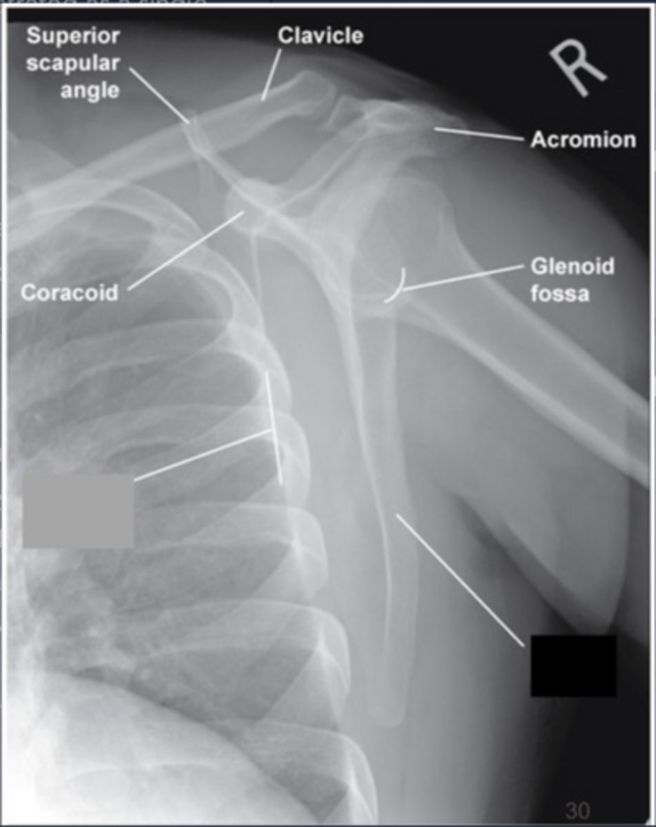
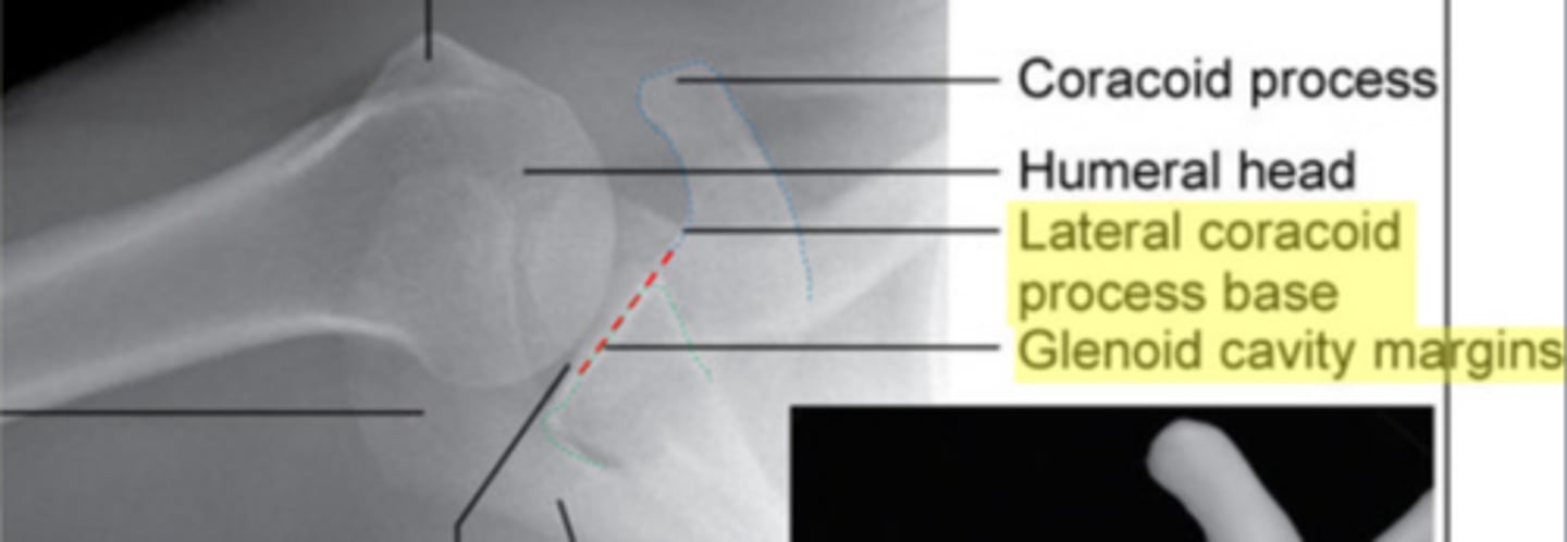
In a properly positioned Y-view, which process is demonstrated closest to the ribs?
Coracoid
In a properly positioned Y-view, which process is demonstrated away from the ribs?
Acromion
If no injury is present, where should the humeral head be demonstrated in the Y-view?
Humeral head and glenoid cavity superimposed
If the patient suffers from an anterior dislocation of the humeral head, where will the humeral head be demonstrated?
The humeral head is under the coracoid process
If the patient suffers from a posterior dislocation of the humeral head, where will the humeral head be demonstrated?
The humeral head is over the coracoid process (beneath the acromion)
How much should the patient's affected arm be abducted for the Inferosuperior Axial projection of the shoulder?
90° away from the body
How much is the central ray angled for the Inferosuperior Axial projection of the shoulder?
15-30°
If the patient's arm cannot be abducted to the recommended amount, how will the central angle be adjusted for the Inferosuperior Axial projection of the shoulder?
Less angle more towards 15°
What is the position of the Humerus for the Inferosuperior Axial Projection?
External rotation
How should the humeral epicondyles be positioned for the Inferosuperior Axial projection of the shoulder?
45° to the floor
When the palm of the hand is facing upward and the humeral epicondyles are 45° to the floor how will the lesser tubercle be demonstrated for the Inferosuperior Axial projection of the shoulder?
In profile anteriorly
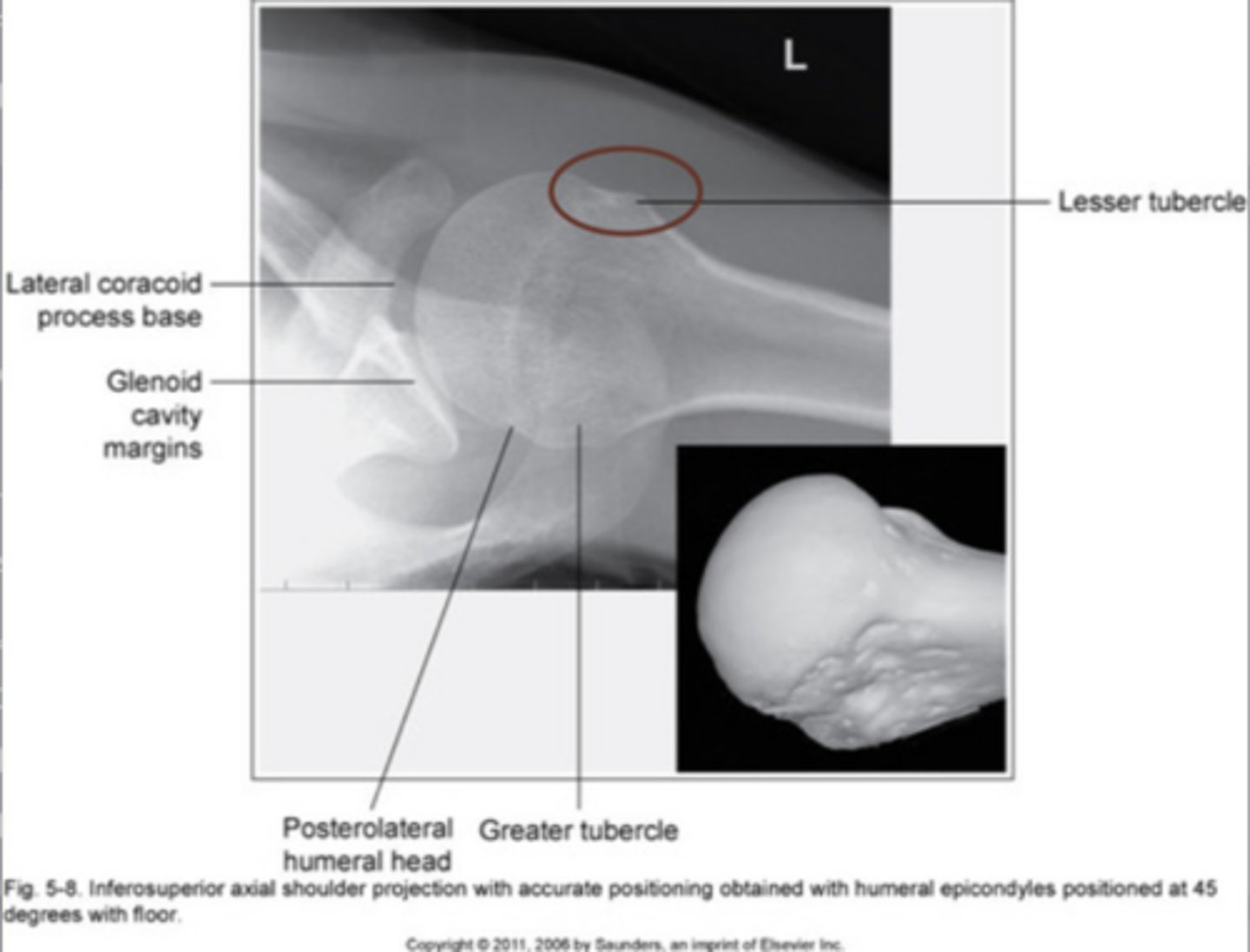
What shoulder be in profile for the Inferosuperior Axial projection of the shoulder?
Glenoid humeral joint
What should be lined up when the proper abduction and CR angle is used in a Inferosuperior Axial projection of the shoulder?
Inferior border of the glenoid cavity and base of the coracoid process
You notice on your image on the Inferosuperior Axial Projection that the glenoid cavity is closed and not in profile and the margin of the glenoid cavity is lateral to the base of the coracoid process, what error occured?
Not enough CR angle (less than 30°)
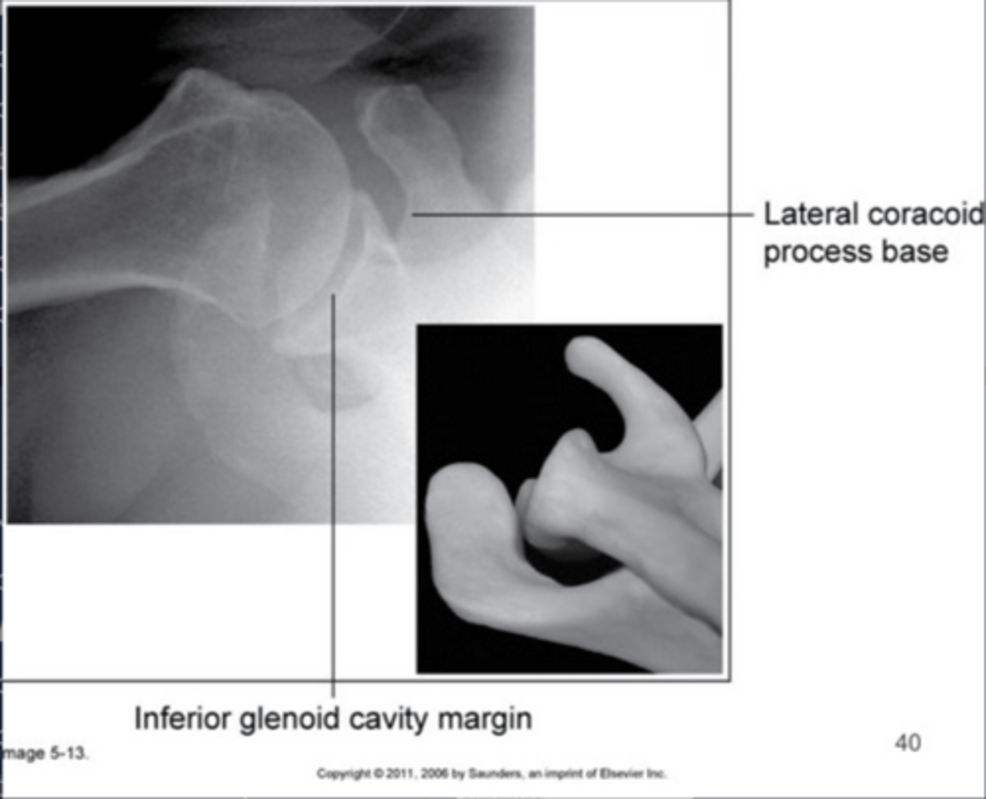
You notice on your image on the Inferosuperior Axial projection that the glenoid cavity is closed and not in profile and the margin of the glenoid cavity is medial to the base of the coracoid process, what error occured?
Too much CR angle (greater than 30°)
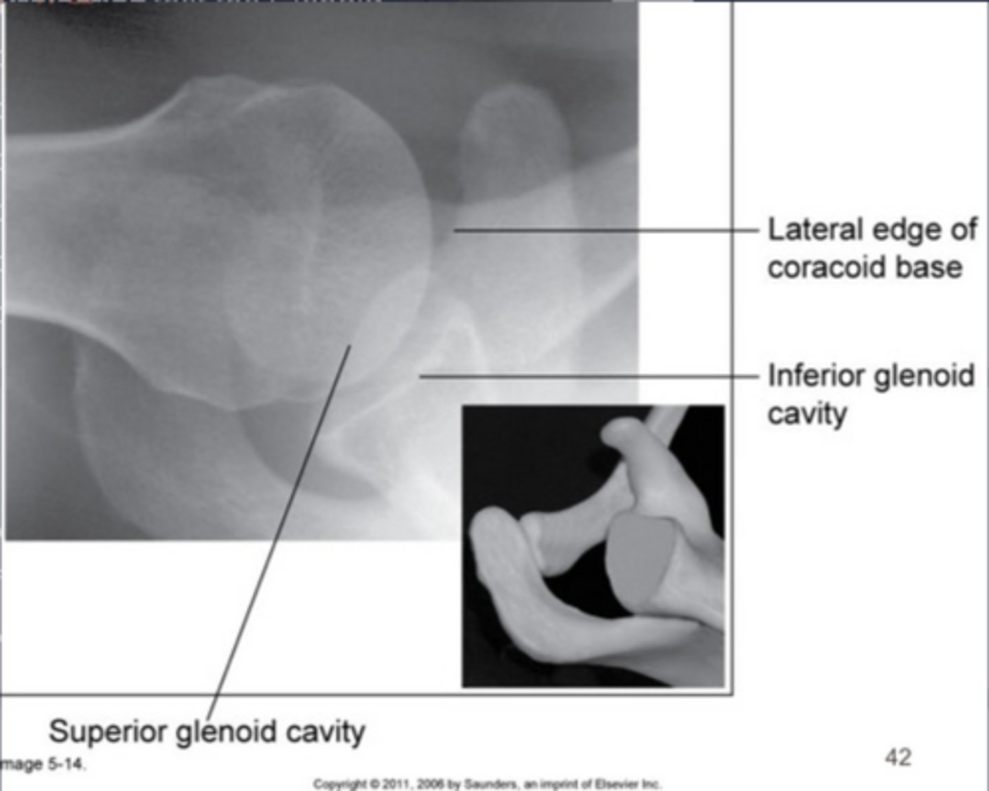
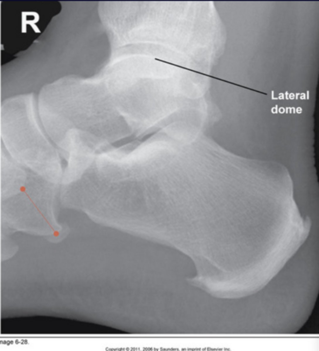
What defect is shown on the Inferosuperior Axial projection of the shoulder?
Hill Sachs defect
CR location for the Inferosuperior Axial projection of the shouder:
Axilla (armpit)
How is the arm positioned for an AP scapula?
Abduct humerus and flex elbow 90° (stop sign)
CR location for an AP scapula:
2" inferior to the coracoid process
How much is the patient rotated for a lateral scapula?
45-60°
CR location for lateral scapula:
Mid medial border of the scapula
How should the arm be positioned if the body of the scapula is of interest for the lateral scapula?
Arm above head
How should the arm be positioned if the coracoid and acromion is of interest for lateral scapula?
Arm reached across to other shoulder
CR location for AP clavicle:
Midshaft of clavicle
CR location for AP Axial clavicle:
Midshaft of clavicle
What is the central ray angel and direction for AP Axial clavicle?
15-30° cephalic
What images are taken to properly demonstrate AC joints?
With and without weights
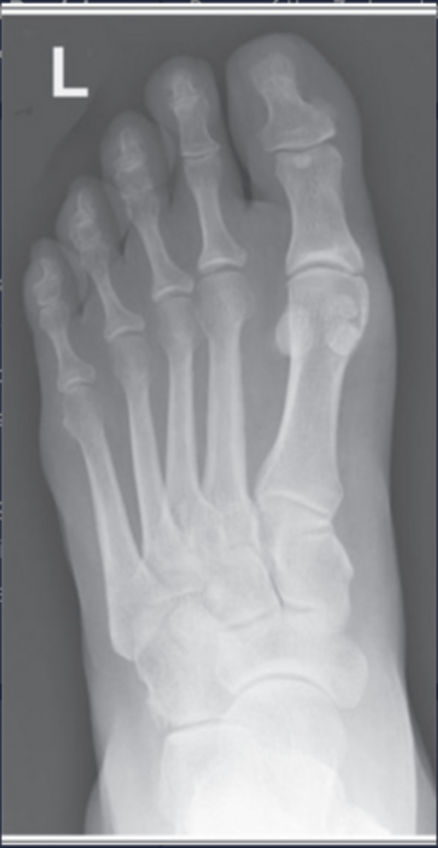
How much is the central ray angled and in what direction for an AP axial projection of the toes?
15° toward heel (posteriorly)
CR location for AP Axial projection of the toes:
MTPJ
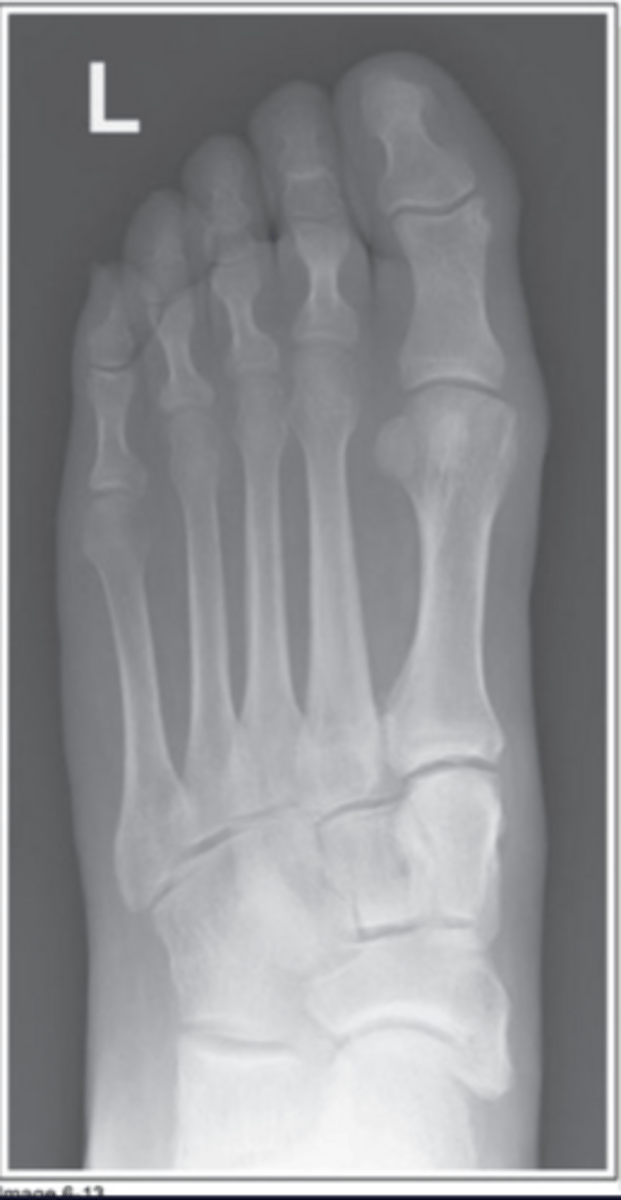
How much is the foot/toes rotated for an AP Oblique projection? Which direction?
30-45° medially rotated
Which direction is the foot rotated if only the 1st or 2nd digit is of interest for an AP Oblique projection?
Medial rotation
Which direction is the foot rotated if only the 4th or 5th digit is of interest for an AP Oblique projection?
Lateral rotation
Which projection should be completed for the 1st or 2nd digit for lateral position?
Lateromedial projection
Which projection should be complested for the 3rd -5th digits for lateral position?
Mediolateral projection
CR location for the 1st digit for lateral projection:
Interphalangeal joint (IPJ)
CR location for the 2nd-5th digits for lateral projection:
Proximal interphalangeal joint (PIPJ)
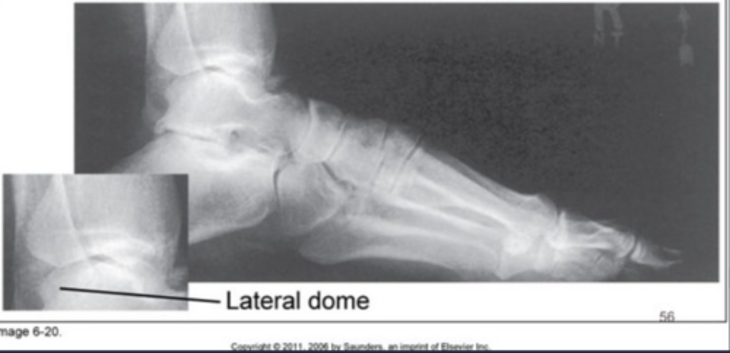
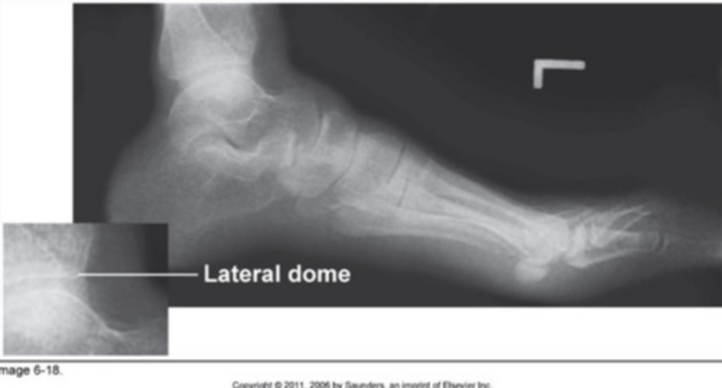
Which projection of the foot will best demonstrate the sinus tarsi?
AP oblique projection
How much is the central ray angled and in what direction for an AP Axial projection of the foot?
10° posteriorly (toward the heel)
How should the calcaneus be demonstrated in relation to the talus on an AP Axial projection of the foot?
3/4" of the calcaneus lateral to the talus
CR location for AP Axial projection of the foot:
Base of the 3rd metatarsal
You notice on your image of an AP Axial projection of the foot that there is greater overlap of the 4th adn 5th metatarsal bases, there is less than 3/4" of the calcaneus sticking out from the talus and navicular tuberosity, what error occurred?
Rotated laterally
You notice on your image of an AP Axial projection of the foot that there is greater overlap of the 1st, 2nd and 3rd metatarsal bases, there is more than 3/4" of the calcaneus sticking out from the talus, what error occurred?
Rotated medially
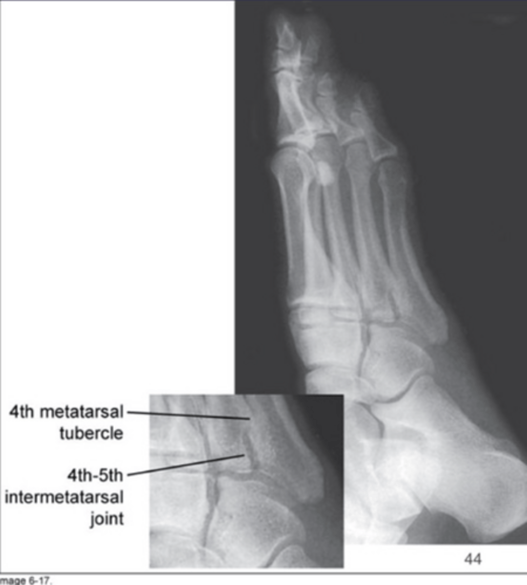
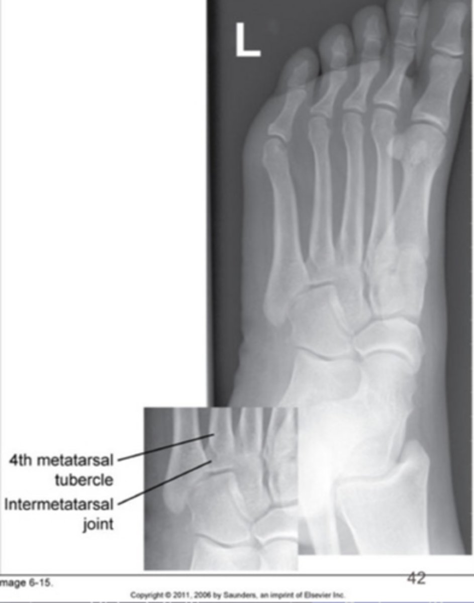
How much is the foot obliqued and in what direction for an AP Oblique projection?
30° medially rotated
What tarsal bone is demonstrated without superimposition for the AP Oblique projection of the foot?
Cuboid
If the foot is over obliqued, how will the cuneiforms be demonstrated?
The lateral cuneiform will superimpose the other cuneiforms
How should the plantar surface of the foot be in relationship to the IR for the lateral projection?
Perpendicular
If the foot is under obliqued, how will the cuneiforms be demonstrated?
Lateral cuneiform slightly superimposed on cuboid
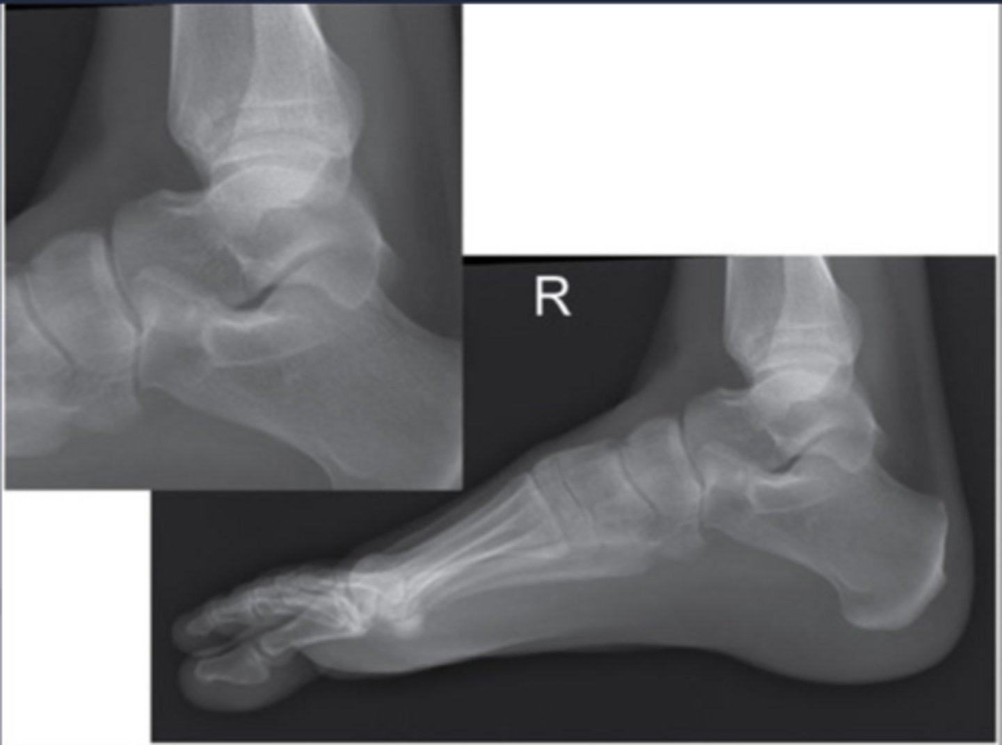
How should the talar domes be demonstrated in a true lateral foot?
Superimposed
In a properly positioned lateral foot, where should the fibula be demonstrated in relation to the tibia?
Fibula in posterior half of tibia
If the fibula is anterior to the tibia for a lateral projection of the foot, what is the positioning error?
Toes elevated
For a lateral foot, when properly positioned and the foot and knee are in the same plane, how much of the cuboid should be seen below the navicular bone?
0.5"
If the fibula is posterior to the tibia for a lateral projection of the foot, what is the positioning error?
Heel elevated
On an image of a lateral foot, you notice that one talar dome is above the other one and there is less than 0.5" of the cuboid below the navicular, what error occurred?
Knee was elevated
On an image of a lateral foot, you notice that one talar dome is above the other one and there is more than 0.5" of the cuboid below the navicular, what error occurred?
Foot elevated
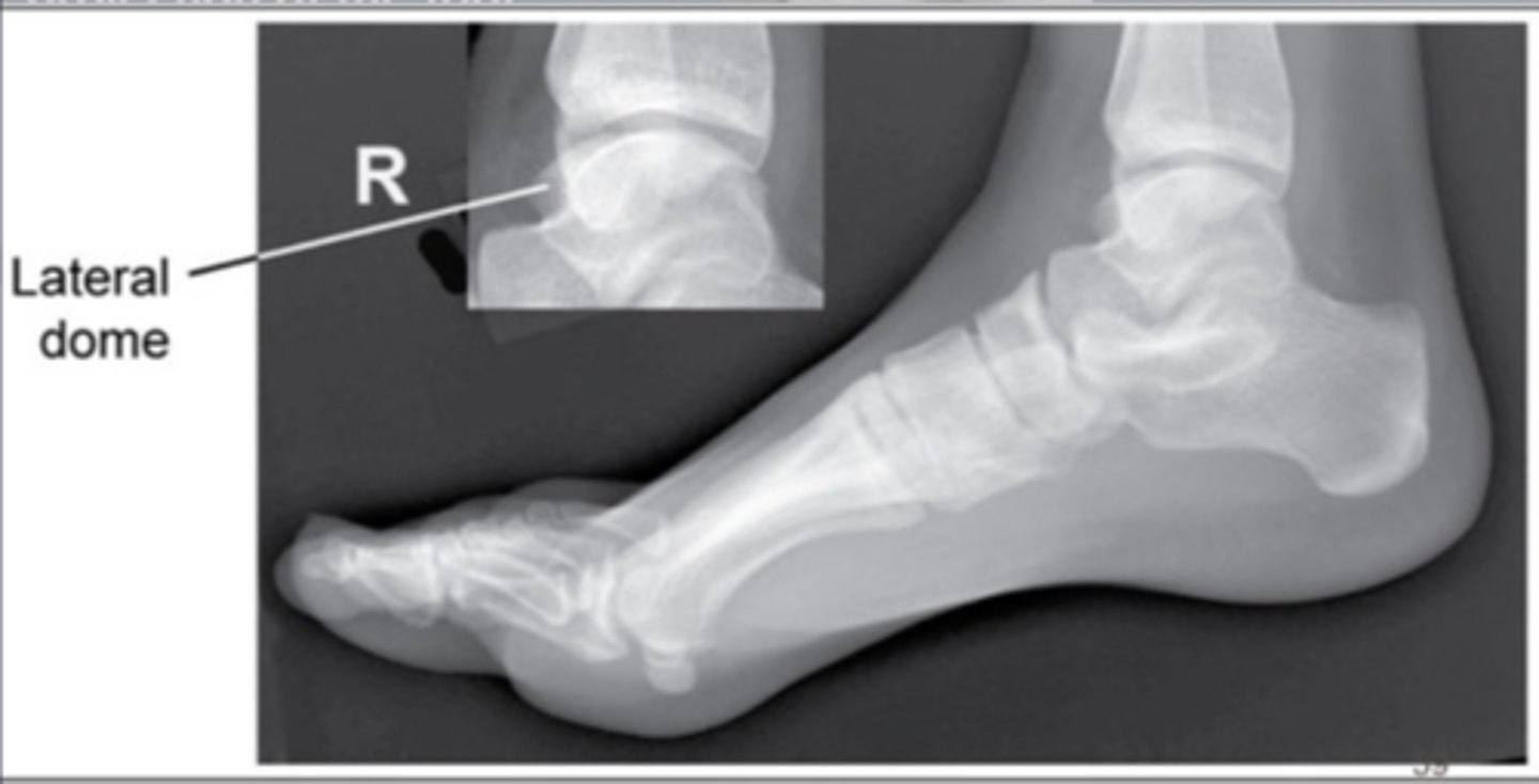
How should the foot be positioned in relationship to the lower leg for the plantodorsal projection of the calcaneus?
Dorsiflex the foot until plantar surface is perpendicular to IR
How much is the central ray angle and in what direction for the platodorsal projection of the calcaneus?
40° cephalic/towards heel
CR location for plantodorsal projection of the calcaneus:
Near the base of the 3rd metatarsal
How should the foot be positioned in relationship to the lower leg for the lateral calcaneus?
Dorsiflex foot 90°
CR location for lateral calcaneus:
1" distal to medial malleolus
In your image of a lateral calcaneus you notice that one talar dome is anterior to another and the fibula is more posterior to the tibia, what error occured?
Heel was elevated
In your image of a lateral calcaneus you notice that one talar dome is above the other one and there is more than 0.5" of the cuboid below the navicular, what error occurred?
Foot elevated
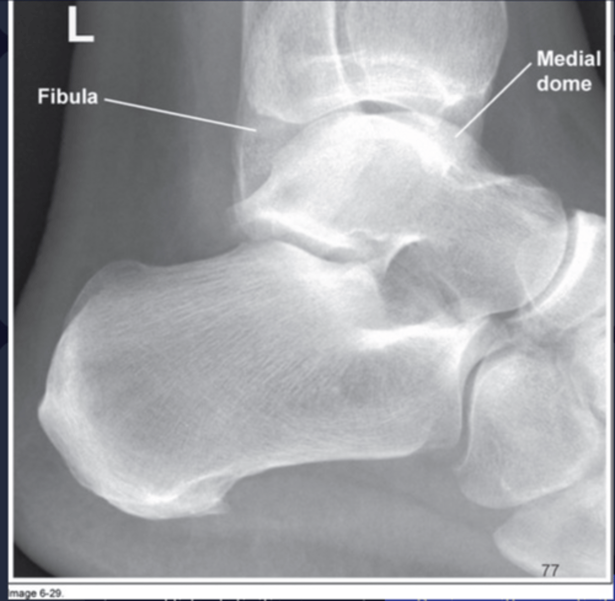
CR location for AP ankle:
Mid-malleolus
How much is the foot/ankle rotated to demonstrate the open tibiofibular articulation?
45° medial rotation
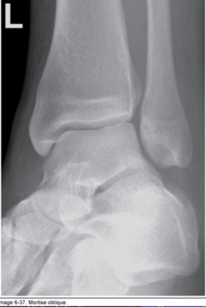
How much is the foot/ankle rotated to demonstrate the open ankle mortise joint?
15-20° medial rotation
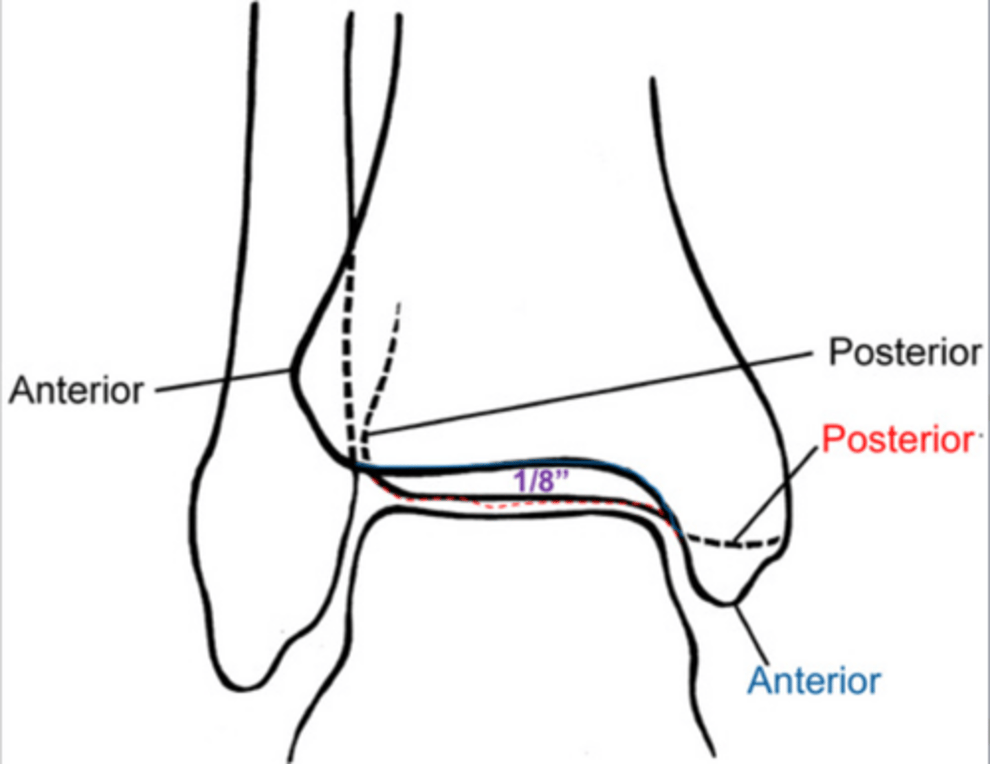
For proper positioning of an AP ankle, how much should the fibula superimpose with the tibia?
1/8"
CR location for an AP Oblique projection of the ankle:
Mid-malleolus
In your image of a mortise AP Oblique ankle you notice that here is still superimposition of the distal fibula over the talus and the lateral mortise is closed, what error occurred?
Under rotated
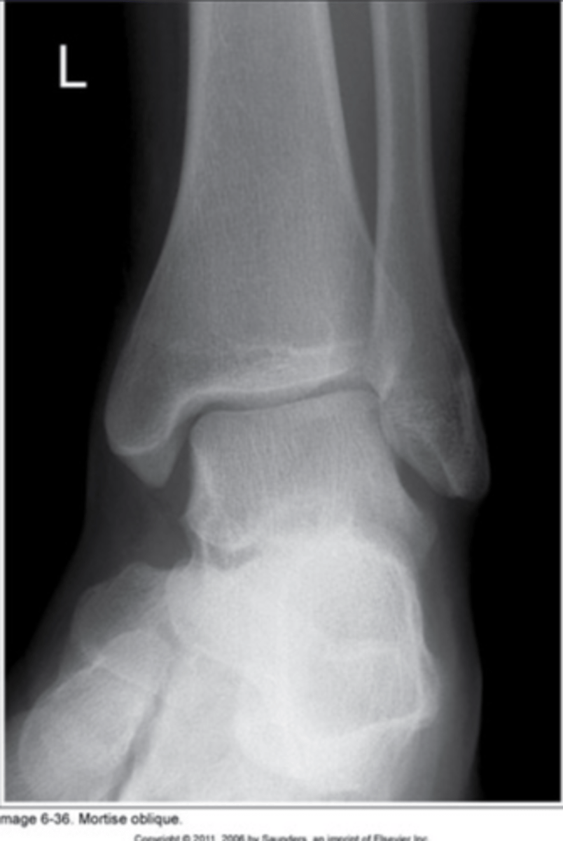
In your image of a mortise AP Oblique ankle you notice that the fibula is completely free of superimposition of the tibia, what error occurred?
Over rotated
CR location for lateral ankle:
Medial malleolus
How should the talar domes be demonstrated in a aproperly positioned lateral ankle?
Superimposed
Where should the distal fibula be demonstrated in relation to the distal tibia in a properly positioned lateral ankle?
Fibula in posterior half of the tibia
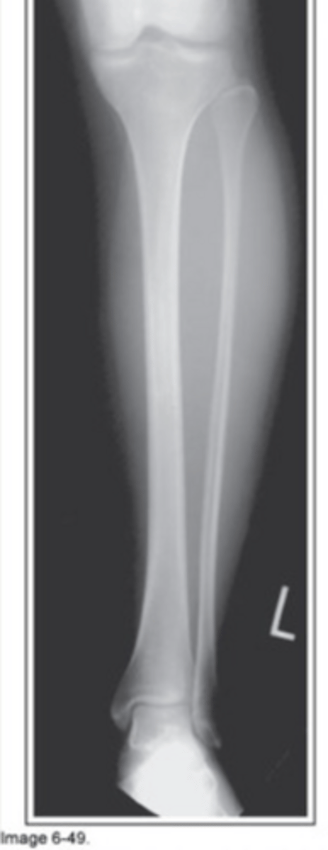
CR location for AP tib/fib:
Mid shaft of tib/fib
How should the femoral condyles be positioned in relation to the IR for a properly positioned AP lower leg?
Parallel
On your image of an AP tib/fib you notice there is superimposition of th shaft of the tibia over the fibula and greater superimposition of the distal tibia over the fibula, what error occurred?
Externally rotated
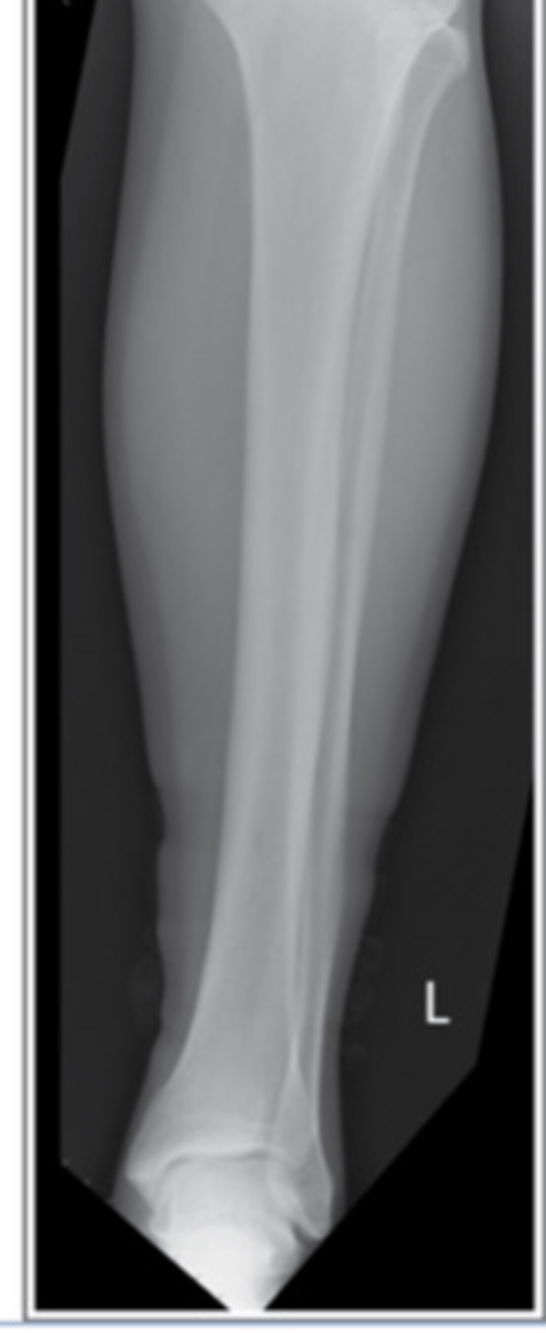
On your image of an AP tib/fib you notice there is seperation between the fibula head and tibia and greater seperation between the shaft of the tibia and fibula, what error occurred?
Internally rotated
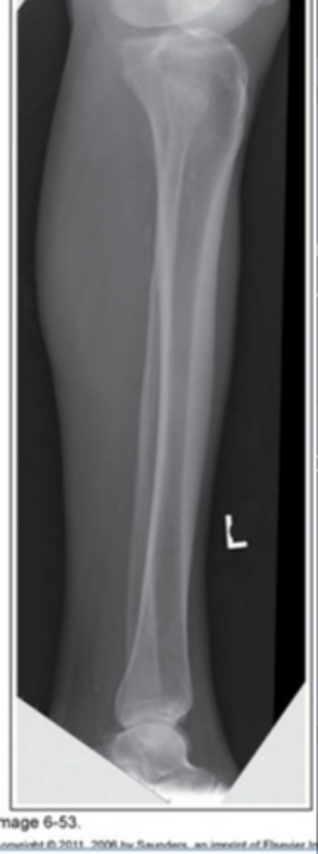
CR location for lateral tib/fib:
Mid shaft of tib/fib
How should the femoral condyles be positioned in relation to the IR for a properly positioned lateral lower leg?
Perpendicular
On your image of a lateral tib/fib you notice that the fibular head is free of superimposition of the tibia and there is greater superimposition between the shafts of the tibia and fibula, what error occurred?
Patella rolled towards IR (anterior rotation)
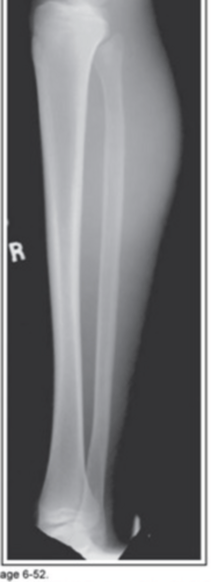
On your image of a lateral tib/fib you notice that the fibular head and a shaft completely superimpsoed by the tibia, what error occurred?
Patella rolled away from IR (posterior rotation)
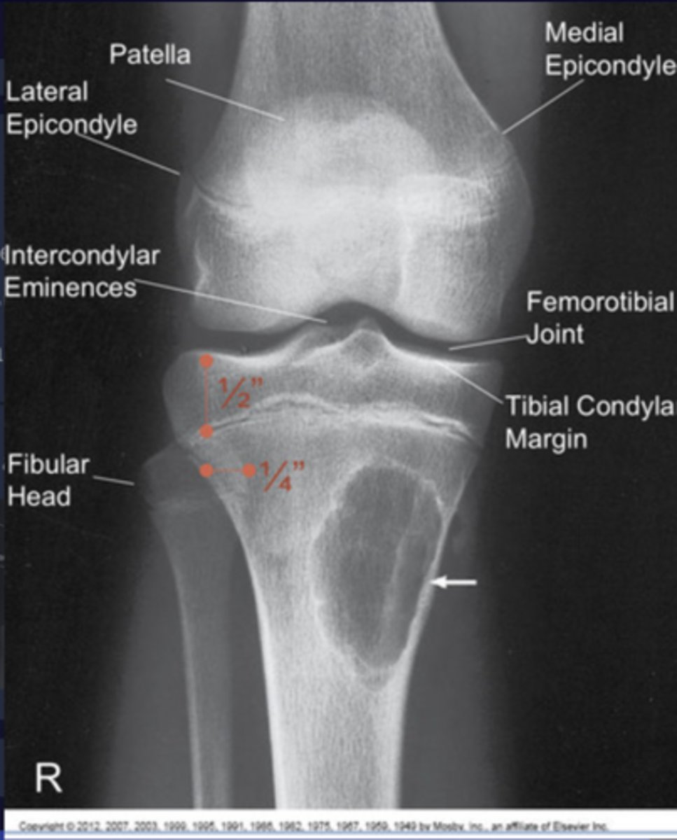
CR location for AP knee:
1/2" below patellar apex
How should the femoral condyles be positioned in relation to the IT for a properly positioned AP knee?
Parallel
To proerly demonstrate an open knee joint, what measurement should be taken to determine the central ray angle/direction?
Between the ASIS and the tabletop
For an AP knee, when the distance from ASIS to tabletop is less than 19cm, the CR is angled _________.
3-5° caudad
For an AP knee, when the distance from ASIS to tabletop is between 19 cm and 24 cm, the CR is angled ________.
Perpendicular
For an AP knee, when the distance from ASIS to tabletop is greater than 24 cm, the CR is angled _______.
3-5° cephalic
What is a varus deformity?
Narrowing of the medial side of the knee
What is a valgus deformity?
Narrowing of the lateral side of the knee
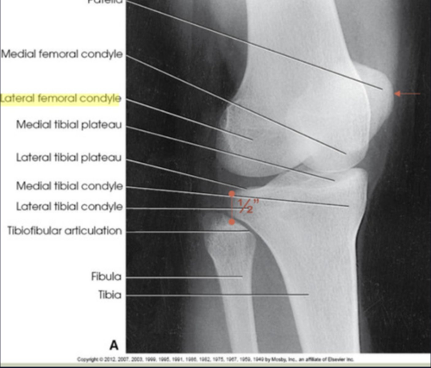
When the proper angle is used for an AP knee, how should the tibia plateau be in relationship to the fibular head?
Tibaia plateau 1/2" proximal to top of fibula head
how much do you rotate for an AP Oblique knee?
45°
You notice on an AP knee that there is less than 1/2" of the tibia plateau proximal to the fibula head, what error occurred?
Too much caudal angle
You notice on an AP knee that there is greater than 1/2" of the tibia plateau proximal to the fibula head, what error occurred?
Too much cephalic angle
CR location for AP oblique knee:
1/2" below patellar apex
Where should the patella be demonstrated when properly positioned for an AP medial oblique knee?
Projects slightly beyond the medial side of the femoral condyle