Excitable Tissues: Neurons
1/40
There's no tags or description
Looks like no tags are added yet.
Name | Mastery | Learn | Test | Matching | Spaced | Call with Kai |
|---|
No analytics yet
Send a link to your students to track their progress
41 Terms
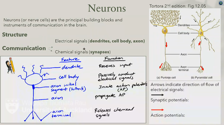
dendrite → receives input
cell body → passively conducts electrical signals
axon initial segment (hillock) → initiate action potentials (AP)
axon → propagates AP
axon terminals → releases chemical signals
outline the structure and function of neurons
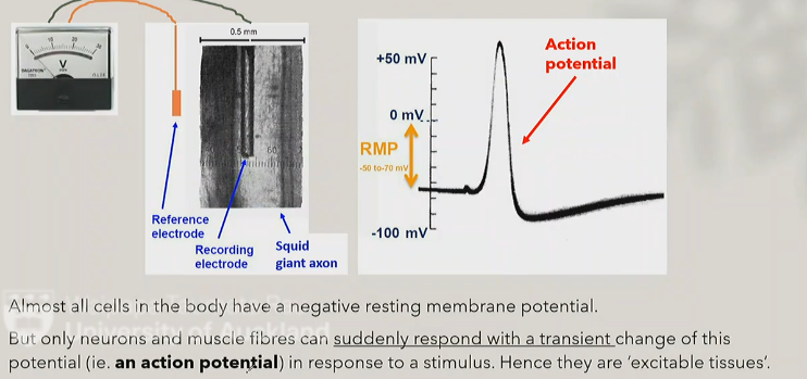
because only they can suddenly response with a transient change to an action potential, all other cells have a negtive resting membrane potential
why are neurons and muscle fibres called excitable tissues
electrical potential difference across cell membrane results from a separation of charge
there are more negative charges inside the cell in comparison to the extracellular fluid
this can be due to:
unequal concentrations of Na+ and K+ inside and outside the cell resulting in the electrochemical gradients driving the movement of these ions → higher K+ concentration inside, higher Na+ concentration outside
unequal permeability (P) of the cell membrane to these ions
what generates the resting membrane potential
non-gated (‘leak’) channels
these are open at rest (on-off state), allows for diffusion of ions
gated channels (voltage-gated, ligand gated, or mechanically gated)
these are closed at rest
in cell membranes of neurons, there are many leak K+ channels, but very few Na+ channels
therefore at rest: PK+/PNa+ = 40/1 (where P is membrane permeabilty)
outline the two main types of ion channels in neurons
an intracellular potential at which net flow of ions is zero according to its electrochemical gradient
what is equilibrium potential
the equilibrium potential can be calculated for each ion by the Nernst equation
Eion = 2.3RT/zF x log[ion]o/[ion]i → 61.5mV x log[ion]o/[ion]i
value for Na+ → quite positive, K+ → quite negative
what is the Nernst equation
a way of calculating the value of the RMP taking into account both the concentration gradients AND the relative permeability of the resting cell membrane to K+ and Na+ ions
Vm = 61.5 mV log (Pk[K+]o + PNa[Na+]o/PK[K+]i + PNa[Na+]i)
PK = 40, PNa = 1
what is the Goldman equation
a brief fluctuation in membrane potential caused by a transient opening of voltage-gated ion channels which spreads, like a wave, along an axon
action potentials occur after the membrane potential reaches a certain voltage called the threshold
what is an action potential
the frequency of action potentials encodes information (a language by which neurons communicate)
action potentials are a key element of signal transmission along (often very long) axons
what is the significance of action potentials
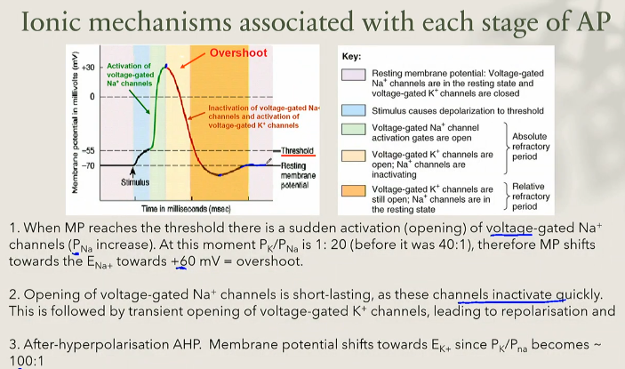
start with resting membrane potential
* = a slow depolarisation evoked by a stimulus
membrane potential reaches threshold, followed by fast depolarisation to ~ +30 mV (‘overshoot’)
when MP reaches the threshold there is a sudden activation (opening) of voltage-gated Na+ channels (PNa increase)
at this moment PK/PNa is 1:20 (before it was 40:1), therefore MP shifts towards the ENa+ towards +60 mV = overshoot.
repolarisation
opening of voltage-gated Na+ channels is short lating, as these channels inactivate quickly
this is followed by transient opening of voltage-gated K+ channels, leading to repolarisation
after-hyperpolarisation
after-hyperpolarisation AHP. Membrane potential shifts towards EK+ since PK/PNa becomes ~ 100:1
1 + 2 = absolute refractory period
3 = relative refractory period
outline the three stages of action potentials (APs)
the voltage-gated Na+ channel has two gates:
activation gate (voltage sensor)
inactivation gate
States of the channel:
State | Activation gate | Inactivation gate | When |
|---|---|---|---|
Resting (RMP) | Closed | Open | At rest, no Na⁺ flow |
Activated (open) | Open | Open | At threshold — Na⁺ flows in |
Inactivated | Open | Closed (blocks pore) | A fraction of a millisecond after opening |
Back to resting | Closed | Open | Once membrane repolarises |
outline the role of voltage-gated Na+ channels in AP
at RMP: activation gate closed, inactivation gate open → channel closed overall
at threshold: activation gate opens → Na+ flows into the cell along both the concentration gradient and the electrical gradient
Na+ influx stops because:
the inside becomes positive (approaches ENa), reducing the driving force for further Na+ entry and
the inactivation gate closes (channel inactivates)
membrane repolarises → channel resets to the resting state (activation gate closes, inactivation gate reopens)
outline the sequence of events that occurs in a voltage-gated Na+ channel in an AP
each action potential is an all-or-none event — once threshold is reached, the AP fires fully; it does not fire “partially”
this contrasts with graded potentials (subthreshold depolarisations/hyperpolarisations), which vary continuously with stimulus strength
AP amplitude is roughly constant (~100mV) and does not depend on stimulus intensity, as long as the stimulus is suprathreshold (above threshold)
what is the all-or-none principle
externally (experimental) — electrical stimulation via electrodes/battery
internally (physiological) — postsynaptic potentials build up at synapses
if a stimulus is large enough to trigger an AP, adjacent voltage-gated channels open in sequence, propagating the signal along the axon
what are the two ways in which action potentials are evoked
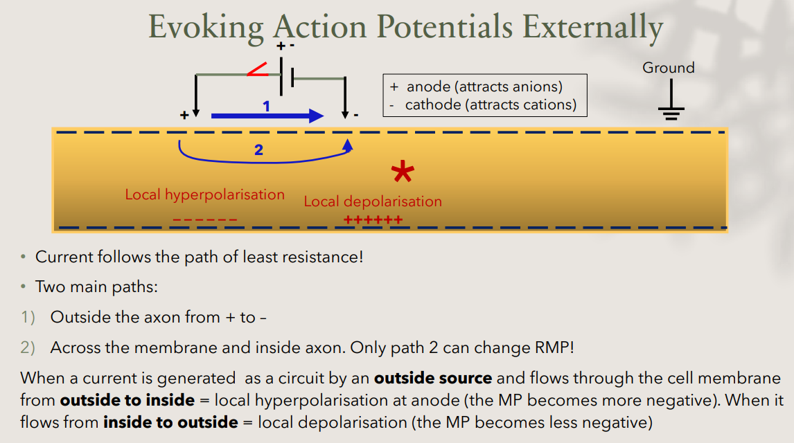
current follows the path of least resistance, two possible paths:
outside the axon, from + to - electrode (does NOT affect RMP)
across the membrane and inside the axon (this is the only path that can change RMP)
effect of current direction across the membrane:
Current direction
Effect
Location
Outside → inside (at the anode, +)
Local hyperpolarisation (MP more negative)
Near + electrode
Inside → outside (at the cathode, −)
Local depolarisation (MP less negative)
Near − electrode
anode (+) attracts anions
cathode (-) attracts cations
outline how APs are evoked externally
APs are first generated at the axon initial segment (axon hillock) — this region has the lowest threshold, making it the trigger zone for APs
depolarisation to threshold is driven by excitatory postsynaptic potentials (EPSPs), which spread passively from the dendrites toward the axon hillock
once an AP is generated at the axon hillock, it is transmitted actively along the axon, away from the cell body
how are APs generated physiologically in CNS neurons
unmyelinated axons: smaller diameter (~1um); transmission of APs, slow, continuous
myelinated axons: larger diameter (5-10um); transmission of APs fast, ‘saltatory’ (in large steps)
two stages of action potential transmission (in both types of axons):
passive spread
generation of action potentials
describe and outline the two types of axons
when (subthreshold) depolarisation occurs at one region of the membrane:
local depolarisation occurs at one point
passive current flow occurs — both inside (axoplasm) and outside (extracellular) the axon
this passively depolarises adjacent parts of the membrane
what is the passive spread of current
an action potential occurs at one point on the membrane
passive current flow spreads to adjacent regions
this depolarises the adjacent membrane to threshold
voltage-gated Na+ channels in the adjacent region open
a new, full-size action potential is generated in that adjacent region
this process repeats — effectively “regenerating” the AP at every point along the membrane
outline the sequence of events in AP transmission in unmyelinated axons
conduction velocity in unmyelinated axons = 1m/sec
although passive current flow itself is fast, the AP must be actively regenerated at every point along the membrane — and this regeneration takes time, slowing overall conduction
why is the speed of conduction in unmyelinated axons slow
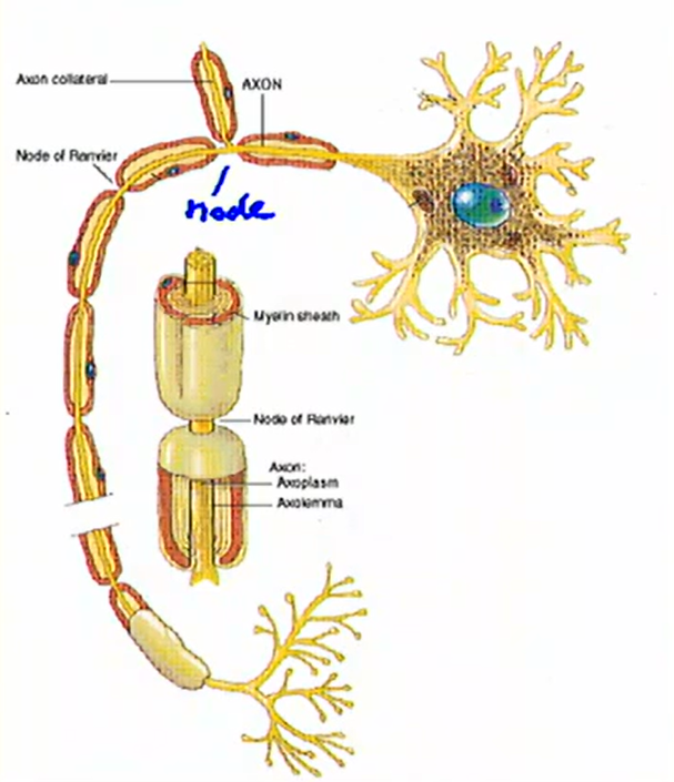
myelin sheath formed:
by oligodendrocytes in the CNS
by Schwann cells in the PNS
note: oligodendrocytes and Schwann cells are two types of glia cells
myelination is discontinuous; interrupted at nodes of Ranvier
outline the structure of neurons with myelinated axons
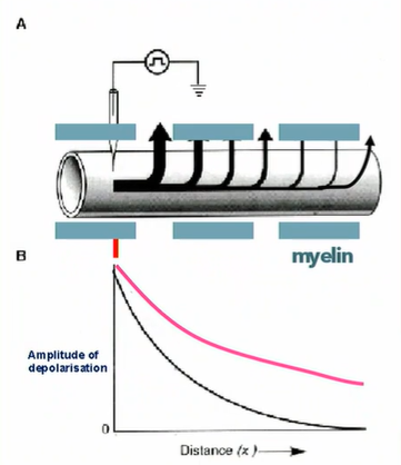
due to the insulating properties of myelin, there is less current dissipation as it flows along the axon
note: passive conduction occurs in both directions (right and left)
outline how myelination increases passive spread of current
myelination increases speed of AP conduction by increasing the efficiency of passive spread, and the fact that APs do not need to be regenerated at every part of the cell membrane
APs are generated only at nodes of Ranvier (current flows passively between nodes)
this process is called “saltatory conduction”
outline how myelination increases action potential conduction velocity
less passive current loss
less time for generation of AP
less energy to maintain gradient ions
what are the benefits of myelination for AP
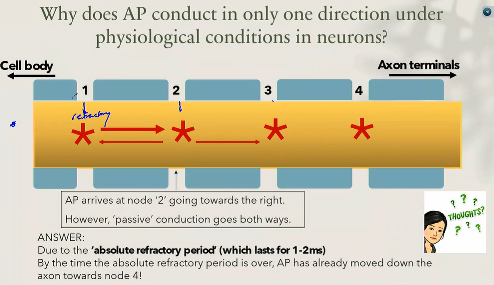
due to the absolute refractory period (which lasts for 1-2ms)
by the time the absolute refractory period is over, AP has already moved down the axon towards node 4
why does AP conduct in only one direction under physiological conditions in neurons
axons and cell bodies of sensory neurons → input
axons of motor neurones → output
neurons forming the ‘autonomic nervous system’
what does the PNS contain
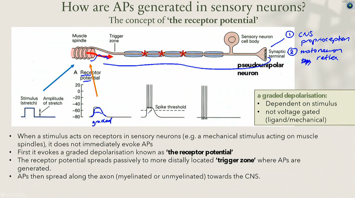
when a stimulus (e.g. a mechanical stretch acting on a muscle spindle) acts on a sensory receptor, it does not immediately trigger an action potential
the stimulus evokes a graded depolarisation called the receptor potential
the receptor potential spreads passively to a more distally located “trigger zone”, where action potentials are generated (if threshold is reached)
from the trigger zone, APs then propagate along the axon (myelinated or unmyelinated) toward the CNS
outline the sequence in which action potentials are generated in sensory neurons
dependent on stimulus strength (not all-or-none)
not voltage-gated — generated via ligand-gated or mechanically-gated channels, depending on receptor type
what are the properties of receptor potential (a graded potential)
the amplitude of the receptor potential
the frequency of the resulting action potentials
what is information about stimulus strength encoded by
action potential arrives at presynaptic terminal
voltage-gated Ca2+ channels open
Ca2+ enters the terminal
synaptic vesicles fuse with the membrane
neurotransmitter (acetylcholine) is released by exocytosis
neurotransmitter (acetylcholine) binds to receptors on the postsynaptic membrane
Na+ and K+ ion channels open, producing a postsynaptic potential
neurotransmitter is removed/inactivated
outline the stages of synaptic transmission
excitatory synapses → depolarisation of the postsynaptic membrane called the excitatory postsynaptic potential (EPSP)
inhibitory synapses → hyperpolarisation of the postsynaptic membrane called the inhibitory postsynamic potential (IPSP)
what are the two main types of chemical synapses in the CNS
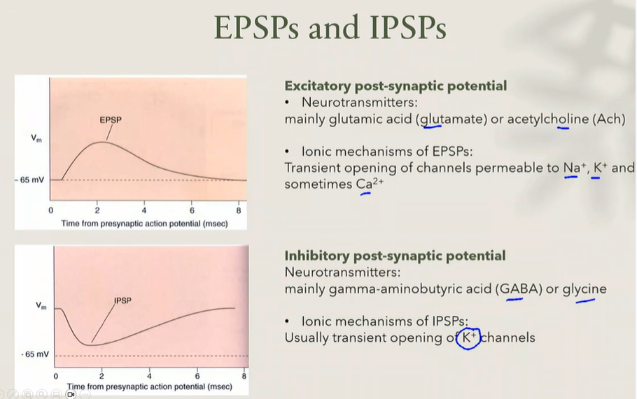
produce excitatory synapses
effect:
depolarisation
membrane becomes less negative
neuron moves closer to threshold
main neurotransmitters:
glutamate → most common excitatory neurotransmitter in the CNS
acetylcholine (ACh) → also excitatory in many situations
ionic mechanism:
EPSPs occur when channels open that allow:
Na+ entry
K+ movement
sometimes Ca2+ entry
result:
positive charge enters cell → depolarisation → EPSP
outline EPSPs
produced by inhibitory synapses
effect:
hyperpolarisation
membrane becomes more negative
neuron moves further from threshold
main neurotransmitters:
GABA → major inhibitory neurotransmitter in the brain
glycine → important inhibitory neurotransmitter in the spinal cord
ionic mechanism:
usually caused by opening K+ channels
result:
K+ leaves cell → cell becomes more negative → hyperpolarisation → IPSP
outline IPSPs
also called classical neurotransmitters
characteristics:
fast acting
millisecond effects
direct action on receptors
examples:
amino acids
acetylcholine
amines
outline small-molecule neurotransmitters
also called neuromodulators
characteristics:
large molecules
slow acting
seconds to minutes
usually indirect effects
examples:
Neuropeptide Y (NPY)
Substance P
Kisspeptin
Enkephalin
outline neuropeptides
type of neurotransmitter
examples:
glutamate → usually excitatory
GABA → usually inhibitory
type of receptor
the same neurotransmitter can produce different effects depending on the receptor present
example:
glutamate
has several receptor subtypes
therefore glutamate can produce different responses in different neurons
number of receptors present
more receptors → stronger response
this leads to synaptic plasticity
the ability of synapses to change strength
long-term potentiation (LTP)
increased synaptic strength
important for learning and memory
long-term depression (LTD)
reduced synaptic strength
describe and outline the three factors that determine synaptic action
diffusion
neurotransmitter drifts away from the synapse
enzymatic degradation
example: acetylcholinesterase
ACh → acetylcholinesterase → breakdown products
reuptake
transporters take neurotransmitter back into the presynaptic neuron
the neurotransmitter can then be recycled
outline the three mechanisms in which neurotransmitters are inactivated
each neuron receives thousands of synaptic inputs
some are:
excitatory (EPSPs)
inhibitory (IPSPs)
the neuron must combine all of these signals
summation is needed as a single synapse produces only a tiny change (~0.1mV) at the axon initial segment, this is far too small to reach threshold
outline what neuronal integration is and why its needed
multiple EPSPs arrive from the same synapse in rapid succession
EPSP + EPSP + EPSP = larger depolarisation
if large enough, action potential is generated
what is temporal summation
EPSPs from different synapses occur simultaneously
EPSP1 + EPSP2 + EPSP3 = larger depolarisation
can bring membrane to threshold
what is spatial summation
cell death caused by excessive neuronal excitiation
mechanism:
excess neurotransmitter release (usually glutamate) → excess activation of glutamate receptors → excess Ca2+ entry → cell damage → apoptosis (programmed cell death)
outline what excitoxicity is