Kines Exam 4
1/82
There's no tags or description
Looks like no tags are added yet.
Name | Mastery | Learn | Test | Matching | Spaced | Call with Kai |
|---|
No analytics yet
Send a link to your students to track their progress
83 Terms
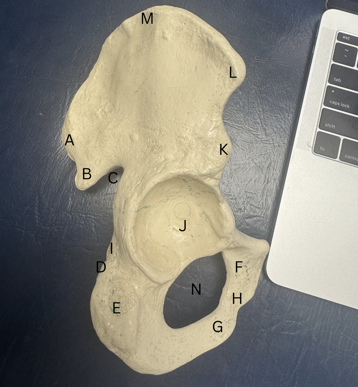
Label these structures from the Innominate bone
A. Posterior Superior Iliac Spine
B. Posterior Inferior Iliac Spine
C. Greater Sciatic Notch
D. Lesser Sciatic Notch
E. Ischial Tuberosity
F. Superior Pubic Ramus
G. Inferior Pubic Ramus
H. Pubic Body
I. Ischial Spine
J. Acetabulum
K. Anterior Inferior Iliac Spine
L. Anterior Superior Iliac Spine
M. Iliac Spine
N. Obturator Foramen
What structures make up the pelvic girdle?
two innominate bones and sacrum
What 2 joints are within the pelvic girdle? What type of joint are they?
Sacroiliac joint- non-axial diarthrodial (gliding)
Pubic symphysis - amphiarthrodial (fibrocartilage)
What are the differences between male and female pelvises?
Females V.S Men
Pelvic arch in females is >90 degrees
men’s width is more narrow
the pelvic inlet or brim is wider/larger in women
the surface markings in women are smoother than men
bones in females are lighter than mens
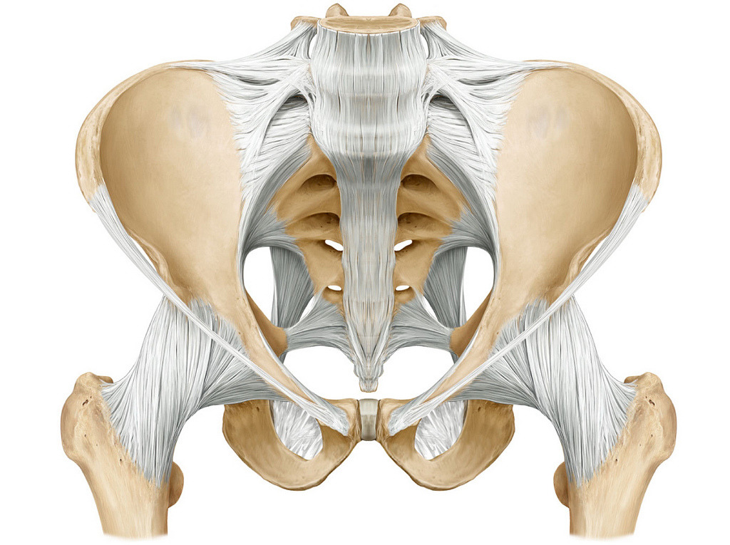
Ligaments of the Pelvic Girdle and the structures they attach
Iliolumbar ligament- ileum to L5
Inguinal ligament- connects ASIS to pubic tubercle
Anterior Sacroiliac ligament- sacrum to ileum in front
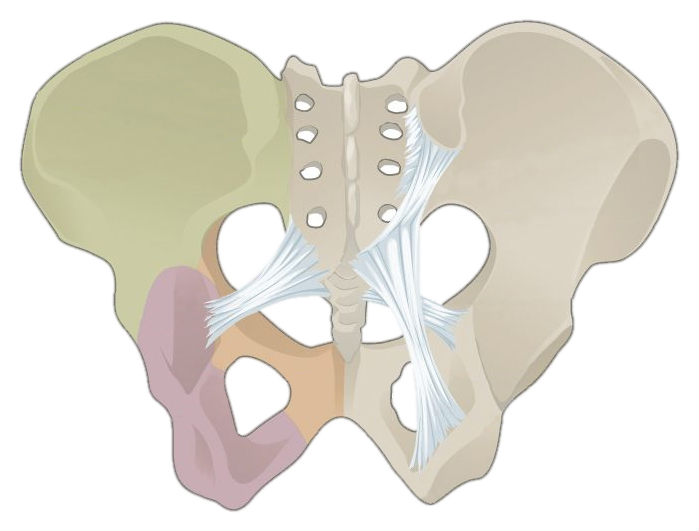
Ligaments of the Pelvic Girdle and the structures they attach
Sacrotuberous ligament- sacrum to ischial tuberosity
Posterior sacroiliac ligament- sacrum to iliac in the back
Sacrospinous- sacrum to ischial spine
Where does the Pelvis tilts when posterior and anterior and what plane of motion?
Posterior- rotates to the back
Anterior- rotates to the front
Sagittal Plane
What are the motions of the Pelvis?
Anterior rotation (forward tilt)-rotation in the sagittal plane in which upper part of the pelvis tilted forward
Posterior rotation (tilt backward)- rotation in the sagittal plane
Left lateral tilt- rotation in the frontal plane in which left hip is lower than right hip
Right lateral tilt- rotation in the frontal plane in which right hip is lower than the left hip
Left rotation- on the transverse plane in which our trunk turns left
Right rotation- on the transverse plane in which our trunk turns right
Why are there more injuries to shoulder than hip
the hip has more muscles to stabilize the joint
the ligaments in the hip are stronger
the acetabulum labrum is deeper for more stability than the glenoid labrum
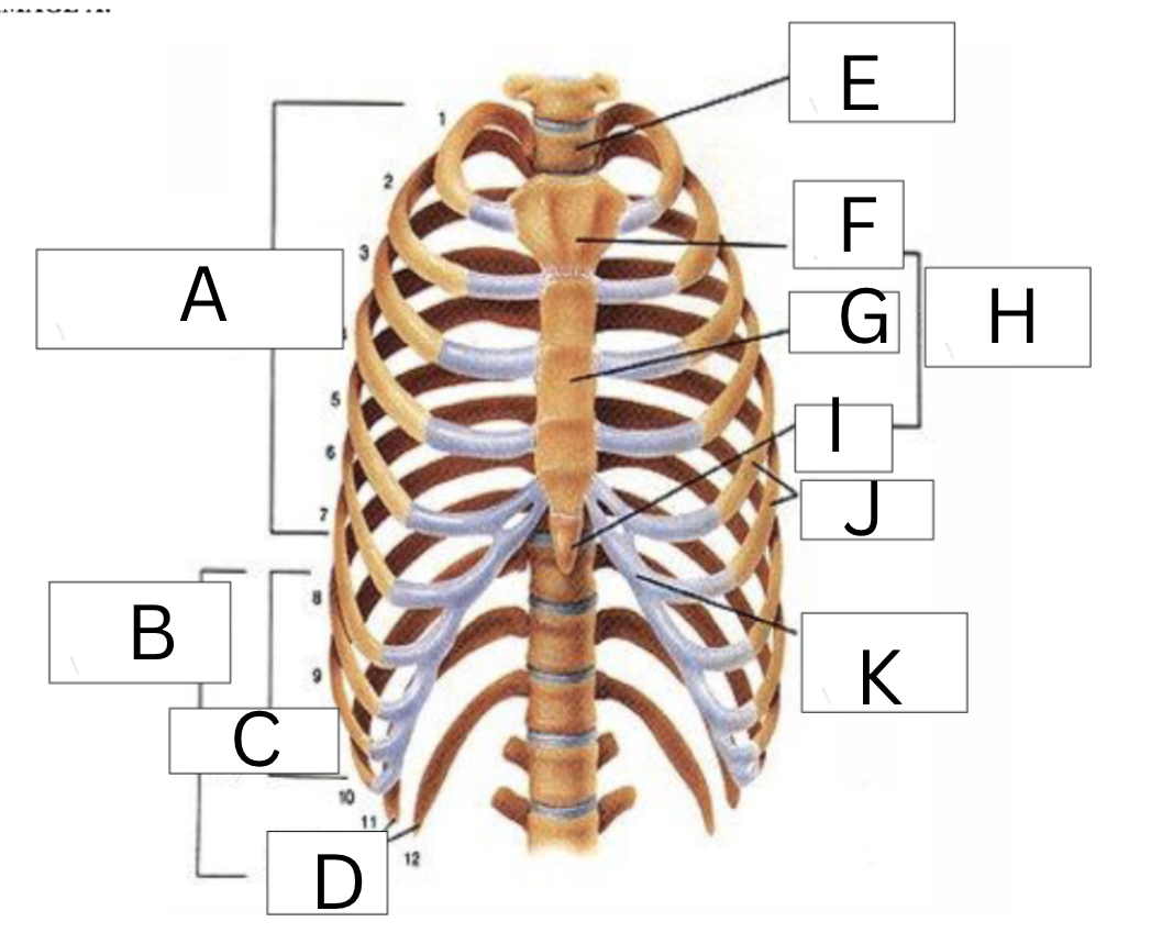
Label the following
A. 7 true ribs
B. 8-12 ribs false and floating
C. 8-10 false ribs
D. 11-12 floating ribs
E. Thoracic vertebrae
F. Manubrium of Sternum
G. Body of Sternum
H. Sternum
I. Xiphoid process of Sternum
K. Intercostal Cartilage
How many attachments in all for ribs ____ to ____ Left and Right
there are 6 attachments for ribs #1-10

What two joints does the vertebrae attach to the ribs? What kind of joints are they
Costovertebral joint- head of rib and vertebral body
Costotransverse joint-rib tubercle and transverse process
Diarthrodial, non-axial, synovial
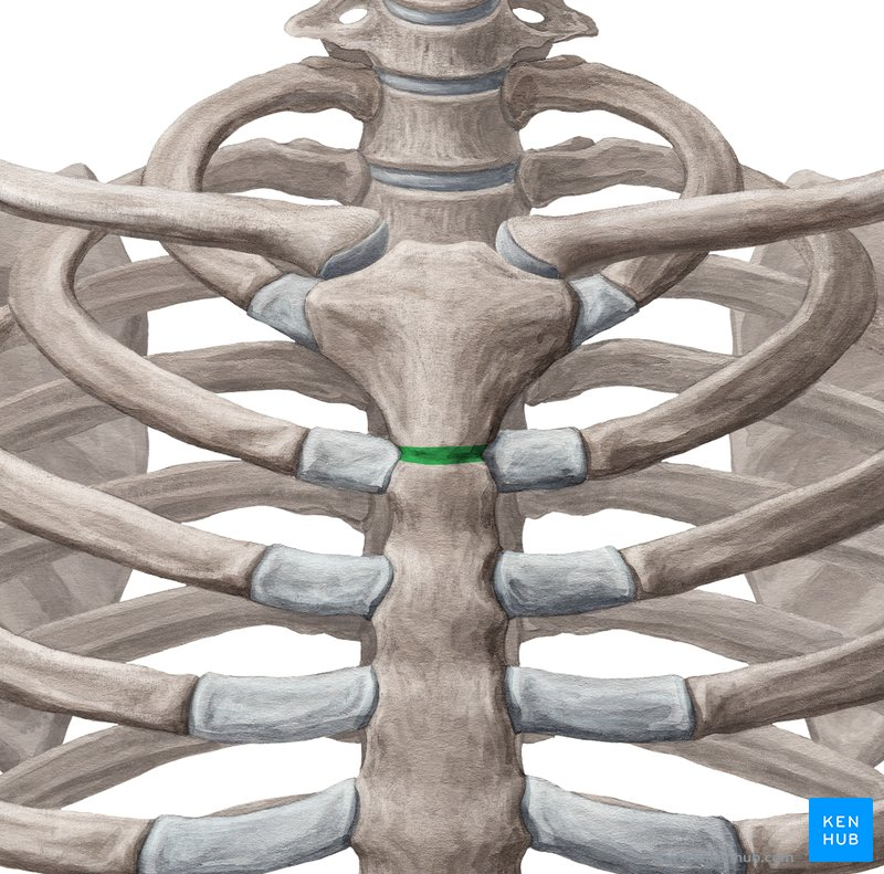
What are the joints of the Sternum?
Manubriosternal Joint- manubrium to body of sternum
Amphiarthrodial, Symphysis, fibrous
What is the joints of the Sternum?
Xiphisternal- xiphoid process to body of sternum
Amphiarthrodial, Synchondrosis, Cartilaginous
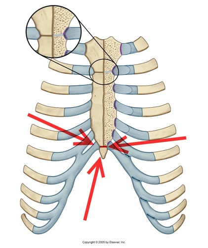
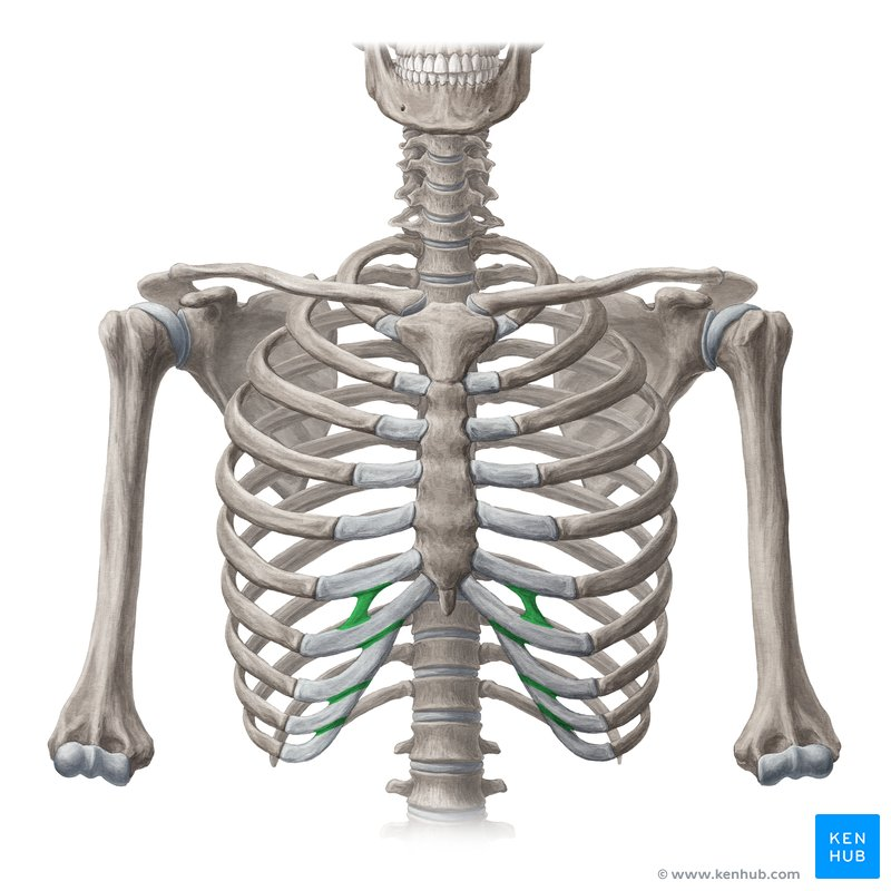
Label this joint of the rib cage?
Costochondral Joint- 10 on R. and 10 on L
Amphiarthrodial, Synchondrosis, Cartilaginous
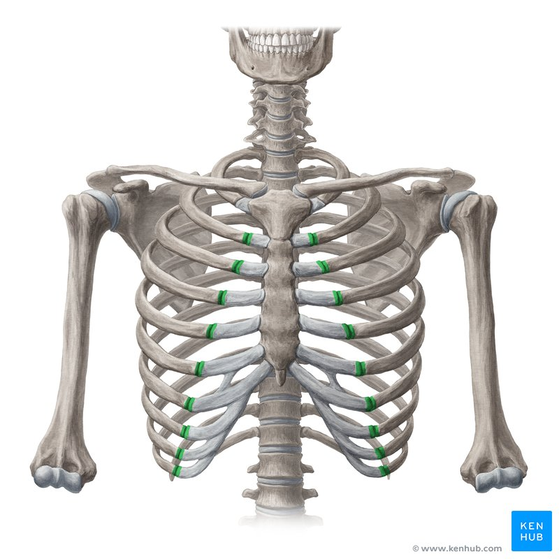
Label this joint of the rib cage
Chondrosternal joint
Diarthrodial, non-axial, synovial
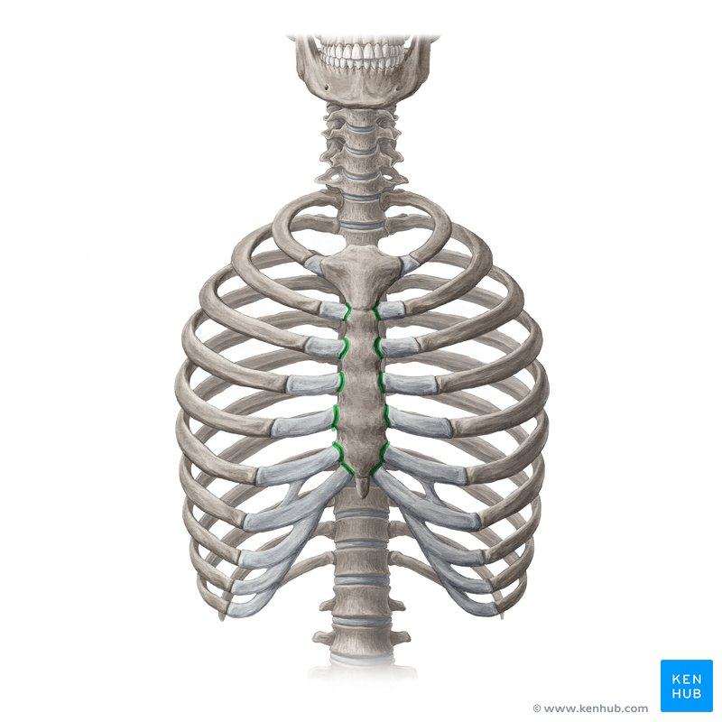
Label the joint of the rib cage
Interchondral Joint
Diarthrodial, non-axial, synovial
What are the muscles of ventilation? What do they do?
External Intercostals- inspiration
Internal Intercostals- expiration
Diaphragm- relaxes for inhalation and contracts for exhalation innervated by nerve C3- if injured, person stops breathing on their own
What are the trunk flexors? What do they do?
Rectus Abdominis- flexes trunk and lateral flexion
External Oblique- flexes trunk, lateral flexion, and when R contracts for left rotation (vise versa)
Internal Oblique-flexes trunk, lateral flexion, and R and L rotation
Transverse Abdominis and Quadratus Lumborum - stabilizes trunk flexors, KEGAL exercise
Psoas Major- flexes hip and trunk
Psoas Minor
What are the Superficial Spinal Extensors?
Spinalis
Longissimus
Iliocostalis
What are the Deep Spinal Erector muscles?
Rotatores
Semispinalis
Multifidus
What joints make up the shoulder girdle?
AC joint-L and R acromioclavicular joint
SC joint- L and R sternoclavicular joint
GH joint- L and R glenohumeral joint
Scapulothoracic joint
What is the conoid tubercle important?
The conoid tubercle is important because it attaches the clavicle conoid ligament (coracoclavicular ligament) to the coracoid.
Where on the clavicle does it fracture the most? Who is more likely to get cosmetic surgery got a healed clavicle bump?
about 80% of fractures occur in the middle third area
females are more likely to request cosmetic surgery than males
Where on the shoulder do we see pain from injury to biceps brachii?
Supraglenoid tubercle which is near acromion process (long head) and Coracoid process (short head) are origin attachments of the muscle
Where on the humerus is there most likely an injury?
the surgical neck, due to the weakest spot on the humerus
What ligaments make up the sternoclavicular joint?
Anterior and Posterior sternoclavicular ligament
Costoclavicular ligament
Interclavicular ligament
What kind of joint is the sternoclavicular joint?
It is diarthrodial, NON-AXIAL MULTI-DIRECTIONAL- since it doesn’t technically flex, extend, etc. It moves though on all planes, with reference to movement of the arm. up/down, retract/protract, etc.
What kinds of joint is the Acromioclavicular joint?
It is diarthrodial, NON-AXIAL MULTI-DIRECTIONAL- since it doesn’t technically flex, extend, etc. It moves though on all planes, with reference to movement of the arm. up/down, retract/protract, etc.
What ligaments make up the glenohumeral joint ?
Acromioclavicular lig.-acromion process and clavicle
Coracoacromial lig.- coracoid process and acromion process
Coracoclavicular lig.- coracoid process and clavicle
Glenohumeral lig.- glenoid cavity with humerus
What is a common injury to the acromioclavicular ligament?
Acromioclavicular sprain
MOI: posteriorly directed force on scapula (FOOSH)
What injury can happen to the glenohumeral joint?
Dislocation
What are the scapulothoracic muscles?
Pectoralis minor, serratus anterior, trapezius, rhomboid major and minor, and levator scapulae and subclavius
What are the Scapulohumeral muscles?
Rotator Cuff muscles (SITS)- Supraspinatus, Infraspinatus, Teres minor, and Subscapularis
Other muscles: Teres major, Teres minor, Deltoid, coracobrachialis, triceps brachii, and biceps brachii
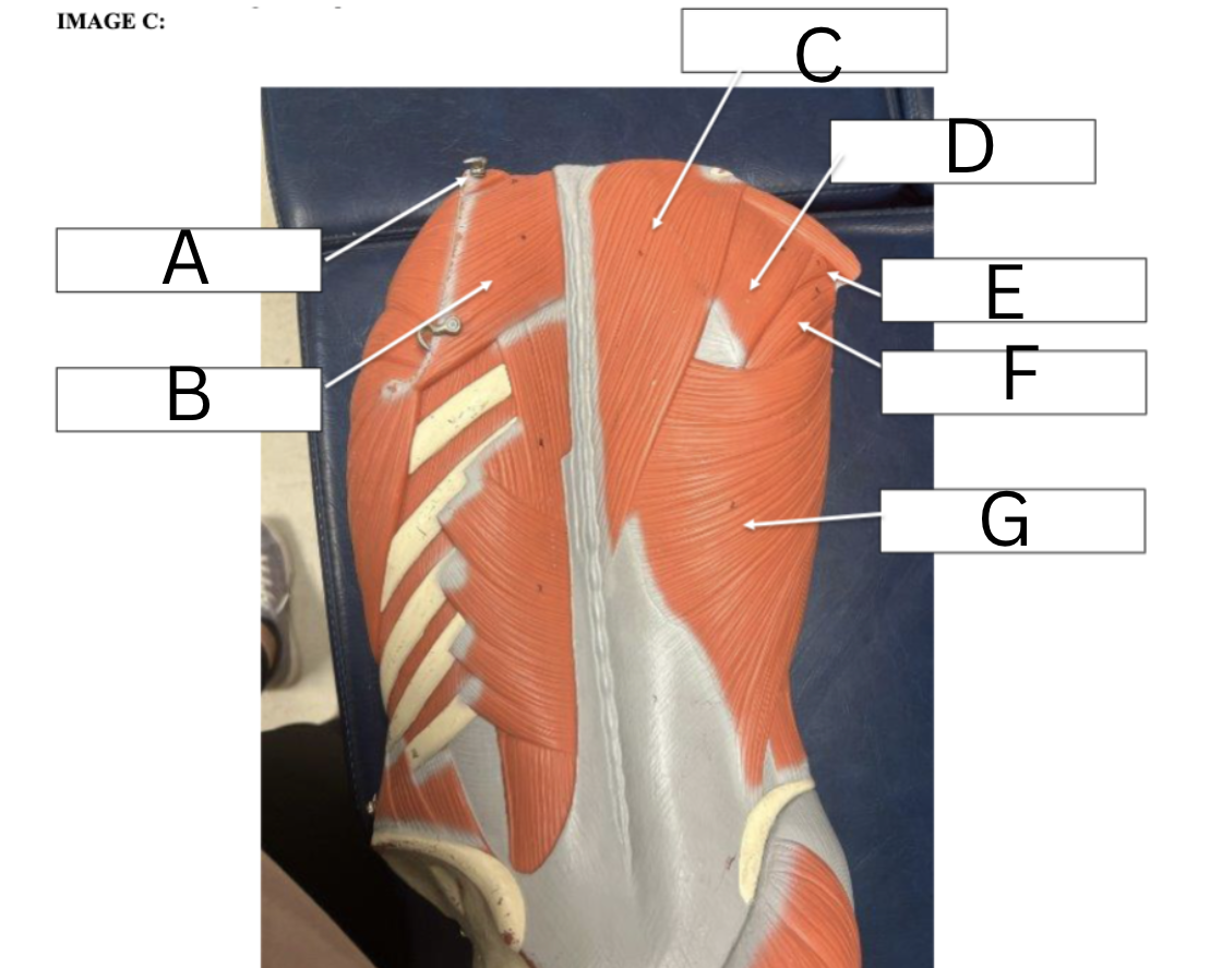
Label the following
A. Supraspinatus- arm abduction
B. Rhomboid major- retracts scapula
C. Trapezius- upper- elevates middle- retract scapula lower- depress scapula
D. Infraspinatus- laterally rotates humerus
E. Teres Minor- lateral rotation of humerus
F. Teres Major- medial rotation of humerus
G. Latissimus Dorsi- adducts arm and medially rotates arm
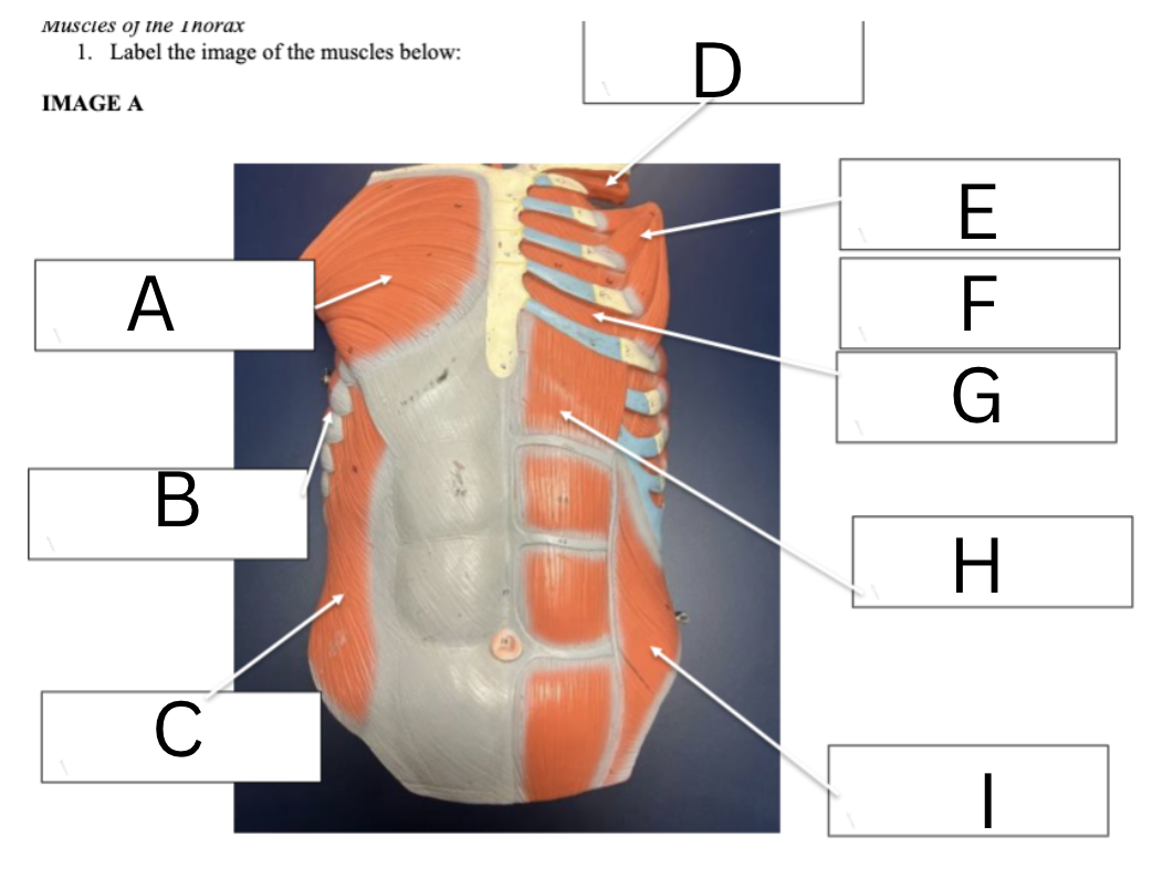
Label the thoracic and abdominal muscles and functions
A. Pectoralis Major- adducts arm
B. Serratus Anterior- protracts scapula
C. External Oblique- trunk rotation/lateral flexion of trunk
D. Innermost Intercostals- assist in depress the ribs for exhalation
E. Outer Intercostals- elevate the ribs for inhalation
G. Inner Intercostals- depress the ribs for exhalation
H. Abdominus Rectus- trunk flexion
I. Internal Oblique- trunk flexion
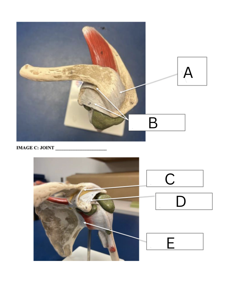
Label the ligaments of the shoulder
A. Acromioclavicular ligament
B. Coracoacromial ligament
C. Coracoclavicular ligament
D. Coracoacromial ligament
E. Glenohumeral ligament
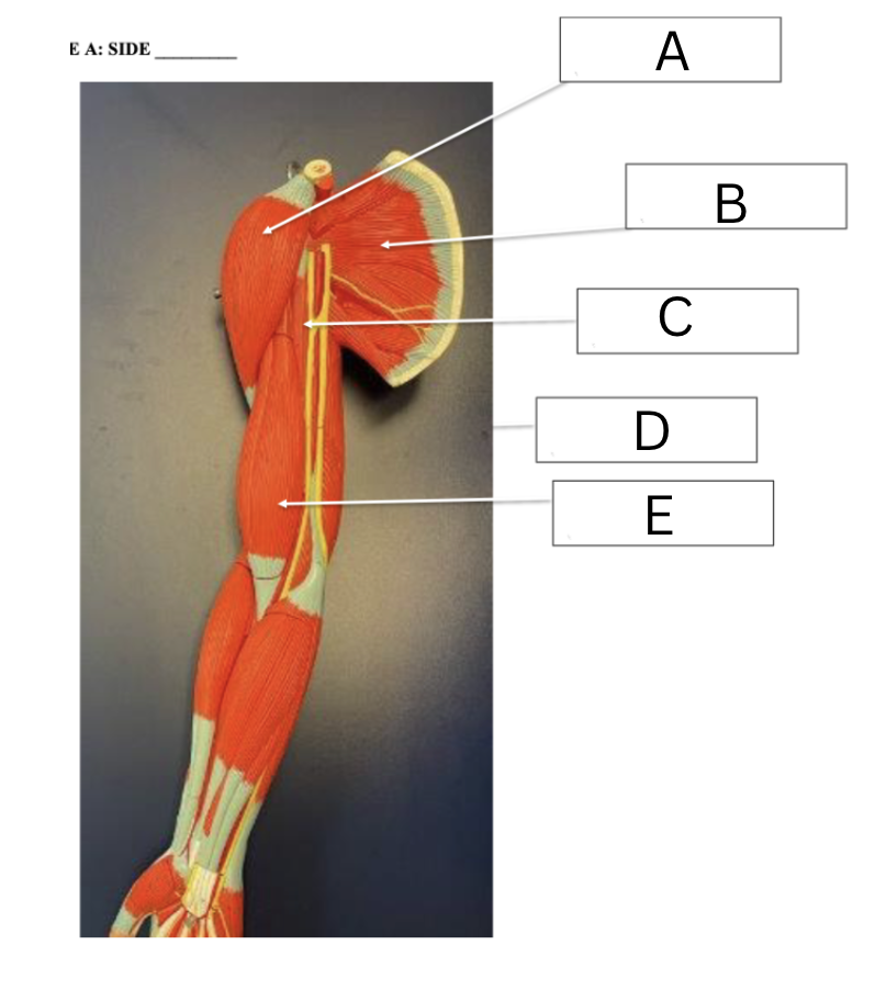
Label the following shoulder and arm muscles
A. Deltoid
B. Subscapularis
C. Coracobrachialis
E. Biceps-short head (biceps long head is lateral to that)
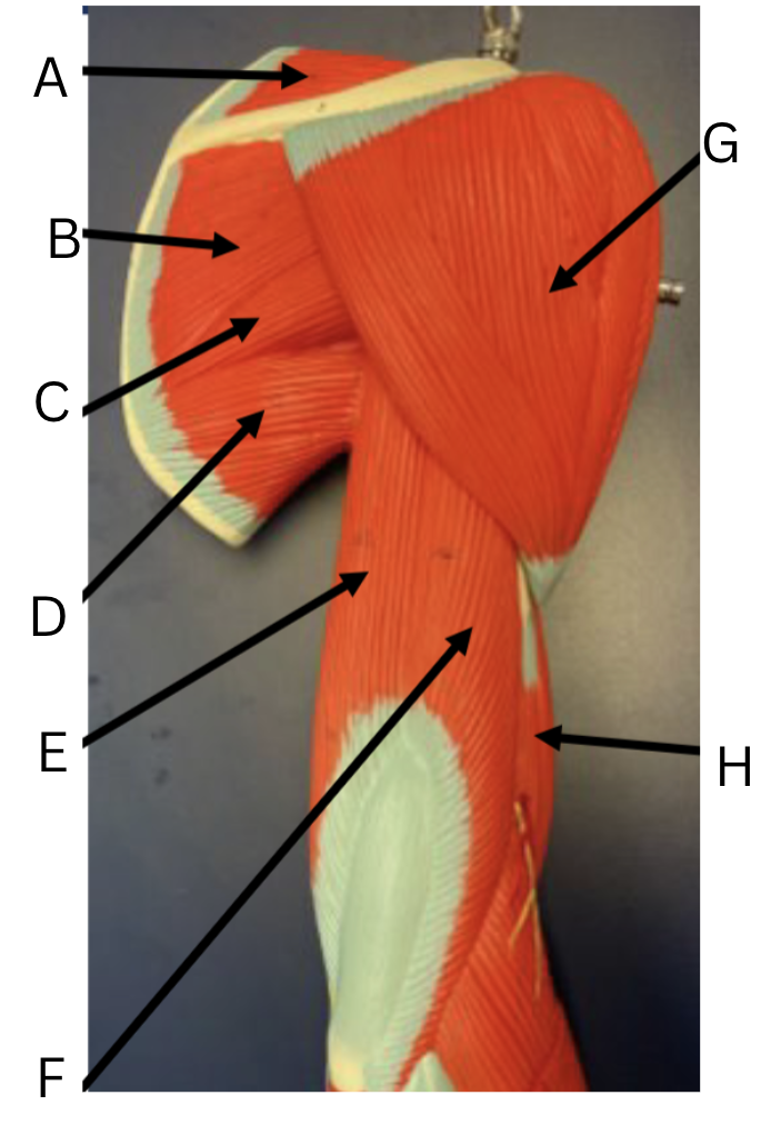
Label the shoulder and arm muscles
A. Supraspinatus
B. Infraspinatus
C. Teres Minor
D. Teres Major
E. Triceps Brachii Long head
F. Triceps Brachii Lateral head (underneath is triceps medial head)
G. Deltoid
H. Biceps brachii (long head)
What is a rotator cuff impingement?
it is the injury of a rotator cuff muscle tendon being squeezed/ pinched leading to irritation, inflammation and pain
most common is the supraspinatus tendon
know the OIF and innervation of Scapulothoracic, Scapulohumeral, and Humerothoracic muscles
Got it!
When the arm/elbow is fully extended and we rotate it, what is it called in terms of rotation?, when the arm/elbow is flexed what is it called in terms of rotation?
Fully extended- medial and lateral rotation of shoulder
Flexed- supination and pronation of forearm
What kinds of joint is the elbow joint? What makes it different to the knee joint?
A diarthrodial, synovial hinge-joint
The knee joint is biaxial do to the “screw-home mechanism” which allows the knee to lock by rotating
Why does the Humeroulnar joint never really get injured during a FOOSH injury?
The strong lock of the olecranon process of the ulna and the olecranon fossa of the humerus allows for a strong stable joint, while the distal wrist and shoulder joints are less stable
What kind of joint is the radiocapitellar joint (radiohumeral)?
Non-axial diarthrodial joint (gliding)
What kinds of joint is the Radioulnar joint?
An amphiarthrodial syndesmosis joint with interosseous membrane.
What happens during forearm supination and pronation?
The radius crosses over the ulna, while the ulna stays in place.
What is unique about the elbow ligaments insertions?
They all come together and insert into the elbow joint capsule
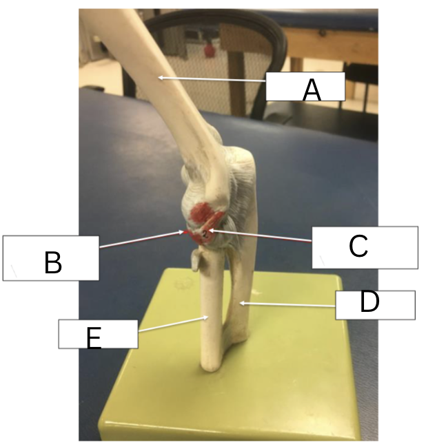
Label the ligaments on the elbow
A. Humerus
B. Annular ligament
C. Radial Collateral ligament/ LCL
D. Ulna
E. Radius
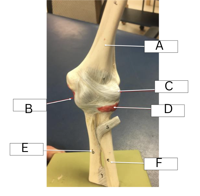
Label the elbow ligaments
A. Humerus
B. Ulnar Collateral Ligament/ MCL
C. Joint capsule
D. Annular Ligament'
E. Ulna
F. Radius
What does the LCL/RCL prevent? MCL/UCL? and Annular ligament?
LCL/RCL: prevents varus forces
MCL/UCL: prevents valgus forces and stabilizes humerus
Annular ligament: hold radial head to ulnar notch
Who is Tommy John? What surgery did he get?
Tommy John was the first MLB player who tore his UCL, he then got this surgery which took his palmaris longus tendon and drilled three holes into the humerus and ulna and threaded the tendon to strengthen it
He then was able to make a full recovery and was able to return to the MLB
Know the OIF and innervation of the Elbow Joint crossing shoulder muscles
Biceps Brachii, Coracobrachialis, and Triceps brachii
Know the OIF and innervation of the Elbow Joint crossing only elbow muscles
Brachialis, Brachioradialis, and Anconeus
Know the OIF and innervation of the Elbow Joint controlling supination and pronation muscles
Pronator teres, Pronator quadratus, Biceps brachii and Supinator
What is Scapulohumeral rhythm?
motions of the scapula and glenohumeral joint that are combined with movement of scapula around rib cage
scapula moves when humerus moves
protaction, retraction, elevation and depression
What plane of movement does the glenohumeral joint during horizontal abduction and adduction occur on?
The transverse plane
Know the OIF and innervation of the wrist flexors and extensors
Flexor Carpi Radialis, Flexor Carpi Ulnaris, Palmaris Longus, Extensor Carpi Radialis Brevis, Extensor Carpi Radialis Brevis, Extensor Carpi Ulnaris
What joint is most responsible for wrist pronation and supination?
The distal Radioulnar joint
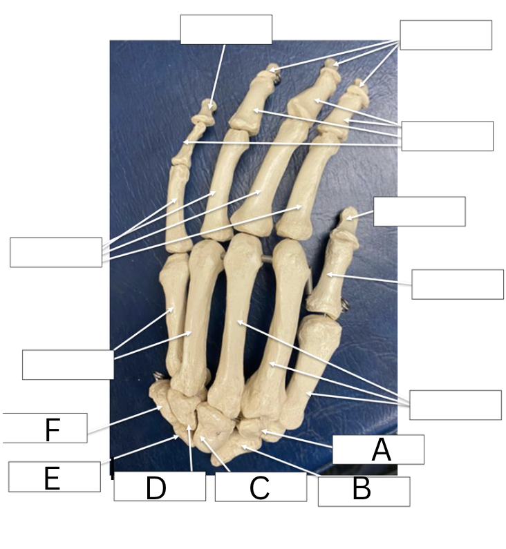
Label the carpal bones only
A. Trapezoid (toward the right is the Trapezium)
B. Scaphoid
C. Capitate
D. Hamate
E. Lunate
F. Triquetrum (pisiform on top)
What is the Roof, Bottom and Contents inside the Carpal Tunnel?
Roof: flexor retinaculum
Bottom: carpal bones
Contents:
4 tendons of Flexor Digitorum Superficialis
4 tendons of Flexor Digitorum Profundus
1 tendon of Flexor Pollicis Longus
Median Nerve
What is Carpal Tunnel Syndrome? What 2 Occupations and Sports experience this syndrome?
It is the compression of the Median nerve within the Flexor Retinaculum, causing pain, tingling, and numbness in fingers
Occupations: Hairdresser, desk job
Sports: Rock climbing, Tennis
What types of joints are in the wrist, carpometacarpal, metacarpophalangeal, and interphalangeal
All diarthrodial-synovial
Wrist- biaxial: flexion/extension and radial/ulnar deviation thumb: opposition/reposition
CMC- non-axial
MCP- biaxial: flexion/extension and abduction/adduction
IP-uniaxial flexion/extension
What are some anterior ligaments of the wrist?
Transverse Carpal ligament (flexor retinaculum)
Palmar radiocarpal ligament
Palmar ulnocarpal ligament
What are some dorsal ligaments of the wrist?
Dorsal Radiocarpal ligament
Dorsal Ulnocarpal ligament
What are the 3 groups of intrinsic hand muscles
Thenar Eminence
Hypothenar
Intermediate
What muscles are in Thenar Eminence group and their innervation?
Abductor pollicis brevis- median
flexor pollicis brevis-median
opponens pollicis-median
adductor pollicis- ulnar
What muscles are in Hypothenar group and their innervation?
abductor digiti minimi- ulnar
flexor digiti minimi- ulnar
opponens digiti minimi- ulnar
What muscles are in the Intermediate group and their innervation?
Dorsal interossei- ulnar
Palmar interossei- ulnar
Lumbricals- two we do not have to know, but they flex and extend phalanges
OIF and innervation of Flexor Digitorum profundus?
O: medial shaft of ulna and interosseous membrane
I: base of distal phalanx 2-5
F: flexions MP joint and DIP
innervation- median
OIF and innervation of flexor digitorum superficialis
O: Medial epicondyle of humerus
I: Intermediate phalanges (2-5)
F: Flexion of phalanges (2-5)
innervation: Median
OIF and innervation of Extensor Digitorum
O: Lateral epicondyle of humerus
I: Middle and Distal Phalanges (2-5)
F: Extension of phalanges (2-5)
innervation: Radial
What is scoliosis? MOI? treatment?
A lateral curvature of the spine >10° (Cobb angle), often with vertebral rotation
MOI: genetics, neuromuscular disorders, adolescent females, postural habits
Treatment: PT for posture, surgical correction
What is Kyphosis? MOI? treatment?
Excessive thoracic flexion (rounded upper back).
MOI: Postural (most common in adolescents), Scheuermann’s disease, Osteoporotic compression fractures, Degenerative disc disease, Trauma
Treatment: Osteoporosis treatment, surgery if deformity, PT for posture
What is Lordosis? MOI? treatment?
Excessive lumbar extension (increased lumbar curve).
MOI: Weak abdominals, Tight hip flexors, Obesity, Pregnancy, Postural habits
Treatment: Stretch hip flexors, Strengthen core + glutes, Weight management, Correct posture, Treat underlying pathology (e.g., spondylolisthesis)
What is Herniated Intervertebral Discs? MOI? treatment?
Nucleus pulposus protrudes through annulus fibrosus → compresses spinal nerve root.
MOI: Flexion + rotation under load (lifting with poor mechanics), Degeneration, heavy lifting
Treatment: NSAIDs, rest, PT, Epidural steroid injections
Surgery (microdiscectomy)
What is Spinal Cord Injuries ? MOI? treatment?
Damage to spinal cord → motor, sensory, autonomic deficits below the level of injury.
MOI: Trauma (MVC, falls, sports), Penetrating injury, Tumor, infection, ischemia
Treatment: Immobilization, High‑dose steroids, Surgical decompression, Rehab, PT/OT, Prevention of complications
Paraplegia vs Quadriplegia
Paraplegia: injury below T1; Affects legs only
Quadriplegia: Injury at or above C5–T1; Affects all four limbs
Sprains/ Strains, Dislocations/Subluxations
Sprains: tear to ligaments
Strains: tear to muscles/tendons
Dislocations: full loss of joint articulation (FOOSH) surgically put back in
Subluxations: partial loss of joint articulation (FOOSH) popped back in easily
Clinically, how can you check the function and sensory of the terminal branches of the brachial
plexus in the upper extremity?
Musculocutaneous Nerve
Motor: Elbow flexion (biceps)
Sensory: Lateral forearm
Axillary Nerve
Motor: Shoulder abduction (deltoid)
Sensory: Lateral shoulder
Radial Nerve
Motor: Wrist extension, finger extension, thumbs‑up
Sensory: Dorsal 1st web space
Median Nerve
Motor: Thumb opposition, “OK sign”
Sensory: Palmar digits 1–3 (tip of index finger = pure median)
Ulnar Nerve
Motor: Finger abduction/adduction (interossei), Froment’s sign
Sensory: Digit 5 + ulnar half of digit 4
Clinically, how can you check the function of the arterial supply in the upper extremity?
Radial pulse (lateral wrist)
Ulnar pulse (medial wrist)
Brachial pulse (antecubital fossa)
Capillary refill <2 sec
Skin temperature & color (cool, pale = poor perfusion)
Compare BP between arms (>20 mmHg difference = vascular issue)
Doppler if pulses are weak or absent
Check for ischemic signs (mottling, delayed healing, trophic changes)
List the Arterial Supply from Heart to Phalanges
Heart (left ventricle)
Aorta
Brachiocephalic
Subclavian
Axillary
Brachial
Ulnar/Radial
Deep Palmar/Superficial Palmar Arches
Digital
The Brachial Plexus “Read The Damn Cadaver Book”
o Roots (C5, C6, C7, C8, T1)
o Trunks (Superior, Middle, Inferior)
o Divisions (3 Anterior, 3 Posterior)
o Cords (Lateral, Medial, Posterior)
o Branches (Musculocutaneous, Ulnar, Radial, Median, Axillary)
draw it out