Anatomy Lecture Test 3
0.0(0)
Studied by 1 personCard Sorting
1/202
Earn XP
Description and Tags
Last updated 3:35 AM on 11/17/22
Name | Mastery | Learn | Test | Matching | Spaced | Call with Kai |
|---|
No analytics yet
Send a link to your students to track their progress
203 Terms
1
New cards
What is a muscle fiber?
muscle cell
2
New cards
What are the 3 types of muscle tissue?
1. Skeletal ─ striated and voluntary
2. Cardiac ─ striated and involuntary
3. Smooth ─ non-striated and involuntary
2. Cardiac ─ striated and involuntary
3. Smooth ─ non-striated and involuntary
3
New cards
What is skeletal muscle?
Skeletal muscle is attached to bones by tendons and is responsible for voluntary movement.
- Multinucleated, striated, unbranched cells.
- All or none contraction
- Multinucleated, striated, unbranched cells.
- All or none contraction
4
New cards
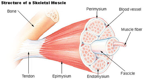
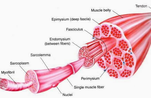
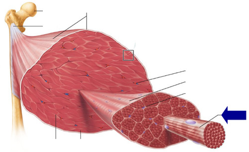
Describe the tissue within skeletal muscle?
Skeletal Muscle has an extensive connective tissue component.
- Optimally arranged for effective transfer of pull of contraction of the muscle attachements.
- Multiple tissue wrappings
- Epimysium
- Perimedium
- Endomesium
- Optimally arranged for effective transfer of pull of contraction of the muscle attachements.
- Multiple tissue wrappings
- Epimysium
- Perimedium
- Endomesium
5
New cards
Epimysium
a sheath of fibrous elastic tissue surrounding a muscle.
6
New cards
Perimesium
the sheath of connective tissue surrounding a bundle of muscle fibers.

7
New cards
Endomysium
Connective tissue surrounding a muscle fiber

8
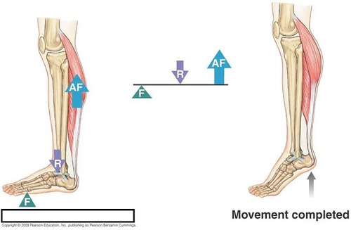
New cards
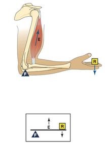
Describe epimysium, perimysium, and endomysium
- The whole muscle is enclosed in the epimysium.
- Partitions extend into the muscle dividing it into bundles of muscle fiber → fascicles
- The fascicles are wrapped in the perimysium
- The perimysium conveys the blood vessels, nerves, and lymphatics from epimysium.
- Delicate, loose connective tissue partitions extend from the perimysium into the individual fascicles.
- Wrap around each fiber in a fascicle
- Contain capillaries and nerve fibers
- This is the endomesium
9
New cards
What is sarcoplasm?
cytoplasm of a muscle cell
- Contains cylindrical striated contractile elements called myofibrils.
- Extend the full length of the fiber
- The bands are made-up of tightly packed segments of myofibrils.
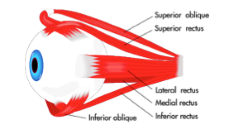
- Contains cylindrical striated contractile elements called myofibrils.
- Extend the full length of the fiber
- The bands are made-up of tightly packed segments of myofibrils.
10
New cards
Describe the anatomy of a muscle
11
New cards
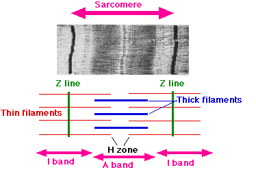
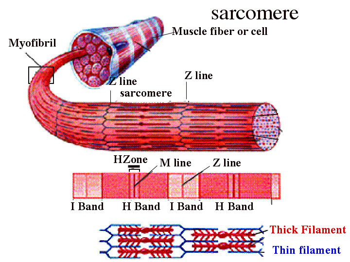
What is inside of myofibrils?
Myofibrils
- A-bands: anisotropic bands (dark bands)
- I-bands: Isotropic bands (light bands)
- these make up skeletal muscle striated
- A-bands: anisotropic bands (dark bands)
- I-bands: Isotropic bands (light bands)
- these make up skeletal muscle striated
12
New cards
Myofibrils
Microscopic protein filaments that make up muscle cells.
13
New cards
What is a sarcomere?
The functional unit of the muscle cell.
The fundamental repeat unit within muscle that is responsible for contraction.
- The contractile elements of the sarcomere are actin (thin filaments) and myosin (thick filaments).
- Cross-bridges are formed between them allowing the actin filaments to slide inward relative to the myosin, thus shortening the length of the sarcomere
14
New cards
Cardiac muscle:
causes the involuntary contractions of the heart.
- Striated and branched cells.
- Contract spontaneously.
- no nerve impulses
- Connected by intercalated discs.
- Striated and branched cells.
- Contract spontaneously.
- no nerve impulses
- Connected by intercalated discs.
15
New cards
intercalated discs of cardiac muscle
Attachment sites between the transverse lines between cardiac muscle cells
16
New cards
Smooth muscle:
is found in the walls of the digestive tract, urinary bladder, and blood vessels.
- Cells are unstriated, spindle shaped, and cause slow, but involuntary movements.
- Control the diameter of lumens.
- Partial contractions.
- Tonic contractions→ maintain tone
- Cells are unstriated, spindle shaped, and cause slow, but involuntary movements.
- Control the diameter of lumens.
- Partial contractions.
- Tonic contractions→ maintain tone
17
New cards
What are tonic contractions?
sustained contractions
- the sustained contraction of different groups of fibers within a muscle to maintain continual muscular tension (tonus)
- necessary for blood vessels that are constantly slightly contracted
- the sustained contraction of different groups of fibers within a muscle to maintain continual muscular tension (tonus)
- necessary for blood vessels that are constantly slightly contracted
18
New cards
Muscle Function
1. Locmotion
2. Produce a great amount of body heat.
3. Digestion and blood circulation and glandular secretion.
2. Produce a great amount of body heat.
3. Digestion and blood circulation and glandular secretion.
19
New cards
What do muscles do?
Muscles can only function by shortening in length.
- They can only contract.
- They can only contract.
20
New cards
How is muscle contraction for striated muscle?
- Striated muscle fibers must fully contract when stimulated.
- An "all-or-none" response.
- Controlled muscle contraction is achieved by varying the number of muscle fibers recruited in a given contraction
- An "all-or-none" response.
- Controlled muscle contraction is achieved by varying the number of muscle fibers recruited in a given contraction
21
New cards
What is the difference between extending and flexing a muscle?
- The actual motion elicited depends on its orientation to the joint.
- If it increases the angle of the joint it when contracted, it is said to extend the joint.
- If it decreases the angle of the joint it when contracted, it is said to flex the joint
Pic just for representation
- If it increases the angle of the joint it when contracted, it is said to extend the joint.
- If it decreases the angle of the joint it when contracted, it is said to flex the joint
Pic just for representation
22
New cards
Synergistic muscles
- Muscles work in conjunction with other muscles in order to elicit a given motion.
- Synergistic muscles cause a similar or ancillary motion when contracted.
nE.g., Brachialis and Biceps brachii work together to flex the elbow.
- Synergistic muscles cause a similar or ancillary motion when contracted.
nE.g., Brachialis and Biceps brachii work together to flex the elbow.
23
New cards
Antagonistic muscles
Antagonistic Muscle is a muscle that opposes the action of another.
- Antagonistic muscles are enlisted for controlled movement.
For example, when the triceps oppose the contraction of the flexing biceps by relaxing, the triceps would be regarded as the antagonistic muscle to the biceps whereas the biceps, the agonist muscle
- Antagonistic muscles are enlisted for controlled movement.
For example, when the triceps oppose the contraction of the flexing biceps by relaxing, the triceps would be regarded as the antagonistic muscle to the biceps whereas the biceps, the agonist muscle
24
New cards
What else do muscles do?
Muscles can be used for stability
- Muscles may also have some muscles acting to stabilize a joint or one end of a joint while the muscles eliciting the muscular action contract.
E.g., muscles in the shoulder and pectoral girdle such as Pectoralis major act to stabilize the shoulder while the elbow is flexed.
- Muscles may also have some muscles acting to stabilize a joint or one end of a joint while the muscles eliciting the muscular action contract.
E.g., muscles in the shoulder and pectoral girdle such as Pectoralis major act to stabilize the shoulder while the elbow is flexed.
25
New cards
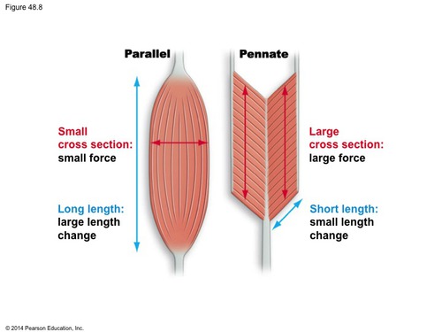
What is the orientation of muscle fibers?
- Muscle fibers are either parallel to the line of pull of the tendon or oblique to the tendon.
- Long parallel fibers occur in muscles that need to be flexed a lot.
- Muscles with a lot of short fibers that are oblique (slanted) to the line of pull (pennate muscles) are adapted to lift heavy weights short distances.
- Long parallel fibers occur in muscles that need to be flexed a lot.
- Muscles with a lot of short fibers that are oblique (slanted) to the line of pull (pennate muscles) are adapted to lift heavy weights short distances.
26
New cards
What are the different shapes of muscles cells?
1) Fusiform
2) Pennate
- unipennate
- bipennate
- multipennate
3) Strap muscles
4) Sheets
5) Sphicters
2) Pennate
- unipennate
- bipennate
- multipennate
3) Strap muscles
4) Sheets
5) Sphicters
27
New cards
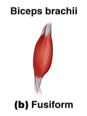
Fusiform muscles cells
(cylindrical) are modified spindles. E.g., Biceps brachii
- Occur in muscles that need to be flexed a lot.
- Fibers are long and parallel to the line of pull.
- They are long, but relatively few fibers.
- The muscle contracts 1/3 to ½ its length.
- Relatively weak.
- Occur in muscles that need to be flexed a lot.
- Fibers are long and parallel to the line of pull.
- They are long, but relatively few fibers.
- The muscle contracts 1/3 to ½ its length.
- Relatively weak.
28
New cards
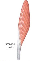
pennate muscles
have short fascicles oblique to the long tendon or tendons running the length of the muscle.
- "feather shaped".
- More fascicles attached directly to tendons than other muscle types.
- Generally more powerful than other muscles.
- tend to be antigravity muscles
- "feather shaped".
- More fascicles attached directly to tendons than other muscle types.
- Generally more powerful than other muscles.
- tend to be antigravity muscles
29
New cards
What are the 3 types of pennate muscles?
unipennate, bipennate, multipennate
30
New cards
unipennate muscle
Direction of pull is toward the side of the tendon.
- all the muscle fibers are on the same side of the tendon
- all the muscle fibers are on the same side of the tendon
31
New cards
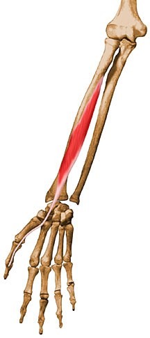
ex of unipennate muscle
Flexor pollicis longus
32
New cards
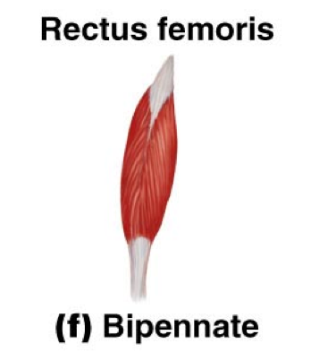
bipennate muscle
muscle fibers on both sides of the tendon; rectus femoris
- Oblique fascicles on both sides of the tendon.
- Equal pull on both sides of the tendon.
- Adapted to lift heavy weight short distances.
- Contract a short distance.
e.g., Rectus femorus or Triceps brachii
- Oblique fascicles on both sides of the tendon.
- Equal pull on both sides of the tendon.
- Adapted to lift heavy weight short distances.
- Contract a short distance.
e.g., Rectus femorus or Triceps brachii
33
New cards
multipennate muscle
- Many oblique fascicles arranged along several tendons.
- Allows movement in multiple
directions in a flexible joint
- Allows movement in multiple
directions in a flexible joint
34
New cards
Strap Muscles
- Fascicles are parallel to the long axis.
- Allow for a wide range of movement, but are not very powerful/strong.
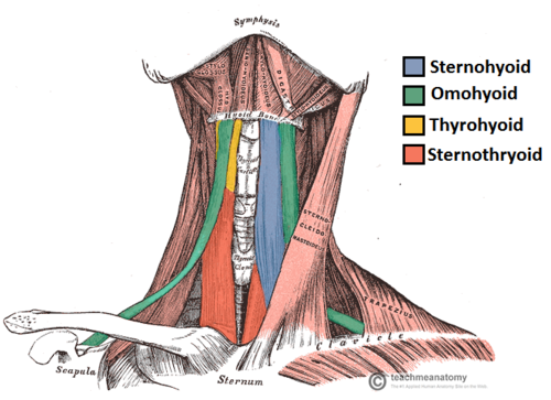
e.g., Sternohyoid
- Allow for a wide range of movement, but are not very powerful/strong.
e.g., Sternohyoid
35
New cards
sheets of muscle
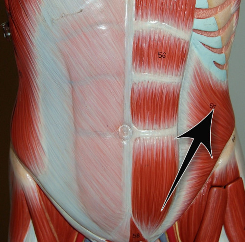
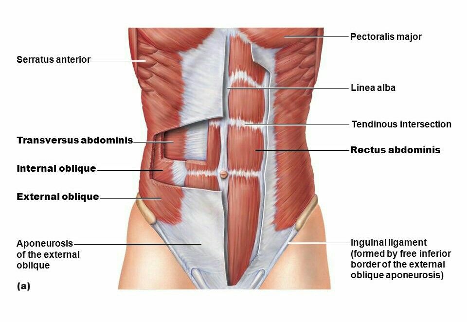
- Similar to strap muscles, but also can be used as constrictors.
e.g., External and internal obliques.
e.g., External and internal obliques.
36
New cards
Valsalva maneuver
Any forced expiratory effort against a closed airway such as when an individual holds his or her breath and tightens his or her muscles in a concerted, strenuous effort to move a heavy object or change positions in bed.
- constricting sheet muscles.
- allows us to compress the abdomen
*don't need to know*
- constricting sheet muscles.
- allows us to compress the abdomen
*don't need to know*
37
New cards
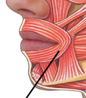
Sphicters
- Fascicles arranged in circular pattern around and opening of a structure.
e.g., Obicularis oris or External anal sphincter
e.g., Obicularis oris or External anal sphincter
38
New cards
Describe muscle structure and attachment
- Muscles attach to bone via tendons.
- Tendons are extensions of the epimysium.
- The tendon is continuous with the periosteum of the bone.
- The periosteum inserts into the CaCO3 of the bone via Sharpey's fibers.
- When a tendon forms a broadsheet it is called an aponeurosis.
- a thin sheath of connective tissue that helps connect your muscles to your bones
39
New cards
sharpey's fibers
connect periosteum to compact bone
- a matrix of connective tissue consisting of bundles of strong collagen fibres connecting periosteum to bone.
- a matrix of connective tissue consisting of bundles of strong collagen fibres connecting periosteum to bone.
40
New cards
aponeurosis
A broad, flat tendon
41
New cards
Origin
the least movable point of attachment.
- The site where bone and muscle are attached, but do not move during contraction.
- The site where bone and muscle are attached, but do not move during contraction.
42
New cards
Insertion
the most moveable point of attachment (moves during contraction).
- Usually insert in the proximal end of the bone to produce rapid movement at the distal end of the bone.
- Usually insert in the proximal end of the bone to produce rapid movement at the distal end of the bone.
43
New cards
Ex of origin and insertion with biceps brachii
Origin ─ Short head: tip of coracoid process of scapulaLong head: supraglenoid tubercle of scapula
Insertion ─ radial tuberosity of the radius
Insertion ─ radial tuberosity of the radius
44
New cards
What are the different types of muscle actions?
The action of a muscle usually refers to its prime action.
Types of actions:
- Flexion and extension
- Plantar flexion and dorsi-flexion.
- Protraction and retraction
- Abduction and adduction
- Inversion and eversion
- Pronation and supination
- Levitation and depression
Types of actions:
- Flexion and extension
- Plantar flexion and dorsi-flexion.
- Protraction and retraction
- Abduction and adduction
- Inversion and eversion
- Pronation and supination
- Levitation and depression
45
New cards
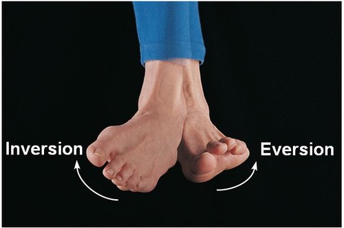
Which actions are the movements of the foot?
- Inversion and eversion
- Dorsiflexion and Plantar Felxion
- Dorsiflexion and Plantar Felxion
46
New cards
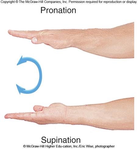
pronation and supination
palms down to palms up
47
New cards
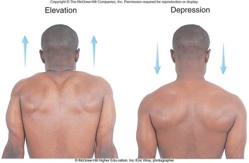
elevation and depresion
elevation (levitation)- moving a sturcture superior
depression - moving a structure down
depression - moving a structure down
48
New cards
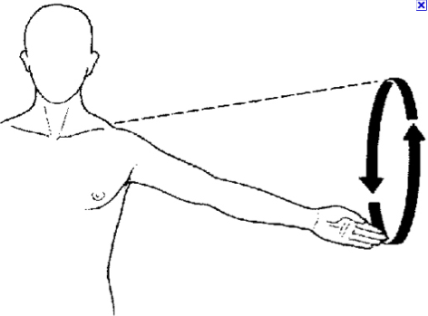
cicumduction
moving arm in loop
49
New cards
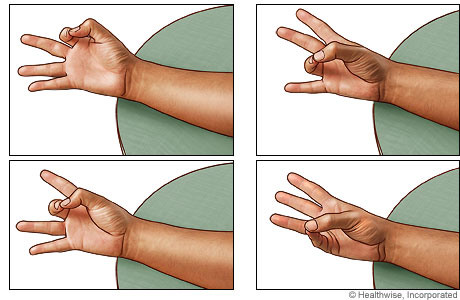
opposition
Movement of the thumb to touch the fingertips
50
New cards
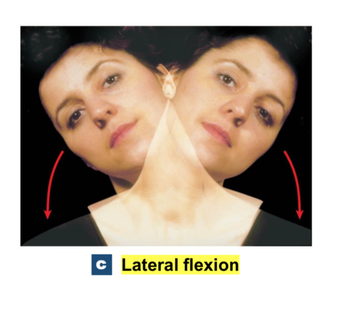
lateral flextion
bending the vertebral column to the side
51
New cards
Describe tension and load
- The strength of the muscle is proportional to its cross-sectional area.
- Tension: the force muscle exerts on something.
- Load: the force a exerted on the muscle..
- Force drops off as the muscle gets shorter.
- Muscles only operate fully to move the lever at a 90 degrees.
- Tension: the force muscle exerts on something.
- Load: the force a exerted on the muscle..
- Force drops off as the muscle gets shorter.
- Muscles only operate fully to move the lever at a 90 degrees.
52
New cards
describe isotonic contractions muscles
- Isotonic muscles produce both work and heat.
- If load and tension are not equal the contractions are isotonic.
- If load and tension are not equal the contractions are isotonic.
53
New cards
Describe isometric contractions
- Isometric muscles just produce a lot of heat.
- If load and tension are equal the contractions are isometric
- If load and tension are equal the contractions are isometric
54
New cards
how do muscles work to be able to accomplish tasks?
Muscles work in conjunction with bones and joints to form a lever system
55
New cards
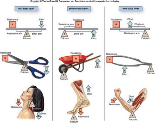
What are the 3 classes of levers?
first class, second class, third class
56
New cards
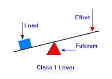
first-class levers
Fulcrum in the middle like a see-saw(nodding the head)
- the fulcrum is betweeen the force and the weight
- the fulcrum is betweeen the force and the weight
57
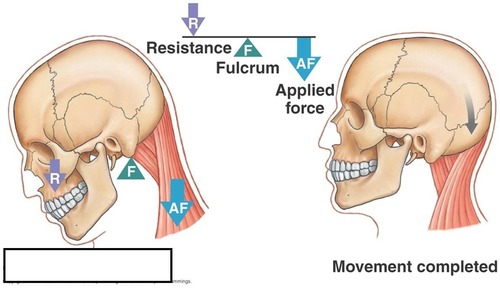
New cards
ex of first class levers
the head
- the occipital condyle is the fulcrum
- the nuchal muscles are the force
- the head is the weight
- the occipital condyle is the fulcrum
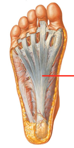
- the nuchal muscles are the force
- the head is the weight
58
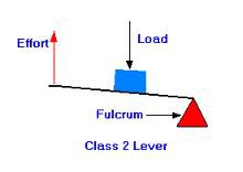
New cards
Second class lever
the weight is between the force and the fulcrum.
59
New cards
ex of second class lever
foot
- metatarsals are the fulcrum
- force is muscles in back of leg
- weight is leg
- metatarsals are the fulcrum
- force is muscles in back of leg
- weight is leg
60
New cards
third class lever
the force is between the weight and the fulcrum.
61
New cards
ex of third class lever
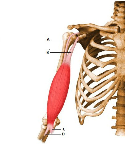
arm
- weight in hand
- elbow joint is the fulcrum
- forearm flexors are force
- weight in hand
- elbow joint is the fulcrum
- forearm flexors are force
62
New cards
What happens when you move your muscles alot?
Hyperactivity causes hyperplasia of muscle fibers (more fibers) and hypertrophy of red muscle fibers.
63
New cards
hypertrophy
increase in cell size
64
New cards
hyperplasia
increase in number of cells
65
New cards
What are the 2 main types of muscle fibers
1) Red muscle fibers are Type I muscle fibers (slow twitch fibers)
2) White muscle fibers are Type II muscle fibers (fast twitch fibers)
- Most vertebrate muscles are a mixture of red and white muscle fibers.
2) White muscle fibers are Type II muscle fibers (fast twitch fibers)
- Most vertebrate muscles are a mixture of red and white muscle fibers.
66
New cards
Red Fibers
- Aerobic (don't fatigue as rapidly)
- Less myofibrils
- More myoglobin
- Contract just a little more slowly
- Very vascular
- A lot of mitochondria
- Good for tonic contractions to maintain posture.
- slow twitch fibers
- very aerobic & needs lots of oxygen
- Less myofibrils
- More myoglobin
- Contract just a little more slowly
- Very vascular
- A lot of mitochondria
- Good for tonic contractions to maintain posture.
- slow twitch fibers
- very aerobic & needs lots of oxygen
67
New cards
white muscle fiber
- More myofibrils
- Less myoglobin
- Contract fast and fatigue fast
- Less vascular
- Less mitochondria
- Basically anarobic
- fast twitch
- Less myoglobin
- Contract fast and fatigue fast
- Less vascular
- Less mitochondria
- Basically anarobic
- fast twitch
68
New cards
type 3 muscle fiber
- fast twitch, fatigue resistant fibers.
- seen in flight muscles of migratory birds
- Contract fast, but not as fast as white fibers.
- Don't fatigue as quickly as white fibers.
- Contain many mitochondria and blood vessels (similar to red fibers).
- Store more oxygen and lipids, and less glycogen than other muscle fibers.
- Particularly prominent in flight muscles of migratory birds.
69
New cards
What muscles have each type of fiber?
- Most muscles have all types of fibers.
- The relative abundance gives the muscle its overall characteristics.
-White meat vs. red meat.
- Twitch times vary with body size and mode of locomotion.
- Faster in smaller animals
- The relative abundance gives the muscle its overall characteristics.
-White meat vs. red meat.
- Twitch times vary with body size and mode of locomotion.
- Faster in smaller animals
70
New cards
What is the embryological origin of muscles?
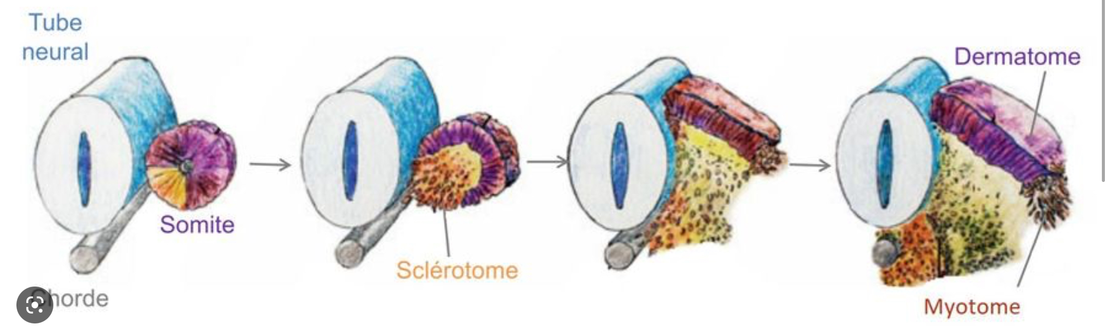
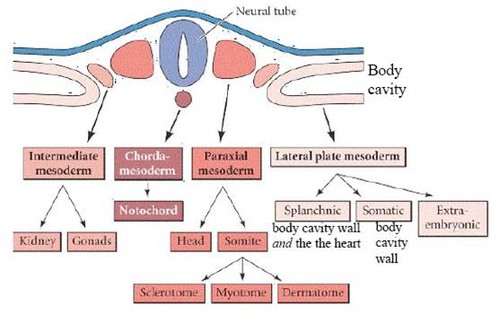
- Muscles develop from myotomes of somites.
- Paraxial mesoderm gives rise to the somites.
- Runs along each side of the neural tube and notochord.
- Segmentation begins in the initial stages of neurulation.
- The segments are called somitomeres.
- Paraxial mesoderm gives rise to the somites.
- Runs along each side of the neural tube and notochord.
- Segmentation begins in the initial stages of neurulation.
- The segments are called somitomeres.
71
New cards
what is a myotome?
the dorsal part of each somite in a vertebrate embryo, giving rise to the skeletal musculature.
72
New cards
How do somites develop?
- somites derived from paraxial mesoderm
- somitomeres develop into somites
- box-like structures
- somitomeres develop into somites
- box-like structures
73
New cards
What are somitomeres?
collections of cells that are derived from the loose masses of paraxial mesoderm that are found alongside the developing neural tube.
74
New cards
Do somitomeres form somites?
Yes but the First 7 somitomeres do not form somites.
- They remain in the head region.
- May contribute to the bones and muscles of the base of the skull.
- Extraoccular eye muscles are thought to form from these cranial somitomeres.
* extraoccular eye muscles move the eye
- alot of chondrocranium will come from
- They remain in the head region.
- May contribute to the bones and muscles of the base of the skull.
- Extraoccular eye muscles are thought to form from these cranial somitomeres.
* extraoccular eye muscles move the eye
- alot of chondrocranium will come from
75
New cards
when/where do somitomeres form somites?
The 8th pair of somitomeres form the first pair of somites.
76
New cards
What are the different types of somites?
Sclerotome: forms bone (,vertebrae, and ribs)
Dermatome: forms the dermis of the skin
Myotome: forms muscle
Dermatome: forms the dermis of the skin
Myotome: forms muscle
77
New cards
Review somite formation photo on slide 57
78
New cards
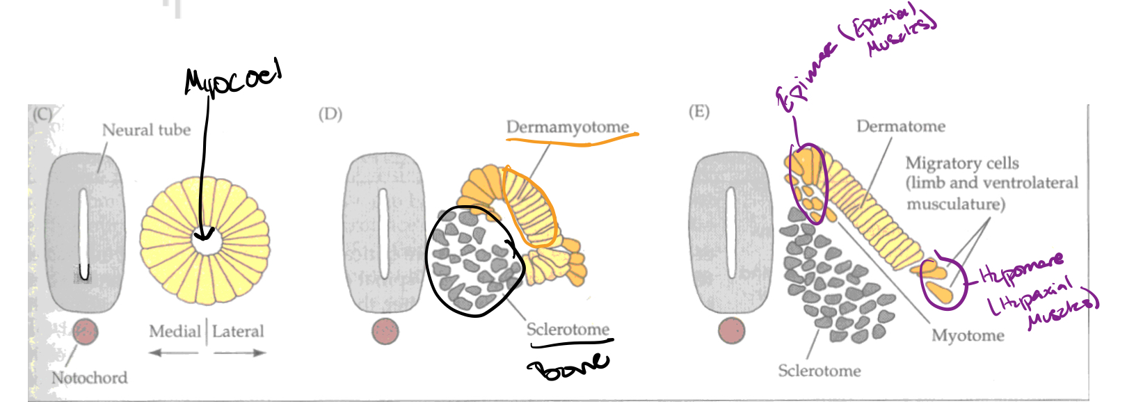
How are myotomes initially interacting in the development of muscle?
- Myotomes are divided into epimeres and hypomeres.
79
New cards
How do te epimeres and hypomeres interact after being developed?
- Epimeres and hypomeres are separated from one another by a lateral septum of connective tissue.
- Epimeres give rise to the epaxial muscles (the "true back"muscles).
- Hypomeres give rise to the hypaxial muscles.
- Virtually all muscles of the body are hypaxial muscles.
- Epimeres give rise to the epaxial muscles (the "true back"muscles).
- Hypomeres give rise to the hypaxial muscles.
- Virtually all muscles of the body are hypaxial muscles.
80
New cards
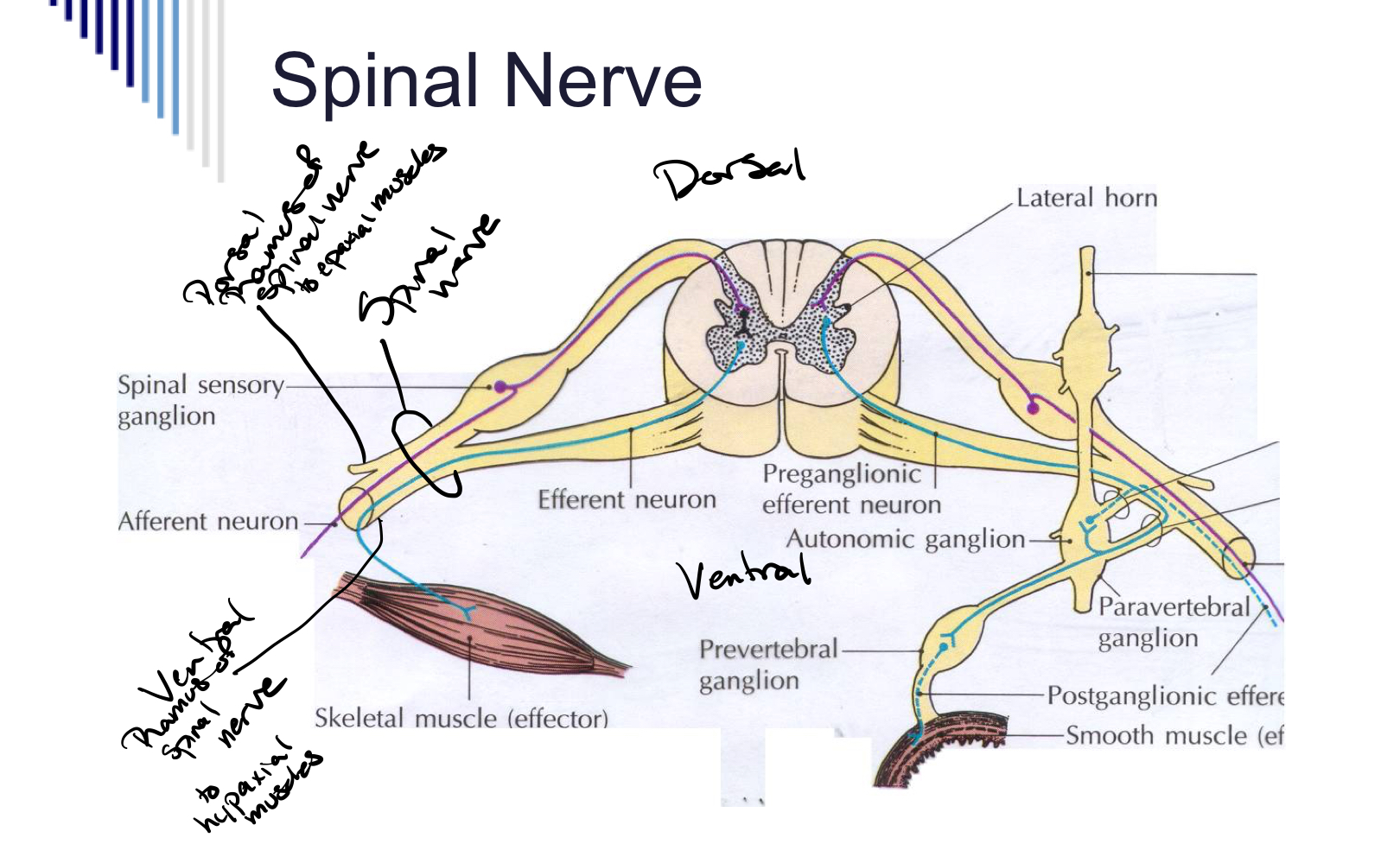
Epaxial muscles
Epaxial muscles dorsiflex the spine and are innervated by dorsal rami of spinal nerves.
81
New cards
hypaxial muscles
Hypaxial muscles ventroflex the spine and are innervated by ventral rami of spinal nerves.
82
New cards
Review slide 61
83
New cards
Extrinsic Muscles of the Eye
Three epaxial myotomes form the extrinsic eye muscles.
- Move the eyes
- Innervated by cranial nerves III, IV, and VI (Occular motor, Trochlear, and Abducens).
- These myotomes are derived from somitomeres (not somites)
- Move the eyes
- Innervated by cranial nerves III, IV, and VI (Occular motor, Trochlear, and Abducens).
- These myotomes are derived from somitomeres (not somites)
84
New cards
Axial Muscles
Muscles of the body wall and spine.
- Derived from epimeres and hypomeres of somites
- Derived from epimeres and hypomeres of somites
85
New cards
Branchial Muscles
Muscles associated with the visceral arches.
- Derived from posterior somitomeres and anterior somites.
- Innervated by cranial nerves V, VII, IX, X, and XI (Trigeminal, Facial, Glossopharyngeal, Vagus, and Spinal Accessory nn)
- Includes muscles of facial expression and muscles of mastication.
- Derived from posterior somitomeres and anterior somites.
- Innervated by cranial nerves V, VII, IX, X, and XI (Trigeminal, Facial, Glossopharyngeal, Vagus, and Spinal Accessory nn)
- Includes muscles of facial expression and muscles of mastication.
86
New cards
Hypobranchial Muscles:
Develop from hypaxial myotomes that stretch under the pharynx.
- Form the coracoarcuals in fishes.
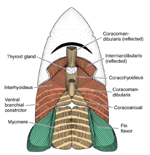
- Form the strap muscles of the neck and tongue in tetrapods.
- muscles after the gills
slide 67 for pictures
- Form the coracoarcuals in fishes.
- Form the strap muscles of the neck and tongue in tetrapods.
- muscles after the gills
slide 67 for pictures
87
New cards
Appendicular Miuscles
Muscles of the appendages.
- In fishes they form from the hypaxial muscles of the body wall. Innervated by ventral rami of spinal nerves.
- In tetrapods they develop as condensations in the mesenchyme of the limb bud; not from myotomes.
- In fishes they form from the hypaxial muscles of the body wall. Innervated by ventral rami of spinal nerves.
- In tetrapods they develop as condensations in the mesenchyme of the limb bud; not from myotomes.
88
New cards
homology
structures inherited from common ancestors
89
New cards
How do we look at the evolution of muslces?
- Homology of muscles can be traced within orders, but it is difficult to establish homologies between classes.
- Can look at position and relationship relative to other muscles and the skeleton.
E.g., supraspinatus is always above the scapular spine.
- Can look at position and relationship relative to other muscles and the skeleton.
E.g., supraspinatus is always above the scapular spine.
90
New cards
Explain the evolutionary difference in muscles on attachment
- Muscle attachments will sometimes change during evolution.
- This alters action.
- Sometimes muscles of similar action fuse.
- More often ancestral muscles split to form several muscles.
- This alters action.
- Sometimes muscles of similar action fuse.
- More often ancestral muscles split to form several muscles.
91
New cards
How do we determine homology of muscles?
- Embryology can be useful in determining homology of muscles.
- Nerve supply is the best indicator.
- Nerve supply is the most conservative aspect of anatomy.
- Does not always stay consistent, so it is not foolproof.
- Nerve supply is the best indicator.
- Nerve supply is the most conservative aspect of anatomy.
- Does not always stay consistent, so it is not foolproof.
92
New cards
What is the difference between fish and mammal muscles
Differentiation from fish to mammals is progressive.
- The number of muscles is related to the precision of movement possible in a particular region.
- Myotomes split when muscles are differentiated.
- Results in changes in the direction of the muscle fibers.
- The number of muscles is related to the precision of movement possible in a particular region.
- Myotomes split when muscles are differentiated.
- Results in changes in the direction of the muscle fibers.
93
New cards
Can we determine homologies of muscles?
- Often get degeneration of certain myotomes during evolution.
- This make homologies of muscles virtually impossible to establish.
- They may even be non-existent
- This make homologies of muscles virtually impossible to establish.
- They may even be non-existent
94
New cards
Which parts of the bodies of fishes and tetrapods typically have enlarged muscles?
- In fishes the axial musculature is propulsive and therefore makes up the bulk of their musculature.
- In tetrapods the limbs take over the propulsive function, therefore the axial muscles diminish and the limb muscles enlarge.
- In tetrapods the limbs take over the propulsive function, therefore the axial muscles diminish and the limb muscles enlarge.
95
New cards
What are the muscles that are only found in mammals?
Diaphragm: made up partly from rectus abdominus m. and partly from the cervical myotomes (the phrenic nerve is a cervical nerve).
- This isn't muscular in reptiles and amphibians. Known as septum transversum in reptiles and amphibians.
Cutaneous trunci mm.: superficial splitting of myotomes derived from the latissimus dorsi m. and pectoralis muscles.
- muscles that move the skin
Platysma: derived from the splitting off of branchial muscles in the facial region.
96
New cards
How do muscles and electricity relate?
- Muscles need to depolarize in order to contract.
- Some fishes have exploited this fact.
- Electric organs are usually derived from muscle cells.
- Some electrical organs may be of glandular or nervous tissue origin.
- Some fishes have exploited this fact.
- Electric organs are usually derived from muscle cells.
- Some electrical organs may be of glandular or nervous tissue origin.
97
New cards
Electrical Fishes
- The electric organ is built like a battery with neurons and electroplaxes.
- It is formed from axial muscle.
- Cells are specialized to develop an ion current flow (they don't contract).
- They discharge in about 3 milliseconds.
- Live in freshwater which doesn't conduct electricity well.
- Therefore need a lot of voltage.
- It is formed from axial muscle.
- Cells are specialized to develop an ion current flow (they don't contract).
- They discharge in about 3 milliseconds.
- Live in freshwater which doesn't conduct electricity well.
- Therefore need a lot of voltage.
98
New cards
electroplaxes
specialized muscle cells
99
New cards
What do fishes use electrical organs for?
1) Fishes use electric organ for feeding, orientation and defense.
- Ampullary organs are receptive to the electrical field in water and therefore can detect objects.
- Objects are detected because they disrupt the electrical current.
- The ampullary organs are derived from the lateral line system.
2) Can generate shocks for predation or defense.
3) Some can recognize members of their own species by the variations in the electrical pulses.
- Specific Mate Recognition system (SMRs)
100
New cards
What is electroplax?
It is the functional unit of an electrical organ.
- These are large, multinucleated cells.
- Have a folded surface on the cell membrane that contains a high concentration of mitochondria.
- Hundreds of thousands of electroplaxes are stacked to form columns.
- These are large, multinucleated cells.
- Have a folded surface on the cell membrane that contains a high concentration of mitochondria.
- Hundreds of thousands of electroplaxes are stacked to form columns.