tissue adaptation
1/16
Earn XP
Description and Tags
Name | Mastery | Learn | Test | Matching | Spaced | Call with Kai |
|---|
No analytics yet
Send a link to your students to track their progress
17 Terms
basic structure of the ECM
fibroblasts in most connective tissue
GAG polysaccharide chains (repeating dissacharides)
GAGs which covently bond to proteins- proteoglycans
fibrous proteins such as collagen
proteoglycans form a gel like substance where fibrous proteins are embedded which resists compressive forces, allow diffusion
glucosaminoglycans
structure
branched or unbranched
hydorphobic or hydrophilic
how do they withstand compression
unbranched polysaccharide chains
repeating disaccharide chains
hydrophilic
porous gels (hydrated) filling up most of the extracellular space
attract cations so water in by osmosis- turgor- withstand compression
collagen
how many polypeptide chains
how many forms
triple stranded helical structure
3 polypeptide chains
29 forms
elastin
hydrophobic or hydrophilic
what is the precursor molecule
how does the precursor form elastin
highly hydrophobic protein
precursor is tropoelastin
tropoelastin is secreted into the extracellular space and assembled into elastic fibres close to the plasma membrane, which then cross link
coiled
stretch and recoil
tendon
collagen
proteoglycans
elastin
tenocytes
ligament
fibrocytes
ecm
lower collagen, more proteoglycan
elastin
cartilage
which collagen
which is the predominant proteoglycan
other molecules
how much percent of water
chondrocytes
type ii collagen mostly
proteoglycan predominantly chondrotin sulphate
hyaluoran
68 percent water
regional variation
ecm of bone
what gives rigidity and compressive strength and where is this deposited?
which gives tensile strength and elasticity
hydroxyapatite gives rigidity and compressive strength
this is deposited on collagen fibres
load bearing
inorganic
type i collagen gives tensile strength and flexibility, elasticy, structural organisation, organic
what is mechanical loading
ecm turnover is triggered by mechanical loading
involves cell signalling
loading increases synthesis of new ecm proteins and degrading enzymes
loading can alter the molecular conformation of proteins changing how enzymes bind and degrade (change in collagen type and organisation)
tissue properties influence how they degrade eg stiffness
wolffs law
increase in loading causes architecture of spongy bone to strengthen and cortical layer strengthening whilst decrease causes bones to weaken and bone tissue to be resorbed
intramembranous ossification
what do mesenchymal cells become and what is secreted
what forms in this area
what aggregates
what happens and what is trapped as a result
what turns into what
wht surrounds blood vessels and what is formed
what is formed on the outside
multipotent mesenchymal cells become osteoblasts which secrete osteoid
blood vessels form in the area
osteoblasts aggregate in the ossification centre
osteoid matrix becomes calcified and hardens trapping osteoblasts
ostoeblasts turn into osteocytes
osteoid surrounds blood vessels forming cancellous bone
mesenchymal cells on the outer surface of newly formed bone forms the periosteum
endochondral ossification
what do mesenchymal cells differentiate into
what happens to them
what is formed
what forms around it
what happens to the cells and what does this form
what happens within and what does this allow
what change occurs and what is brought over
what is deposited and what does this lead to
what does this form and what is brung in as a result
what structure does this form
mesenchymal cells differentiate into chondroblasts that secrete ECM
chondroblasts become encased forming chondrocytes
forms a hyaline cartilage model
perichondrium forms around it
chondrocytes increase in size and some burst releasing cell contents triggering calcification
chondrocyte cells die within the calcifying matrix forming cavities for osteoblasts to move into
perichondrium changes to periosteum. it contains blood vesels containing nutrients which diffuse into the cartilage precursor and bring osteoblasts
osteoblasts deposit bone around the diaphysis preventing nutrients from diffusing into the hyaline cartilage leading to chondrocyte death at the centre
this forms cavities where blood vessels can penetrate bringing in osteogenic cells
this forms the medullary cavity, osteoblasts begin depositing bone into the spaces which is the primary ossification centre
secondary ossification centres develop in each end of the long bone
thin cartilage called the epiphyseal growth plate develops between primary and secondary
chondrocytes in the plate continue proliferating and also forms new cartilage which turns into bon
in adulthood chondrocytes in the grwoth plate stop dividing until the physis itself ossifies. then cartilage is only found at the articular surface of joints.
nutritional and hormonal influences
which 2 salts and what do absorption of these depend on
vitamins
which hormones are needed for calcium metabolism regulation
which hormones for bone growth
dietary calcium and phosphate salts
absorption depends on calcitriol (a hormone only made in the presence of vitamin D )
Vitamins C, A, K, and B12
calcitonin and parathyroid hormone - calcium metabolism regulation
insulin, growth hormone, thyroxine- bone growth
oestrogen- growth plate closure (also testosterone) osteoblast activity
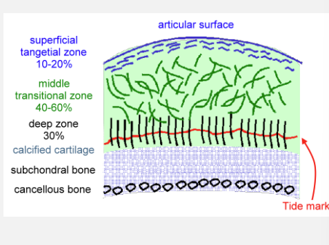
supericial tangential zone
85% collagen; collagen fibres orientated tangential to surface.
Greatest ability to resist shear stresses
middle transitional zone
Transition between the shearing forces of surface to compression forces in deep layer; collagen arranged obliquely;
composed largely of proteoglycans
deep radial
Collagen fibers attached radially (vertical) into the tidemark - distributes loads and resists compression; high PG content
regions