Week 2 - Anatomy ( + Textbook Chapter 3)
1/99
There's no tags or description
Looks like no tags are added yet.
Name | Mastery | Learn | Test | Matching | Spaced | Call with Kai |
|---|
No analytics yet
Send a link to your students to track their progress
100 Terms
Human Anatomy
study of the structures that make up the human body and how those structures relate to each other
helps us understand movement
what is the basic function of: bones, joints, and muscles
bones - provide structural frame to support and protect
joints - provide mechanism for movement to occur
muscles - cross joints and supply power to create movement
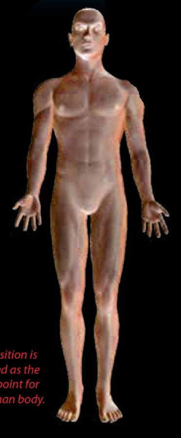
Anatomical Position
Universally accepted position as the starting reference point for describing the human body
standing upright
facing forward
arms hanging at the sides
palms facing forward
legs feet toes parallel
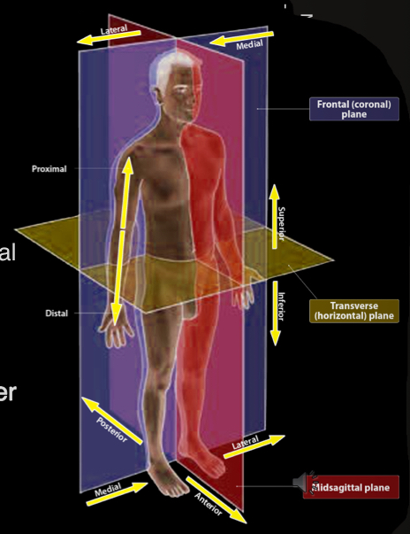
Sagittal Plane (and Midsagittal plane) and movement
vertical plane divides the body into right and left
midsagittal plane (or median plane) - sagittal plane that divides the two sides equally
movement - forward roll, running, cycling
Frontal plane (AKA Coronal Plane) and movement
vertical plane dividing the body into anterior and posterior sections
movement - side stepping, cartwheel, jumping jack
transverse plane (AKA Horizontal Plane)
divides the body into upper and lower sections
movement - twist in diving, spin in figure skating or pirouette in ballet
Planes of the body
nose is medial to ears
ears are lateral to eyes
knee is distal to hip
knee is proximal to toes
hamstrings are posterior to quad muscles
abdominal muscles are anterior to back muscles
ribs are superior to hips
ankles are inferior to hips
skin is more superficial than muscles
organs are deeper than skin
Supine
position of laying on your back
Prone
position of laying on your front
What are the joint movement pairs
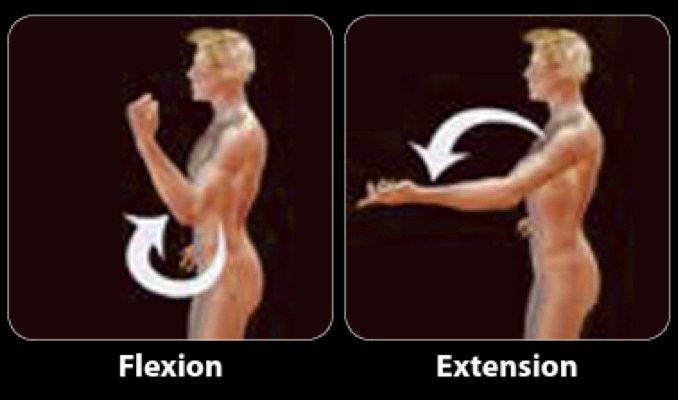
Flexion-Extension
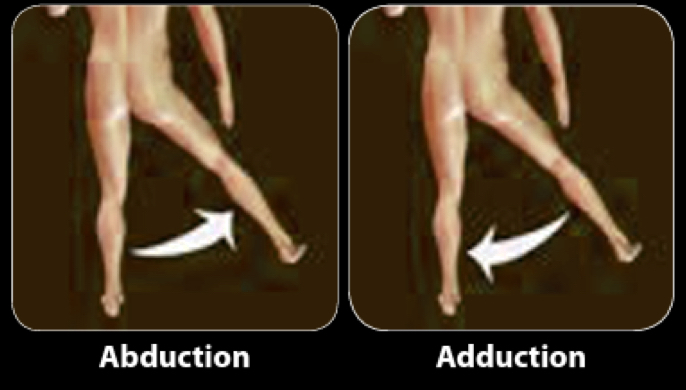
Abduction-Adduction
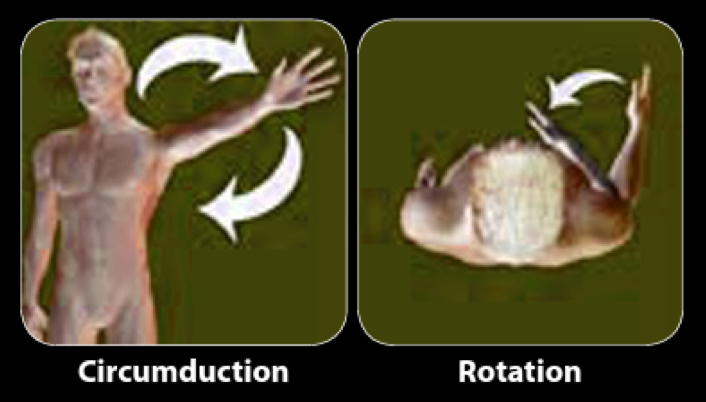
Circumduction-Rotation
Pronation-Supination
Inversion-Eversion
Dorsiflexion-Plantar Flexion
Flexion-Extension
Flexion - reduces joint angle
lifting a weight in a bicep curl
extension - increases joint angle
lowering a weight in a bicep curl
Abduction-Adduction
abduction - movement away from midline
adduction - movement towards the midline
Circumduction-Rotation
Circumduction - circular movement by combining flexion-extension with abduction and adduction
rotation - bone rotates along its longitudinal axis to create internal or external rotation
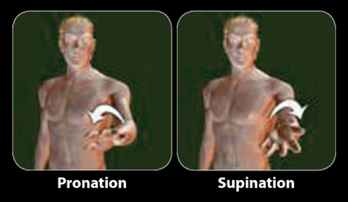
Pronation-Supination
describes movement in relation for forearm and hand
ex. holding a bowl of soup
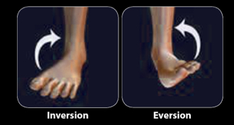
Inversion-Eversion
describes movement in the ankle relative to the sole of the foot
inversion - when the sole is turned towards the median plane of the body
eversion - when its turned award from the median plane
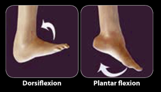
Dorsiflexion and Plantar Flexion
flexion and extension of the ankle
How many bones make up the axial and appendicular skeleton
206
Axial skeleton
80 bones
composes head, spine, ribs, sternum
stabilizes body
Appendicular skeleton
126 bones
composes shoulder, pelvis, arms, and legs
responsible for large portion of movements
Bones
living tissues with blood supply and nerves
are porous with varying degrees of porosity
compact, spongy
composed of calcium carbonate, phosphate, collagen, and water
Compact (Cortical) Bone
bone that has low porosity
found in long bones (arm and leg bones)
required to be stronger to resist greater stress
Spongy (Cancellous) Bone
Bone that has high porosity with more non-mineralized tissue
characteristic honeycomb structure
provides more flexibility
found in areas where shock absorption and a better ability to change shape are important
Epiphyseal growth plate
remains at the ends of long bones so that growth in length can occur
this enables an individual to reach a mature height
Characteristics of long bones (Like the femur)
proximal epiphysis (end)
diaphysis (shaft)
Epiphyseal Line
when growth ceases, the epiphyseal growth plate ossifies and becomes an epiphyseal line
Periosteum
thin membrane that covers bones (except where they form joints)
Effect of fitness on bone
when bone is subjected to regular, weight bearing or load stimulus, they become denser
bone responds to the force exerted on it
when not exposed to regular load, density decreases
other factors impact bone density including but not limited to:
age
gender
Bone Shapes
Long
Short
Flat
Sesamoid
Irregular
Long Bone Shapes
Appendicular Skeleton
ex. Radius, ulna, humerus, femur, tibia, fibula, metatarsals, metacarpals, phalanges
Short Bone Shapes
Appendicular Skeleton
ex. Carpals, tarsals
Flat Bone Shapes
protect underlying organs and provide areas for muscle attachment
Appendicular skeleton
ex. Scapula, Clavicle
Axial Skeleton
Ribs, sternum
Frontal, Parietal, occipital, mandible
Seismoid Bone Shapes
shaped like a pea and found in tendons of the knee, hand, and foot
Appendicular Skeleton
Patella
Irregular Bone Shapes
Axial Skeleton
Vertebrae, facial bones of skull
Appendicular Skeleton
Pelvis
Ligaments
fibrous tissues that connect bones
3 types of joints
Fibrous - don’t move
ex. skull
Cartilaginous - absorb shock, have little mobility
ex. intervertebral joints, pubic symphysis
Synovial - most common, allow greatest amount of movement
have a joint cavity with synovial fluid to lubricate and cushion the joint
Joint Capsule
surrounds the joint space and helps provide support
Synovial Membrane
lines the joint capsule
secretes the lubrication fluid
cartilage
reduces friction between the articulating bone surfaces
covers the ends of the bones that form synovial joints
Types of synovial joints
Hinge (Ginglymus)
Pivot
Plane (Gliding)
Condyloid (Knuckle)
Saddle
Ball and socket
Hinge (Ginglymus) Synovial Joint
allows for movement in one plane only
ex. elbow, fingers, knee
Pivot Synovial Joint
one bone rotates around one axis
ex. atlantoaxial joint of neck, forearm during pronation-supination
Plane (Gliding) Synovial Joint
gliding action is the only movement allowed
forward-backward, side to side
ex. acromioclavicular joint of the shoulder, facet joints of the vertebrae, wrist
Condyloid (Knuckle) Synovial Joint
flexion-extention, abduction-adduction, circumduction all possible
ex. metacarpophalangeal joints (except the thumb)
Saddle Synovial Joint
flexion-extension, abduction-adduction, circumduction all possible
ex. carpometacarpal joint of the thumb
Ball and Socket Synovial Joint
movement in all planes, greatest range of motion of any joint type
ex. shoulder, hip
types of muscles (and their movement)
skeletal muscles - voluntary skeletal movement
smooth muscle - involuntary, slow and uniform, fatigue-resistant
cardiac muscle - involuntary, intrinsic beat
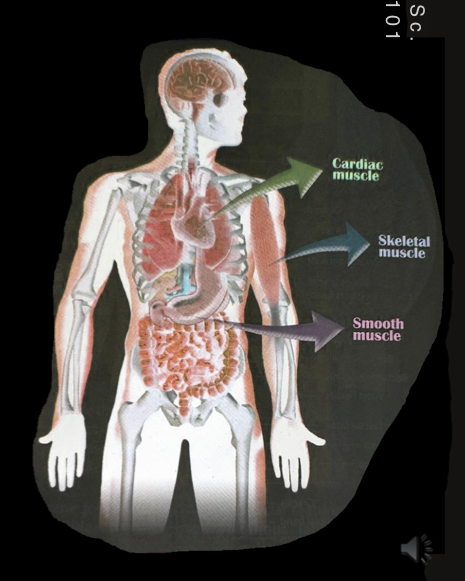
Skeletal Muscles
over 600 muscles
attached to bone
contraction is responsible for supporting and moving the skeleton
voluntary skeletal movement
tendons attach muscle to the bone
have fascia
How does muscle movement occur
movement happens when muscles attached to bones contract to move a joint
can only happen when a muscle crosses a joint
Smooth Muscle
forms the walls of blood vessels and body organs
like the respiratory tract, iris of the eye, and gastrointestinal tract
involuntary movement
slow and uniform
fatigue-resistant
Cardiac Muscle
provides contractile activity of the heart and has its own intrinsic beat
involuntary movement
Origin (Proximal attachment)
muscle attachment closer to the center of the body
attached to more stationary structures of the skeleton
Insertion (Distal attachment)
muscle attachment away from the center of the body
attached to more mobile structures of skeleton
tendons
fibrous tissues at end of muscles
fascia
fibrous material that surround muscle and other structures for support and protection
Axial Skeleton Components
Skull
curved flat bones from the calvaria (vault to protect the brain)
calvaria - frontal, parietal, temporal, occipital, sphenoid bones
facial bones - nasal, lacrimal (drainage for tears), zygomatic (cheek), maxilla (upper jaw), and mandible (lower jaw)
Concussions
brain injury from violent shaking of the head or impact to the head
can result from the direct trauma or impact and/or when the brain bounces against the inside of the skull
Application of bones in Sports
Calvaria bones can be broken with trauma and/or in sports
concussions
sport regulating bodies have now put protocols in place to help players and coaches identify signs of concussion
and protocols for safe return to play
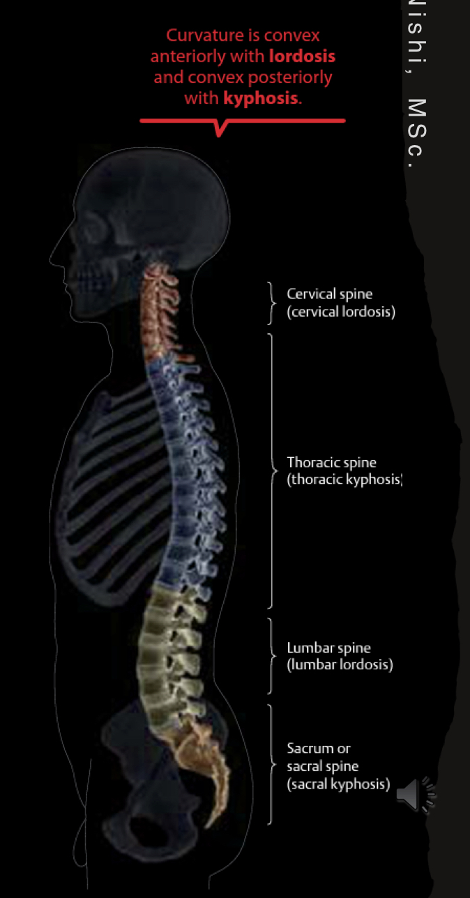
Vertebral Column Components
33 bones
Cervical C1-C7
(C1 = axis) (C2 = atlas)
Thoracic T1-T12
Lumbar L1-L5
Sacrum - found at middle line of the buttocks
Coccyx - tailbone made of fused vertebrae
spinal cord (nerves) - run through the vertebrae, through vertebral column
vertebral column curvature - lordosis and kyphosis
Sternum
midline breastbone and made up of manubrium, sternal body, and xiphoid process
Ribs and Sternum
Protect the heart and lungs
12 ribs
true ribs - upper 7 rib pairs
attached to sternum and vertebrae
false ribs - pairs 8-10
attach to sternum indirectly
floating ribs - pairs 11 and 12
don’t attach to sternum
what do bone and costal cartilage in ribs do
allow chest to expand
sternocleidomastoids
allow for head and neck movement
Erector spinae
maintain erect position
hold ourselves up
4 types of anterior abdominal muscles
internal obliques
external obliques
transverse abdominis
rectus abdominis - six pack from the tendinous intesections that allows for:
trunk flexion
side bending
forced expiration
Appendicular skeleton components
Pectoral girdle (chests
pelvic girdle (hip)
upper limbs
lower limbs
Pectoral girdle
includes clavicle and scapula
joints of pectoral girdle
sternoclavicular joint
acromioclavicular joint
glenohumeral joint
muscle groups of appendicular skeleton
anterior muscle group
posterior muscle group
rotator cuff muscle group
lateral group
Anterior Muscle group of appendicular skeleton
pectoralis major
pectoralis minor
serratus anterior
Posterior Muscle group of appendicular skeleton
trapezius
latissimus dorsi
teres major
levator scapulae
rhomboids
Rotator Cuff Muscle Group of appendicular skeleton
subscapularis
supraspinatus
infraspinatus
teres minor
Lateral Group of Appendicular Skeleton
deltoid
anterior, middle, and posterior fibres)
Bones of the upper limb
humerus (arm)
radius and ulna (forearm)
radius is on thumb side and can rotate crossing over the ulna
Joints of Upper Limb
glenohumeral joint (main joint of the shoulder)
elbow joint
3 joints of the elbow and their types of movement
humeroradial joint - flexion-extension
humeroulnar joint - flexion-extension
proximal radioulnar joint - pronation-supination
Carpus
the wrist
formed of 8 carpals (She Likes To Play, Try To Catch Her)
scaphoid
lunate
triquetrum
pisiform
trapezium
trapezoid
capitate
hamate
Joints of the wrist and hand
Radiocarpal joint (between radius and carpals)
carpometacarpal joints
midcarpal joints
intercarpal joints
intermetacarpal joints
metacarpophalangeal (MCP) joints - knucles
interpalangeal joints
Proximal Interphalangeal joint (PIP)
Distal interphalangeal joint (DIP)
Anterior compartment of arm muscles
coracobrachialis
biceps brachii
brachialis
Posterior compartment of arm muscles
triceps brachii
Forearm muscles
flexor-pronator group
extensor-supinator group
brachioradialis
Joints of the pelvic girdle
pubic symphysis
sacroiliac joints
Lower limb bones
femur
patella
tibia
fibula
tarsals
metatarsals
phalanges
Quads (Quadriceps Femoris)
anterior compartment of legs
run from femur to tibia and extend the knee
4 parts:
rectus femoris
vastus lateralis
vastus medialis
vastus intermedius
sartorius
runs from ilium to tibia
allows for hip flexion
abduct the thigh
cross your legs
medial compartment of leg muscles
Adductor muscles
Pectineus
adductor longus
adductor brevis
adductor magnis
posterior compartment of leg muscles
flex the knee, extend the hip
hamstrings - run from ischial tuberosity to fibia/tibia
biceps femoris
semitendinosus
semimembranosus
glutes - extend the hip
gluteus maximus
gluteus medius
gluteus minimus
Anterior compartment of lower legs
tibialis anterior - dorsiflexion of ankle
lateral compartment of lower leg muscles
fibularis longus and fibularis brevis - plantarflexers since they pass behind the lateral malleolus
posterior compartment of lower leg muscles
calf muscles
gastrocnemius
soleus
plantaris merge into the achilles ton
act to plantarflex the ankle
gastrocnemius cross behind the knee so some knee flexion
deep group of muscles
flexor hallucis longus
flexor digitorum longus
tibialis posterior
popliteus
toe flexion
Ligaments of the Kneejoint (tibiofemoral joint)
Anterior Cruciate Ligament (ACL)
Posterior cruciate ligament (PCL)
medical collateral ligament (MCL)
ACL and PCL crisscross and provide stability forward and back
Talocrural Joint (ankle)
talus is between the medial and lateral malleolus
Transverse tarsal joint
talus and calcaneus bones
Stress fracture
special type of bone fracture from repeated low-magnitude/low-impact forces
sprain
stretch or tearing of ligaments
ex. ACL tear
strain
stretch or tearing of muscle or tendon
Separated shoulder
sprain in acromioclavicular joint
tendinitis
acute tendon inflammation
tendinosis
chronic, repetitive strain of the tendon
Jumper’s knee
patellar tendinosis or patellar tendinitis
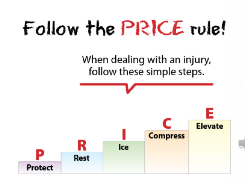
how to deal with an acute injury?
Follow the PRICE rule