unit 11 - pulp anatomy - periodontal configuration
1/70
There's no tags or description
Looks like no tags are added yet.
Name | Mastery | Learn | Test | Matching | Spaced | Call with Kai |
|---|
No analytics yet
Send a link to your students to track their progress
71 Terms
pulp chamber, pulp cavity or coronal pulp are terms used to designate what?
the area of the crown filled with soft tissue
root pulp, radicular pulp, pulp canal and root canal are used to describe what
the portion of the root filled with soft tissue
does the pulp cavity fill the entire tooth or is it split
fills entire cavity
descriptive only
the crown and root portion that contain the pulp tissues have been divvied into what
pulp chamber - crown
root pulp/canal - root
how has the pulp space been divided
arbitrarily
(split by convenience of description not reality)
what is the dental pulp
soft tissue
where is the dental pulp
internal cavities of the tooth
the outline/shape of the pulp tissue generally corresponds to what
the tooths external outline form
the dental pulp originates from what
the mesenchyme
the dental pulp have 4 functions what are they
formative
nutritive
sensory
defensive
what is the pulps initial function and when is it carried out
Dentine formation during the developmental period
what does the complex sensory system within the dental pulp control
blood flow
the complex sensory system is also responsible for mediating what
the sensation of pain
what represent the defensive response of the pulp to irritation
formation of reparative or tertiary dentine
reparative dentine is formed in response to what type of irritation
mechanical
thermal
chemical
bacterial
reactive dentine is usually limited to what
the area of pulpal irritation
what might be difficult or impossible to distinguish
reactive changes from purely ageing related changes
what is used for diagnosing and treating pulpal diseases
radiographs or digital radiography
what must be visualised when using radiographs
morphological features of the pulp chambers and root canals
what has to happen to visual the features
3D features are compressed into 2D radiographic images
radiographic views from a facial orientation show what type of view
a monoplane, buccolingual view of hard tooth structures and radiolucent spaces for the pulp canals
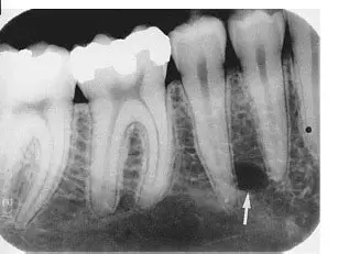
what colour is the pulp and hard tissue (enamel)
pulp = darker
hard tissue = lighter
what does the size of the pulp chamber depend on
the age of the tooth and its history of trauma
what is formed continuously throughout the life of the tooth
secondary dentine
under what conditions is secondary dentine formed continuously
as long as the vitality of the tooth is maintained
is secondary dentine formation uniform
no
READ - it will make everything make sense
done
where do odontoblasts produce greater quantities of secondary dentine
adjacent to the floor and the root of the pulp cavity
where do odontoblast produce less secondary dentine
adjacent to the walls of the pulp cavity
how does the pulp cavity size compare between young individuals and adults
larger in young individuals than adults
what can initiate a different type of dentine formation
severe traumatic injuries
what is irritation-induced dentine also called
reparative dentine
reparative dentine may be formed in response to what
carious process, abrasion , attrition, and operative procedure
is reparative dentine protective or harmful
protective, but is detrimental in later years
why might reparative dentine be detrimental in later years
because a finite amount of space is present within the pulp cavity
true or false
you should compare the pulp cavity inn a given tooth to others
true
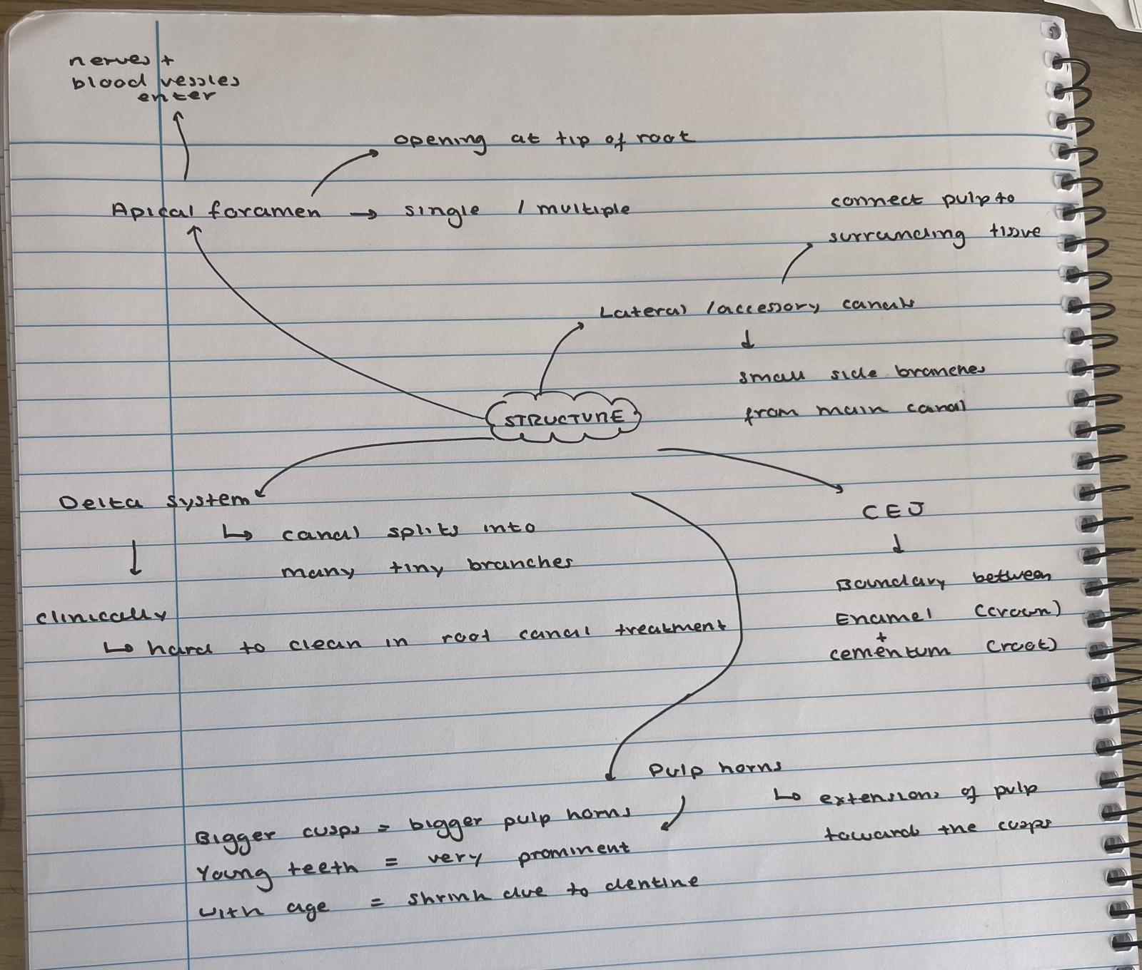
what supplies the internal contents of the pulp cavity
the neurovascular bundle
where does the neurovascular bundle entre
through the apical foramen or foramina
what is the size of the apical foramen during early root development
larger than the pulp chamber
what happens to the apical foramen at completion of root formation
it becomes more constricted
can a root have multiple apical foramen
yes
if the apical foramen are large enough the space that leads to the main root canal is called what
supplemental or lateral canal
what is the supplemental or lateral canal
a space leading to the main root canal if the openings are large enough
what is the delta system
when the root canal breaks into multiple tiny canals
why is it called a delta system
because of its complexity
demarcation of pulp cavity and canal
what is the CEJ
cementoenamel junction
demarcation of pulp cavity and canal
does the CEJ correspond exactly to the transition from pulp chamber to the root canal
no
demarcation of pulp cavity and canal
how is the demarcation mainly based
macroscopically
demarcation of pulp cavity and canal
how may the demarcation be visualised
by exploring the CEJ and noting the density difference on radiographs
demarcation of pulp cavity and canal
what covers the external surface of dentine
enamel
demarcation of pulp cavity and canal
what does dentine make up part of
the pulp chamber
demarcation of pulp cavity and canal
how does pulp in the chamber compare microscopically to pulp in the root canal
more cellular
demarcation of pulp cavity and canal
what shape are odontoblasts in the coronal pulp chamber
cuboidal
demarcation of pulp cavity and canal
what happens to the shape of odontoblasts towards the apex
flatten out
demarcation of pulp cavity and canal
is the transition between pulp chamber and root canal sharply demarcated microscopically
no
demarcation of pulp cavity and canal
is the transition sharply delineated macroscopically
no
what are pulp horns
projections or prolongations in the pulp chamber of roots
pulp horns correspond to what
the crowns major cusps or lobes
what determines the prominence of pulp horns
the prominence of cups or lobes
when are pulp horns more prominent
in young individuals
why do pulp horns become less prominent with time
due to formation of secondary dentine
read
done
what must the clinician be aware of during operative procedures
the location and size of the pulp cavity
why must clinicians know pulp location and size
to prevent unnecessary encroachment on the pulp
what else must the clinician known the location of
the mandibular canal and nerve
what procedures require thorough knowledge of the pulp cavity
endodontic procedures
what errors may result in the loss of a tooth
perforation
failure to locate all canals
perforation of the root surface
what must the clinician known for endodontics
size and location of the pulp chamber and expected number of root and canals
can radiographs always detect accessory root or canals
no
what may indicate the presence of additional canals
shape of the crown
what must the clinician recognise during endodontic procedures
internal signs of additional canals