RT209 OTHER RADIOGRAPHIC EQUIPMENT
1/75
There's no tags or description
Looks like no tags are added yet.
Name | Mastery | Learn | Test | Matching | Spaced | Call with Kai |
|---|
No analytics yet
Send a link to your students to track their progress
76 Terms
Film Cassette Changers
Used in rapid and serial exposure
Cut Film Changer
Maximum of 6 per second
Roll Film Changer
Maximum of 12 per second
6 per second
Cut Film Changer has a maximum of??
12 per second
Roll Film Changer has a maximum of??
Manual Cassette Changer
Up to 1 exposure per second
1 exposure per second
Manual Cassette Changer is up to??
cerebral angiography
Manual Cassette Changer Usually used in ______________________ where blood is flow is slow
3 to 5 cassettes - holder - large spring
Manual Cassette Changer _________________ held together in a ________ by a __________
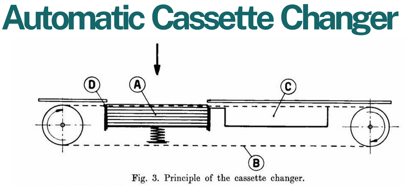
Automatic Cassette Changer
Designed to accomplish rapid serialization of examination
3 exposure per second
Automatic Cassette Changer is up to??
A = Supply of Unexposed Cassette
B = Conveying Chain
C = Box for Exposed Cassette
D = Driving pin, moving the Cassette to the Box
Automatic Cassette Changer Components:
Photofluoroscopy Unit
The photography of the image obtained on a fluoroscopic screen.
permanent record
Photofluoroscopy Unit is a _______________ is made through the fluoroscopic unit
Cinefluoro Unit
A specialized x-ray machine that uses a diagnostic technique in which a camera is used to record moving images of internal body structures produced through fluoroscopy
Radiographic Units
Fluoroscopic Units
Two types of Mobile Equipment:
Direct Power
Battery Power
Capacitor - Discharge
High Frequency
The Four Components of Radiographic Unit:
can vary in the radiation output for a given exposure technique.
Mobile x-ray units / Mobile Radiographic Imaging
care - transporting - manipulating - patient’s bedside
Mobile Radiographic Imaging In addition, _____ must be exercised in _____________ and ___________ the mobile unit at the ______________.
Fluoroscopic Units
Usually used to aid the physician during surgery or angiographic studies
Physician during surgery
Angiographic studies
Fluoroscopic Units Usually used to aid the _______________ and _______________
Chest
Panoramic X-Ray
Bone Densitometry
Mammography
Four modalities that uses Dedicated Units:
Chest Unit
A dedicated chest unit is designed to image the thorax in the upright position
Panoramic X-ray
(Panorex) designed to image curved surfaces, typically the mandible of the teeth.
Panorex
Panoramic X-ray is also known as?
dual-energy x-ray absorptiometry (DXA or DEXA)
Bone Densitometer Also known as??
Bone Densitometer
enhanced form of x-ray technology that is used to measure bone loss.
specialized procedure - ionizing radiation - information - skeletal bones
Bone Densitometer Bone:
a _____________ using ________________ to provide _____________ on the condition of the ____________
anatomic regions - two different x-ray energies - soft tissue attenuation
Bone Densitometer:
The _______________ are scanned with ______________________ to isolate bone from __________________
DXA scintillation detectors - x-ray photons - proportionally - visible light - measured - sent - data analysis
Bone Densitometer:
The ____________________ absorb the __________ and convert the energy _____________ to __________, which is ___________ and _____ to a computer for ____________.
T – Score
Z – Score
Two score types of Bone Densitometer
T – Score
Indicates fracture risk
Z – Score
May signify the need to evaluate patient for secondary causes of osteoporosis
secondary - osteoporosis
Z – Score May signify the need to evaluate patient for __________ causes of _______________
Central device
Peripheral device
2 Types of DXA Equipment:
Mammography
Is a specialized radiographic imaging procedure of the breast
Low
Mammography uses ____ kVp.
24 to 34 kVp
Mammography:
Low kVp is used (____________)
0.1 to 0.3 mm
Mammography:
Focal spot size used is ______________
Mammography:
Exit port is made up of _____________
Exit port
is made up of beryllium
Focal spot size
used is 0.1 to 0.3 mm
Low kVp
is used (24 to 34 kVp)
tube anode constructed molybdenum and rhodium
a molybdenum or rhodium-composed tube filter
a beryllium port window
smaller focal spot sizes for improved resolution
compression for imaging a more uniform breast
the ability to magnify areas of the breast
Dedicated Mammography Units
Unique features if a dedicated mammography unit include the ability to produce low kVp photons by using a:
Mammography and Xeroradiography
First attempted in 1920
Robert Egan
demonstrated a successful mammographic technique
Late 1950’s
YEAR when Robert Egan demonstrated a successful mammographic technique
Wolf and Ruzicka
xeroradiography developed by?
1960’s
YEAR when xeroradiography developed by Wolf and Ruzicka
Xeroradiography
The making of radiographs by a dry, totally photoelectric process, using metal plates coated with a semiconductor, such as selenium
Selenium
Xeroradiography uses a semiconductor such as??
Immobilizes breast
Less thickness tissue
Uniform thickness
Reduces scatter radiation
Position closer to image receptor
Thinner tissue
Purpose of Compression during Mammography:
tungsten (Z = 74)
molybdenum (Z = 42)
rhodium (Z = 45)
Target Composition Made of
20 to 35 kVp
Target Composition Range of kVp use is from??
74
tungsten (Z = ___)
42
molybdenum (Z = ____)
45
rhodium (Z = ___)
Target Composition
Made of tungsten (Z = 74), molybdenum (Z = 42) or a rhodium (Z = 45)
Range of kVp use is from 20 to 35 kVp
Focal Spot
Circular or rectangular
(0.6/0.3), (0.5/0.2), (0.4/0.1)
Circular
Rectangular
Focal spot can be ___________ or ____________
(0.6/0.3)
(0.5/0.2)
(0.4/0.1)
Focal Spot (__________), (__________), (__________)
30 to 60 microns of molybdenum or rhodium
Filtration Should be filtered with??
X-ray tube window
is made up of beryllium (Z=4), or borosilicate
0.1 mm Al equivalent
Inherent filtration =
3 micrometer
If molybdenum target is used, molybdenum filtration of ____________ is used
50 micrometer
If rhodium target is used, rhodium filtration of _____________ is used
Moving Grids
Grid ratio
Grid frequency
Grids:
4:1 or 5:1
Grid ratio =
30 lines per centimeter
Grid frequency =
Film – Screen system
Single emulsion film
Film:
The Cylinder (or cone)
Tube
Dental X-ray Unit:
The cylinder
is affixed to the tube head and is used to align the tube head with the patient and the X-ray film
Tube
contains all the components necessary to generate x-rays
Cone
The Cylinder also known as??
Cone
The Cylinder is also known as??