Biology Midterm Study Set: Key Terms & Definitions
1/123
There's no tags or description
Looks like no tags are added yet.
Name | Mastery | Learn | Test | Matching | Spaced | Call with Kai |
|---|
No analytics yet
Send a link to your students to track their progress
124 Terms
A pathologic lesion found frequently in 30-year-old black women that requires a radiographic image and historical data for diagnosis is termed
periapical cemento-osseous
Regional odontodysplasia is a rare, localized developmental anomaly of the dental hard tissues of a group of contiguous teeth. Many cases are probably misdiagnosed as malformed teeth or odontomas.
Ghost teeth
personal history contributes to diagnosis found in black women 30-40 years old - teeth are vital
cemento-osseous dysplasia
*Describing the base of a lesion that is flat or broad instead of stemlike
*picture of a fibroma with a ______ base
Sessile
What is the radiographic appearance of periapical cemento-osseous dysplasia in its earliest stage?
Radiolucent
Radiopaque
Radiolucent and radiopaque
Cotton-wool radiolucencies
Radiolucent
What is an area that is usually distinguished by a color different from that of the surrounding tissue? It is flat and does not protrude above the surface of the normal tissue.
Macule (ex: a freckle)
*A circumscribed, elevated lesion that is more than 5 mm in diameter. Usually contains serous fluid, and looks like a blister.
It is associated with ____.
Bulla (plural)/
erythema multiforme
acute self-limited disease that affects skin and mucous membranes
erythema multiforme

a freckle is a _____
Macule
A small, circumscribed lesion usually less than 1 cm in diameter. It is elevated or protrudes above the surface of normal surrounding tissue.
Papule
It is a small bump.
*picture is of Linhen planus
Variously sized circumscribed elevations containing pus
Pustules
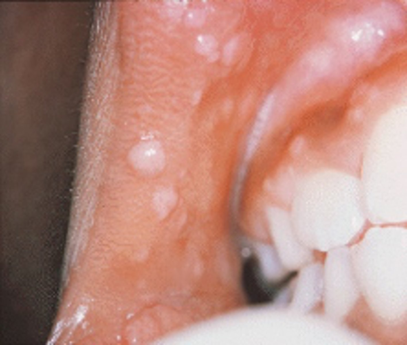
condyloma acuminatum (genital warts)
Pink, papillary lesion(s), more diffuse than papilloma
lesion that appears as a result of human papilloma virus; on the skin, lesions appear as cauliflower-like warts, and on mucous membranes, they have a flat appearance; also known as venereal or genital warts
a lesion with borders that are not well defined, making it impossible to detect the exact parameters of the lesion, treatment can be difficult depending on biopsy results
Diffuse
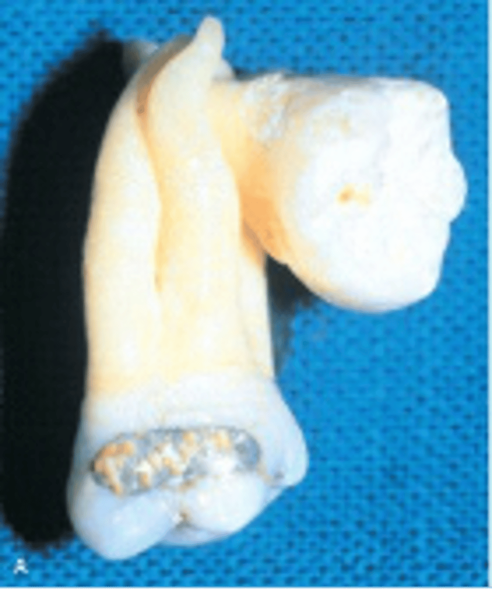
Supernumerary tooth between two permanent maxillary central incisors
Mesiodens
Localized Juvenile Spongiotic Gingivitis
Distinct subtype of gingivitis that does not respond to local plaque control. Appears as papillary or velvety, red nodule that bleeds easily
loss of tooth surface in the cervical area, caused by tooth grinding compression forces
Abfraction
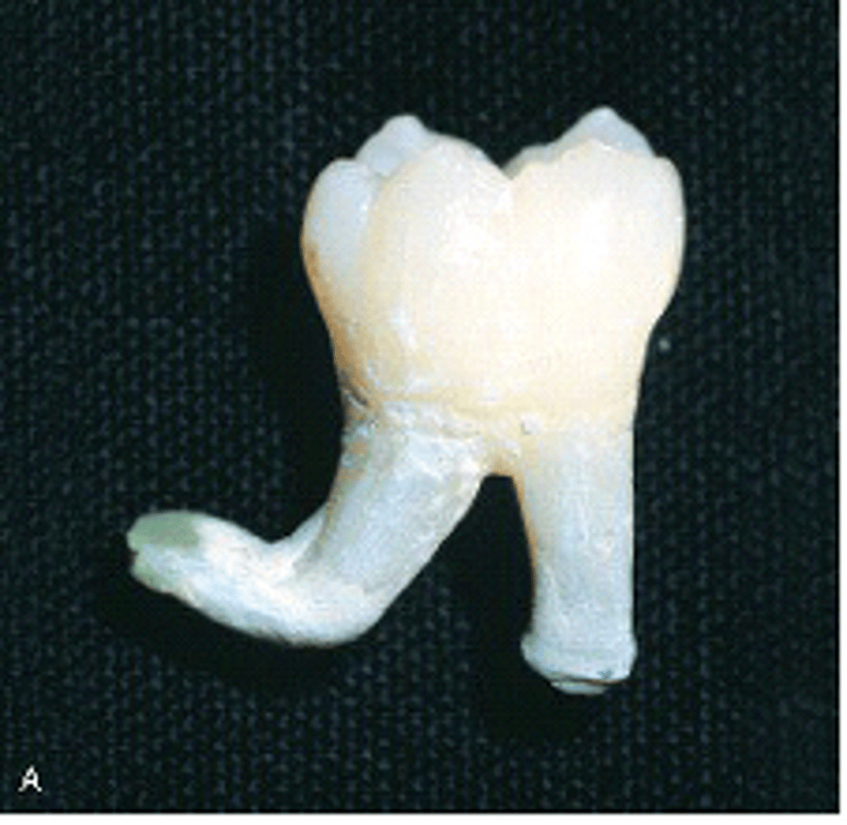
an abnormal bend or curve, as in the root of a tooth
Dilaceration
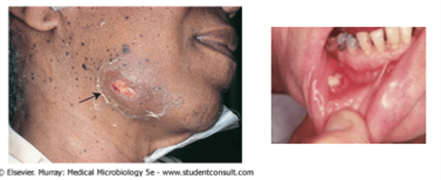
Actinomycosis (lumpy jaw)
Draining abscesses, "sulfur granules"
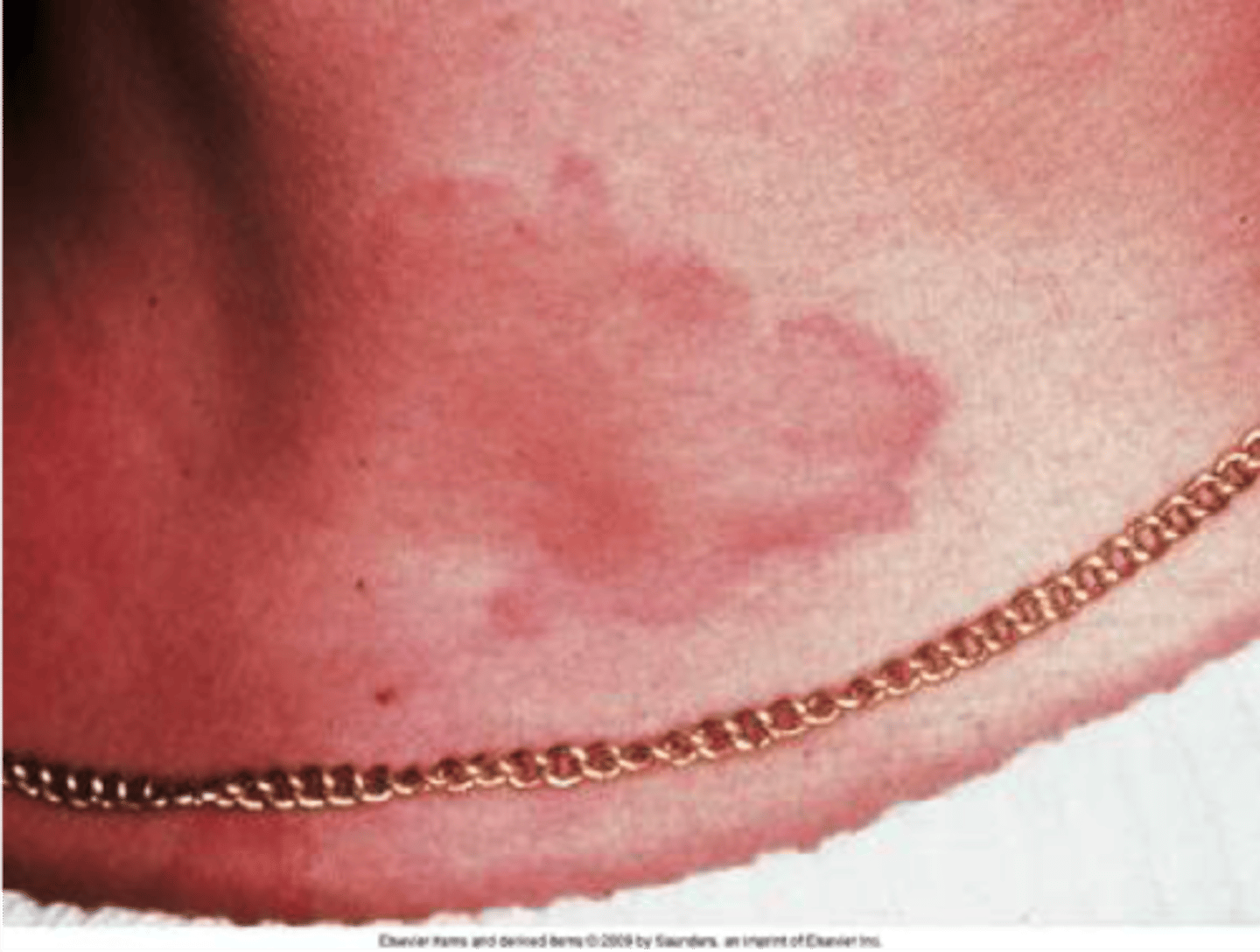
multiple areas of well demarcated swelling of skin
may be itching
Urticaria (Hives)
lesions caused by diffuse swelling as a result of increased permeability of deeper blood vessels
Angioedema
Impetigo
Vesicles or bullae, more often on skin of face and extremities
Mucosal ulceration with bullae
Pemphigus Vulgaris
severe progressive autoimmune disease
intraepithelial blister formation resulting from acantholysis
Pemphigus Vulgaris
Syphilis
Primary (chancre), secondary (mucous patch), tertiary (gumma)
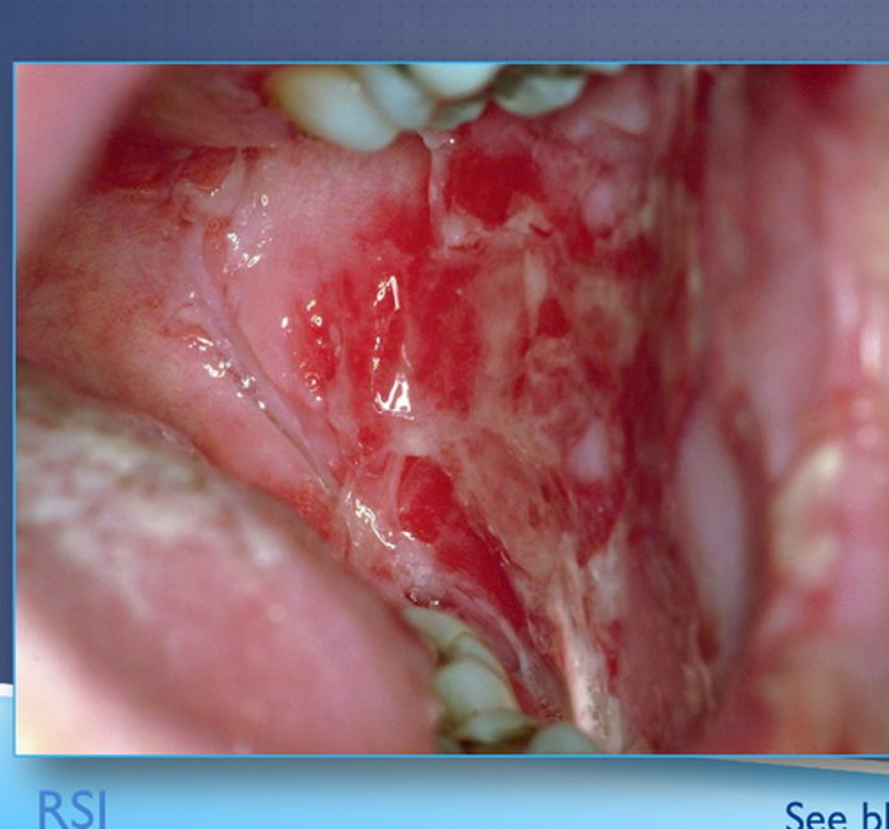
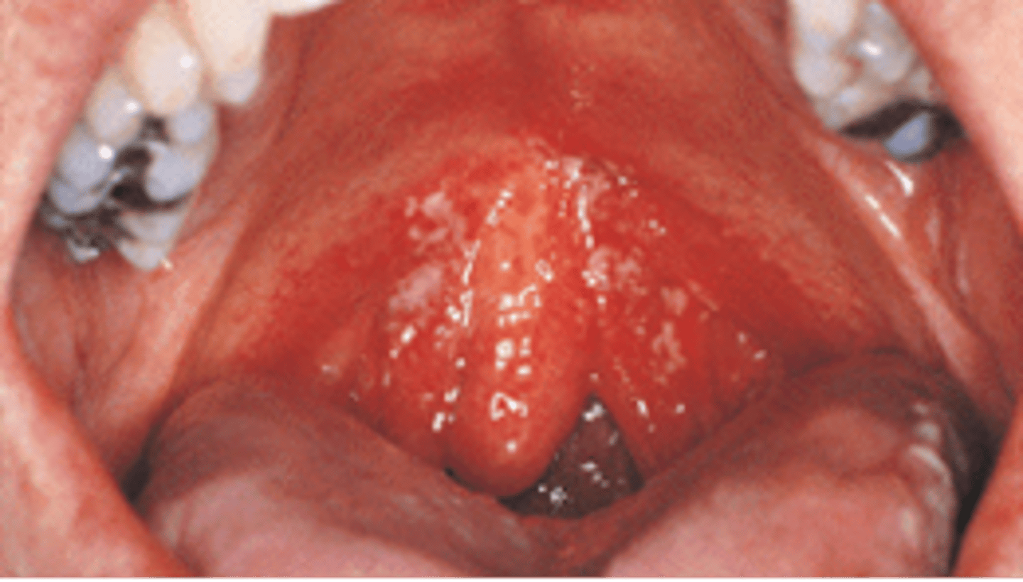
Primary herpetic gingivostomatitis
Multiple tiny vesicles that progress to form painful ulcers
Tuberculosis
Granulomatous lesions with primary infection to the lungs
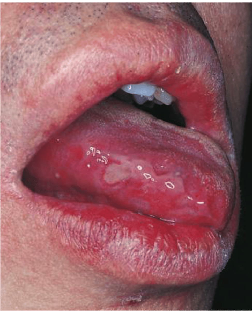
white, plaquelike lesion on the oral mucosa that cannot be rubbed off or diagnosed as a specific disease
Leukoplakia
process by which parts of a whole join together, or fuse, to make one
Coalescence
Verruca vulgaris (common wart)
White, papillary exophytic lesion resembling a papilloma
chronic recurrent autoimmune disease
primarily oral ulcers
no sex predilection
Behçet syndrome
Hairy leukoplakia
Irregular corrugated white lesion most commonly occurring on the lateral border of the
tongue
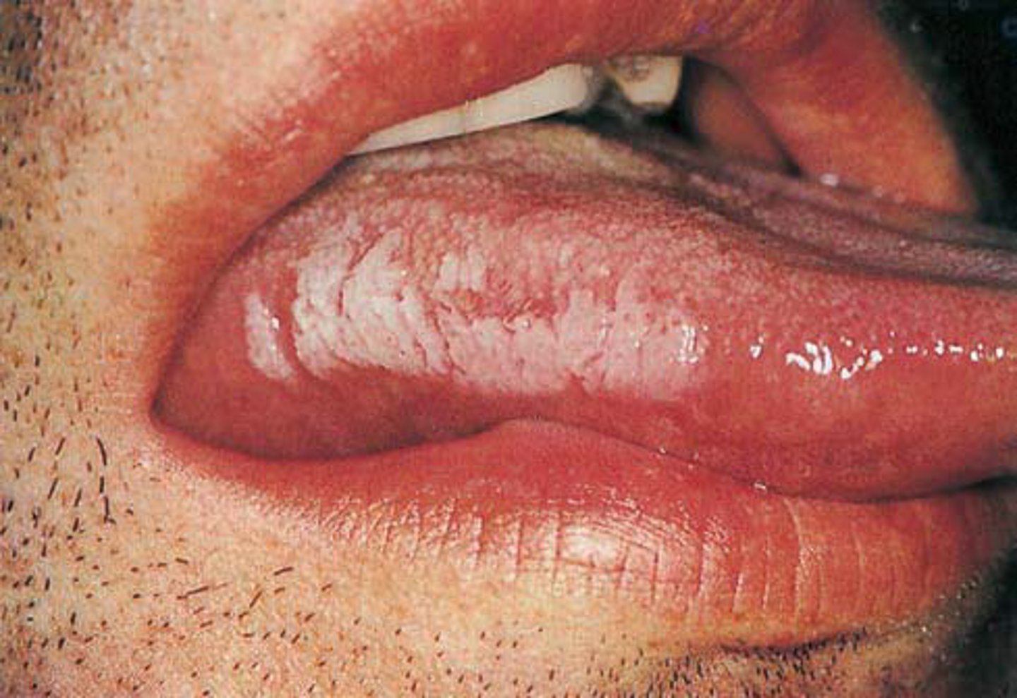
Hairy Leukoplakia
a white rough patch that arises on the LATERAL tongue. Usually seen in immunocompromised and is due to EBV induced squamous cell hyperplasia. NOT pre-malignant.
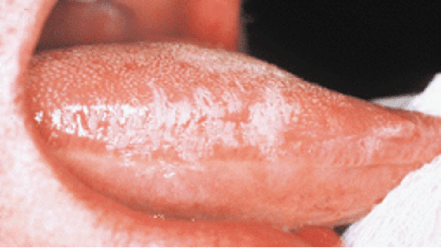
Oral hairy leukoplakia
is a white, non-removable lesion of the lateral tongue caused by Epstein Barr Virus.
-Elongated filiform papillae that are white
-Result of either an increase in keratin or a decrease in normal desquamation
-Tell patient to brush gently to remove debris
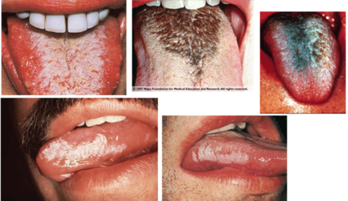
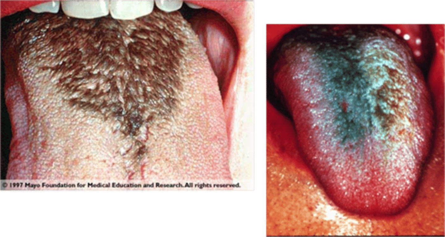
White Hairy Tongue

-Papillae are brown to black b/c of chromogenic bacteria
-Tobacco, foods, hydrogen peroxide, alcohol, chemical rinses contribute
-Tell patient to brush tongue gently with TB to remove debris
Black Hairy Tongue
-Appears as flat or slightly raised oval or rectangular erythematous area in the center of the tongue
-Can be associated with chronic infection with Candida albicans
-No treatment is needed but an antifungal can be used
Median Rhomboid Glossitis
What is a small, circumscribed lesion usually less than 1 cm in diameter? It is elevated or protrudes above the surface of normal surrounding tissue.
It is a small bump.
*picture is of Linhen planus
papule
is a cutaneous condition characterized by white to pinkish papules that occur diffusely in the oral cavity. It is caused by the human papilloma virus types 13 and 32.
Heck Disease
A benign lesion typically associated with pipe and/or cigar smoking; may also occur with cigarette smoking
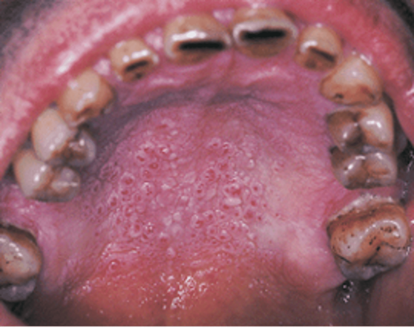
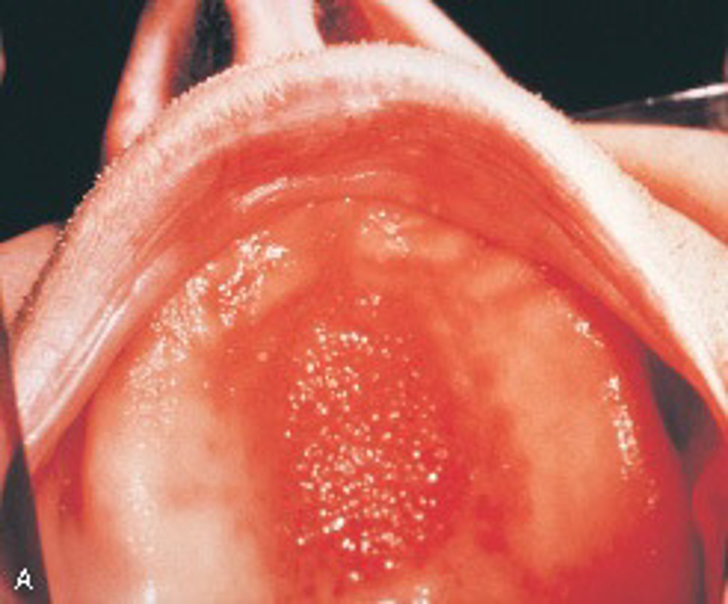
- Appears as multiple keratotic circular papules on the palate.
- Due to an irritation of the minor salivary glands from the intense heat of smoking.
Nicotinic Stomatitis
a palpable cystic midline mass in the neck due to incomplete closure of the thyroglossal duct.
Thyroglossal duct cyst
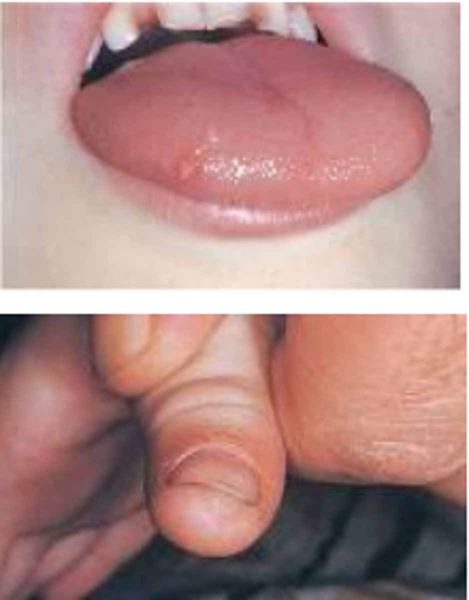
developmental anomaly characterized by a shortened lingual frenum that limits movement of the tongue.
Can cause speech problems, periodontal defects, and problems with breast feeding.
More common in males
Ankyloglossia (tongue tie)
Union of teeth by cementum
Concrescence
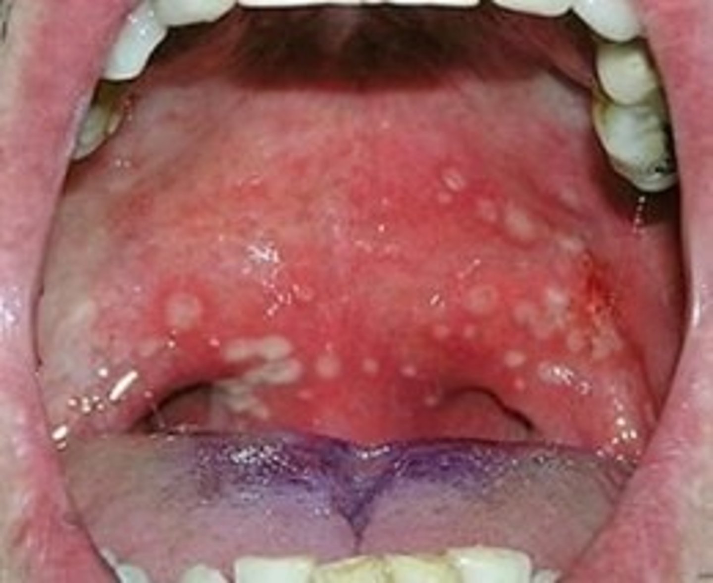
Herpangina
Vesicles on the soft palate along with fever, malaise, sore throat, dysphagia, and
erythematous pharyngitis
Hand, Foot, and Mouth Disease (HFMD)
Painful vesicles and ulcers that can occur anywhere in the mouth; present in epidemic
form in children younger than 5 years
Acute lymphonodular pharyngitis
Hyperplastic lymphoid tissue of the soft palate or tonsillar pillars appears as yellowish or
dark pink nodules
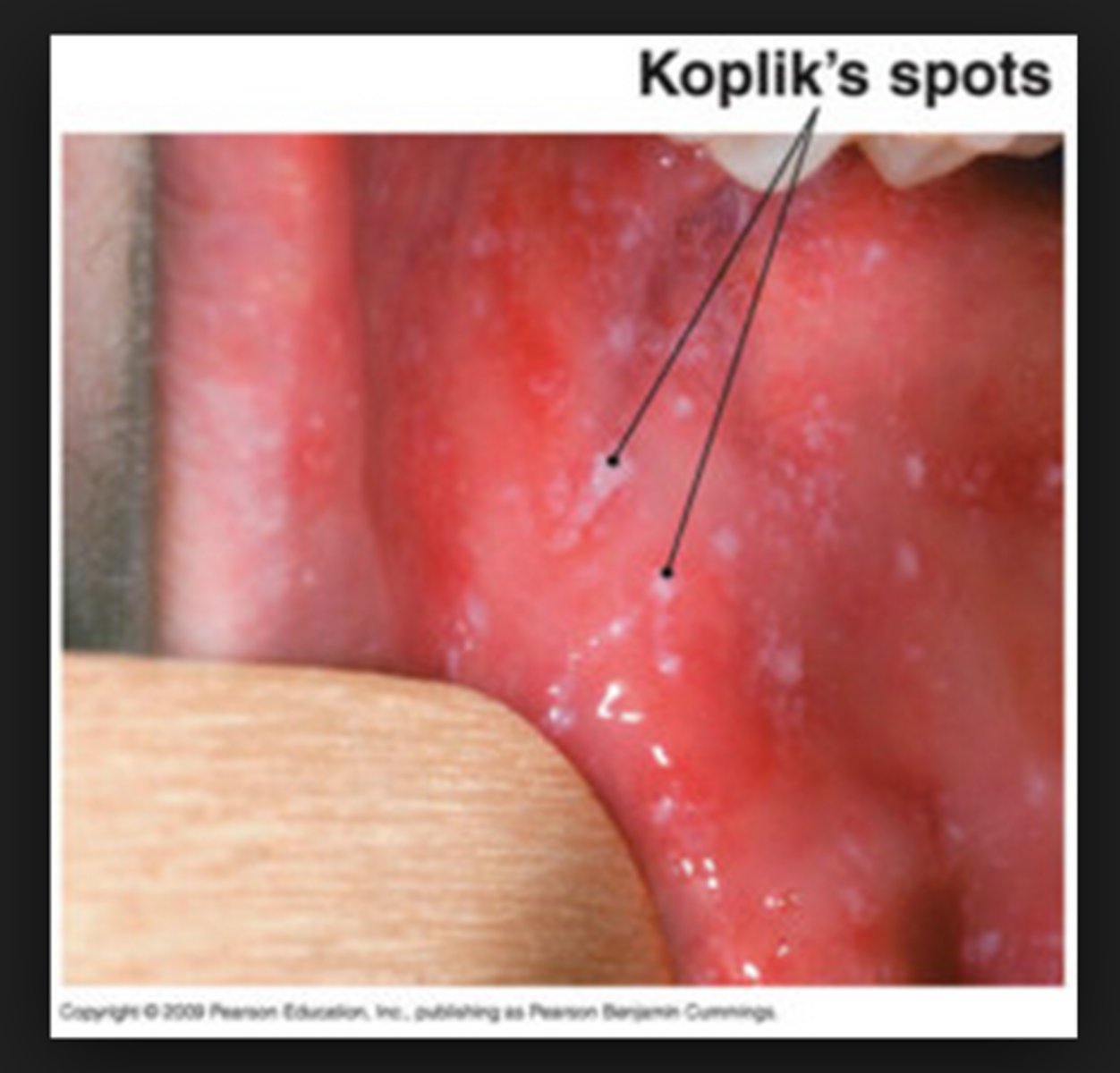
Measles (Rubeola)
Koplik spots in the oral cavity and skin rash that results from a paramyxovirus
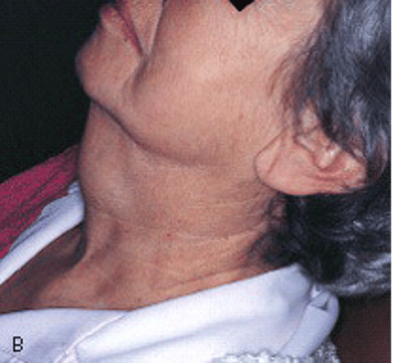
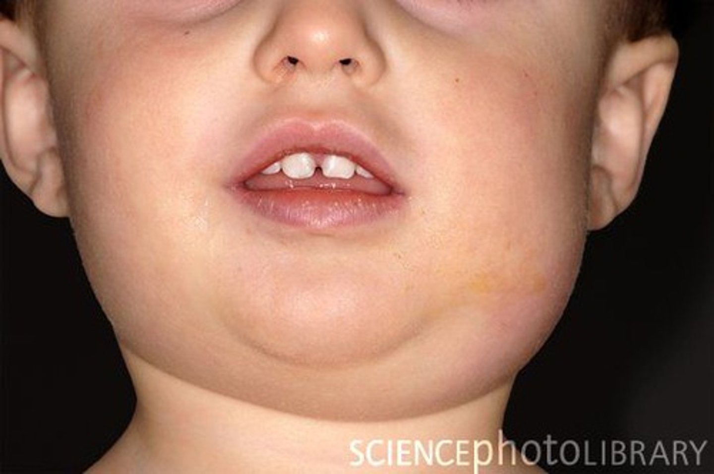
Mumps
Epidemic parotitis characterized by bilateral swelling of the parotid glands
Pemphigus Vulgaris
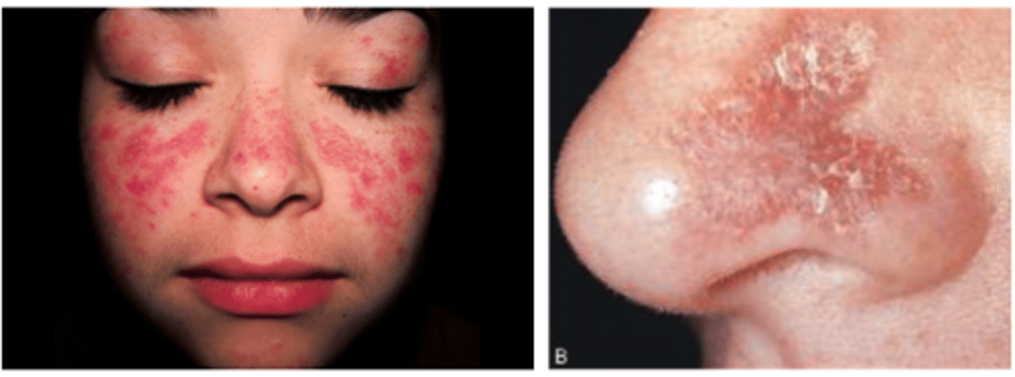
*Systemic lupus erythematosus (SLE)
butterfly rash on the bridge of the nose
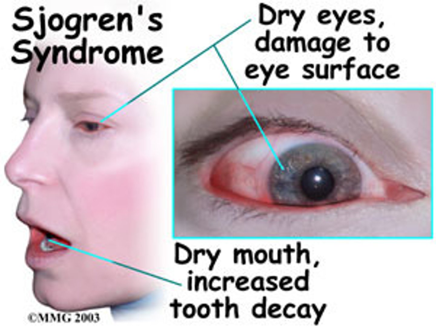
Sjögrens syndrome
Xerostomia
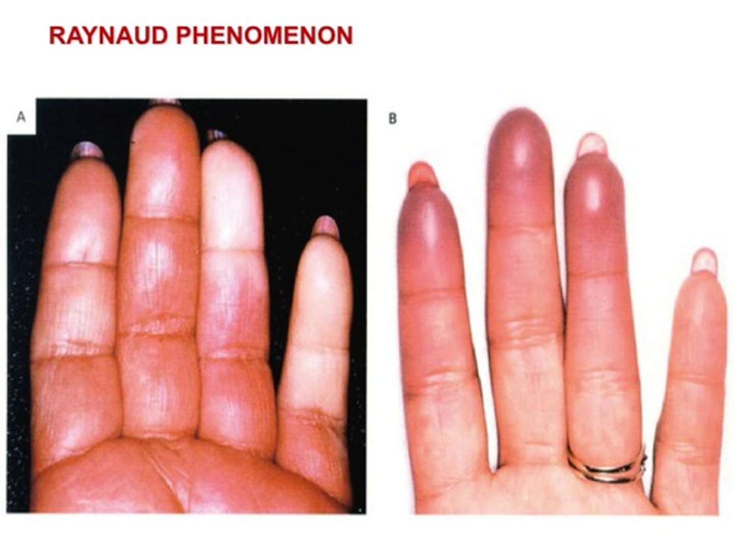
Raynaud Phenomenon
Circulatory disorder
-Dorsal surface of the tongue has deep fissures or grooves
-Cause is unknown, genetics potentially
-Seen in 5% of pop
-Inform patient to brush tongue gently with TB to remove debris
-No treatment necessary
Fissured Tongue
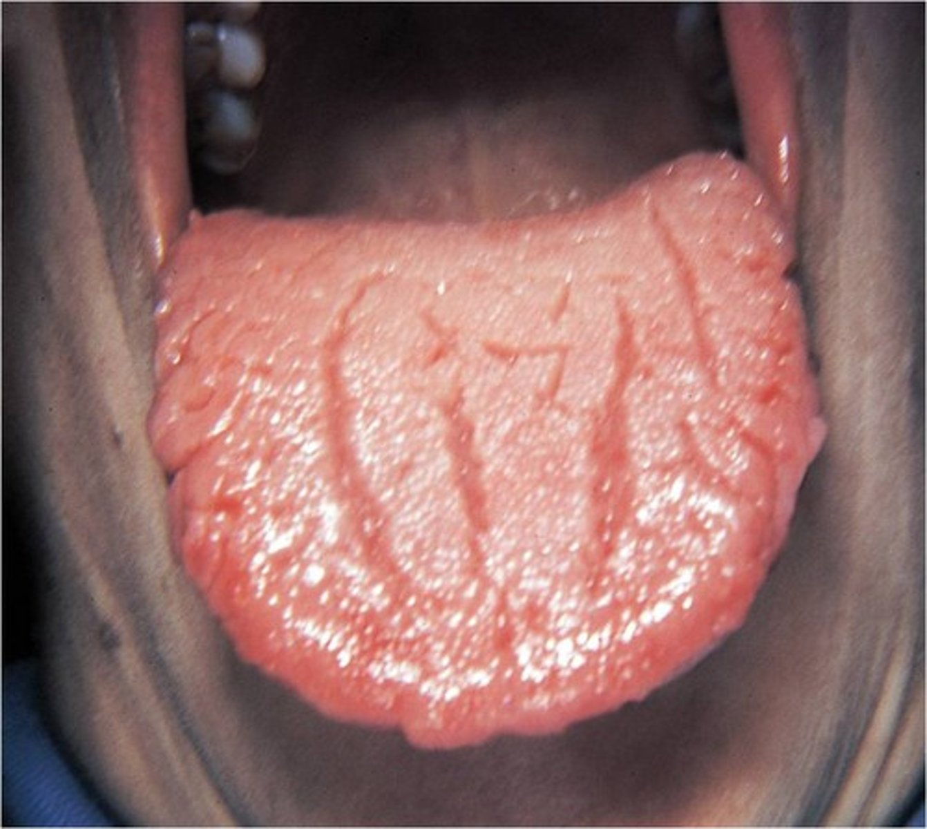
Your patient comes in, and while you are doing the intraoral exam, you notice their tongue. What is the pathological name for the condition of this tongue?
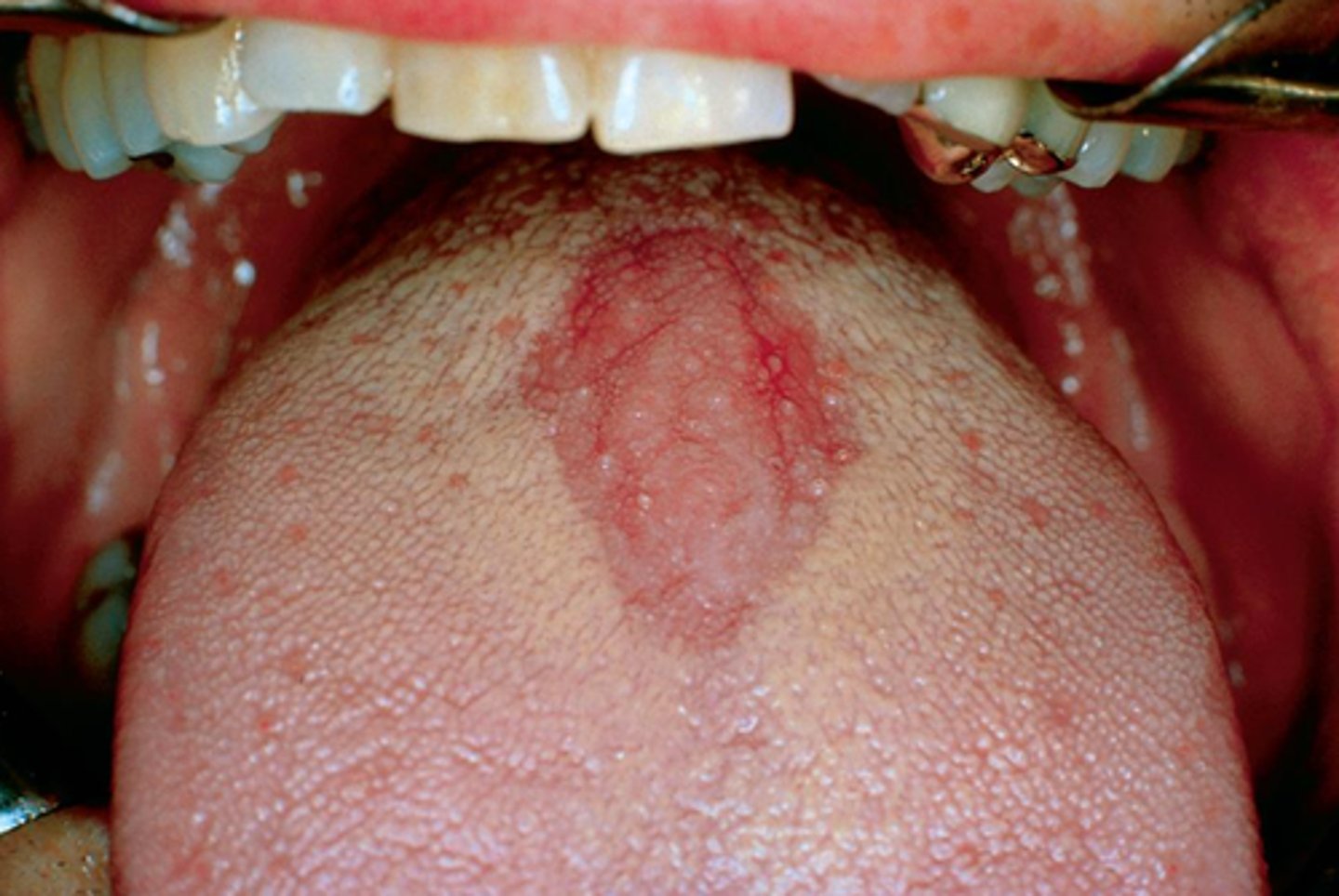
-Appears with erythematous patches surrounded by white or yellow border
-Includes areas devoid of filiform papillae and distinct presence of fungiform papillae
Benign Migratory Glossitis (another name for geographic tongue)
Erythema Migrans

displays diffuse areas devoid of filiform papillae with erythematous patches surrounded by a yellow or white perimeter
Red flat areas of the dorsal tongue with yellow-white borders best describe which of the following conditions?
Geographic tongue or Erythema migrans

Also referred to as Geographic tongue (benign migratory glossitis), involves 2/3rds of the dorsal and lateral borders of the tongue
Erythema migrans (Geographic tongue (migratory glossitis))
Median rhomboid glossitis
is a form of candidiasis that manifests of a red, rhomboidal shaped patch on the mid-dorsal tongue.
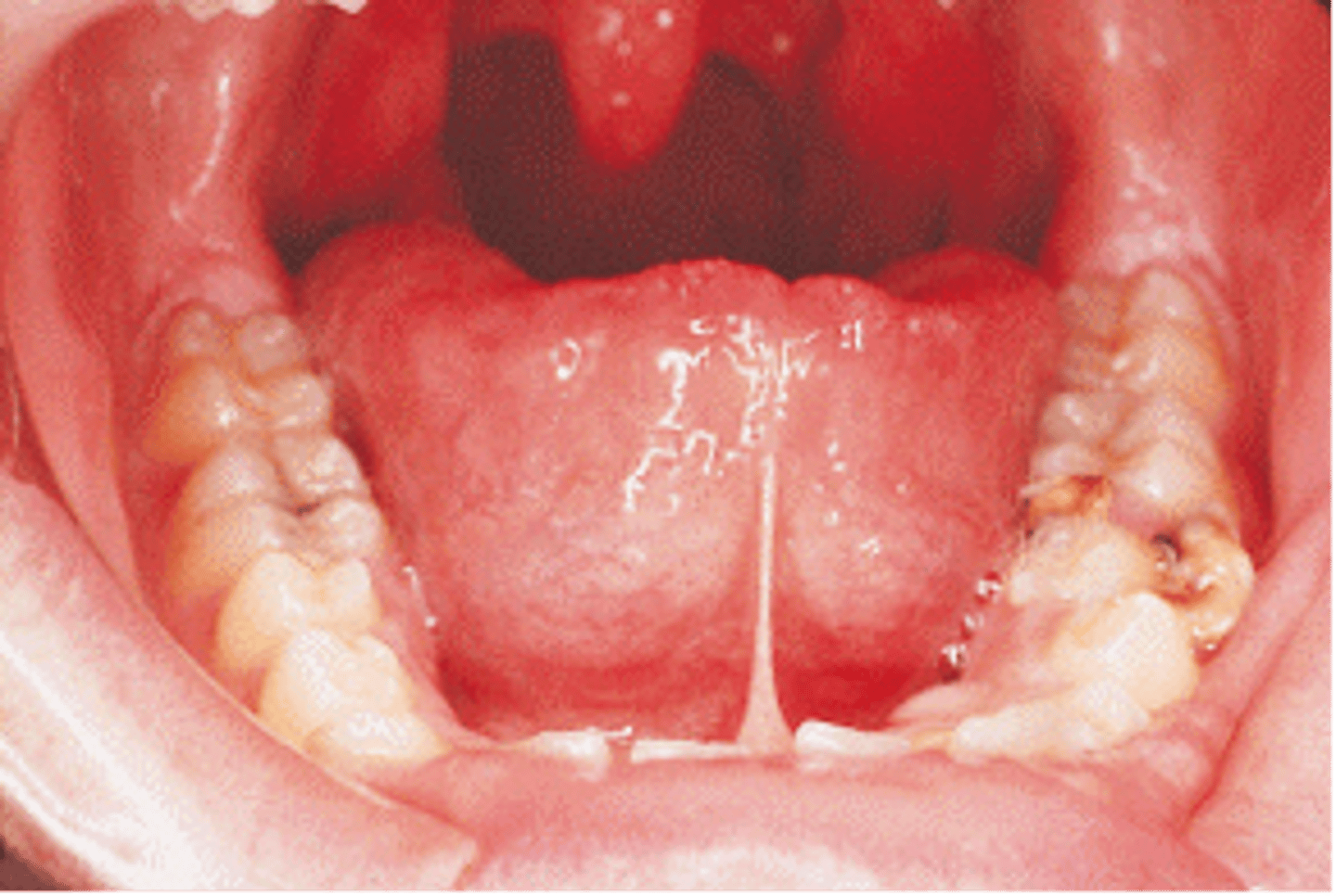
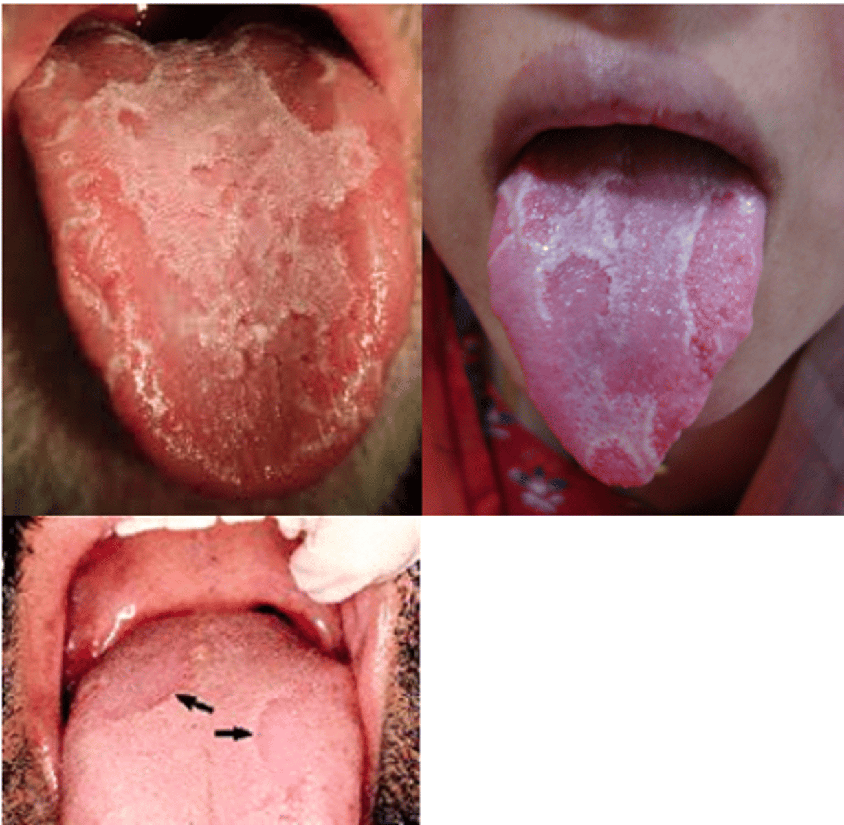
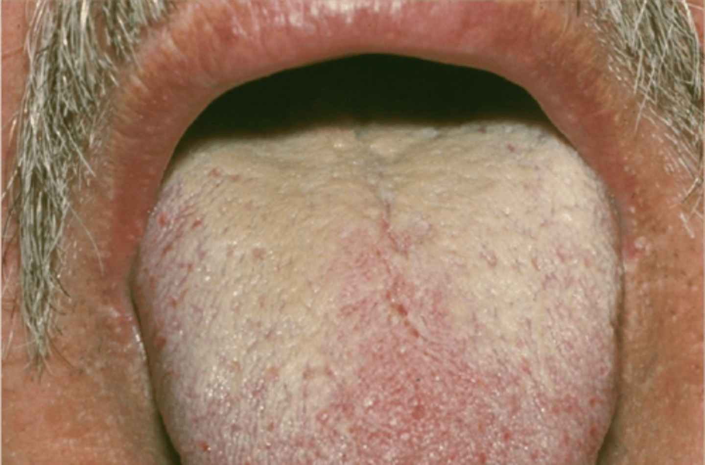
White coated tongue
is the result of elongation of the filiform papilla. O
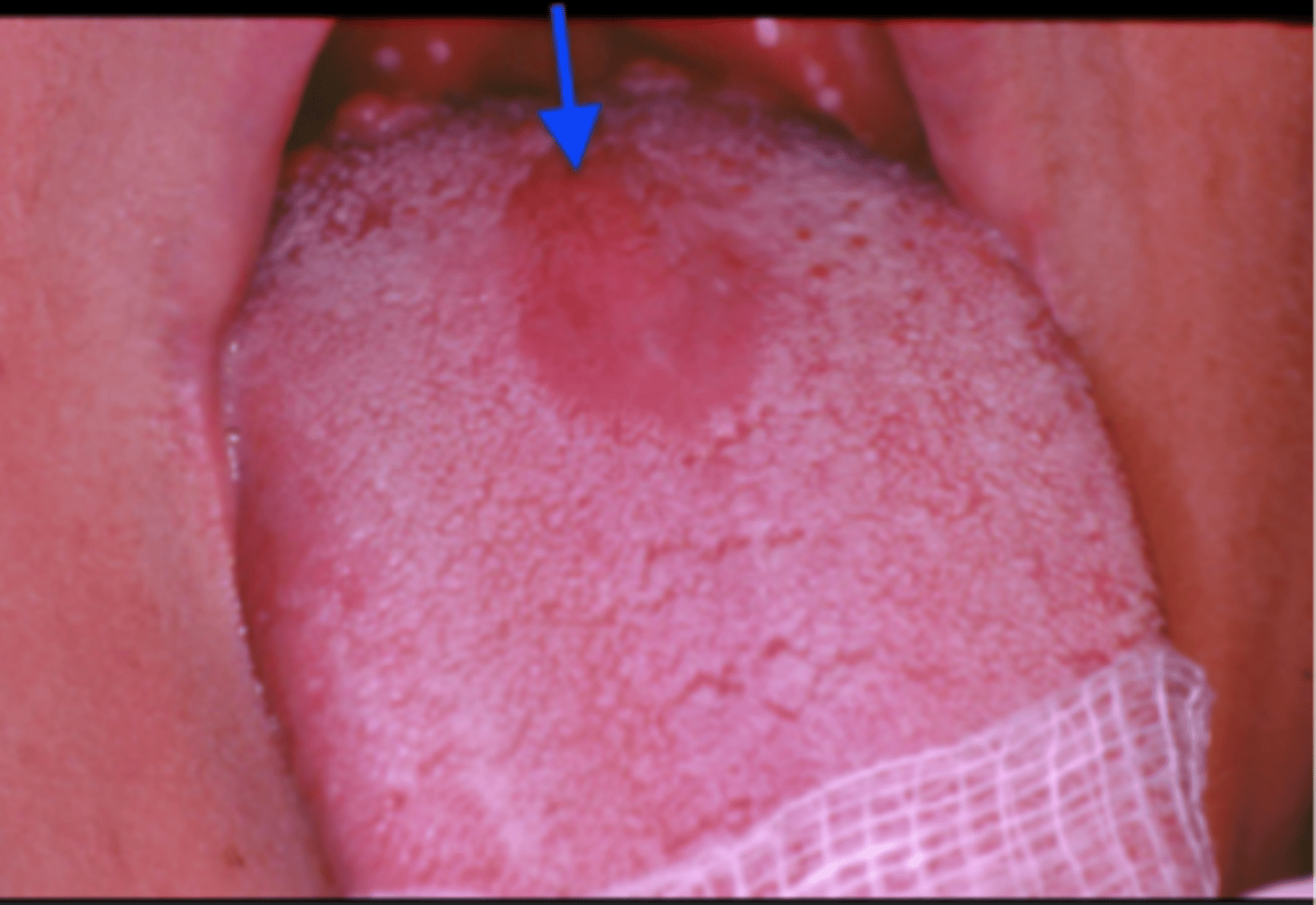
The dorsal tongue lesions marked by the arrows are best diagnosed as
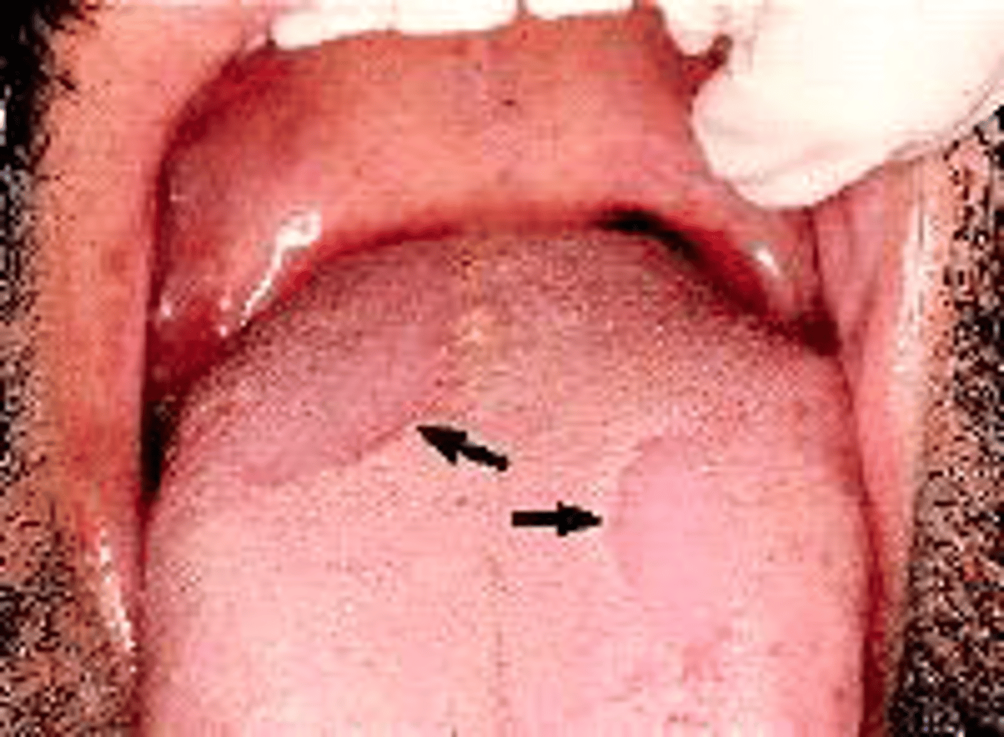
trauma to a minor salivary duct.
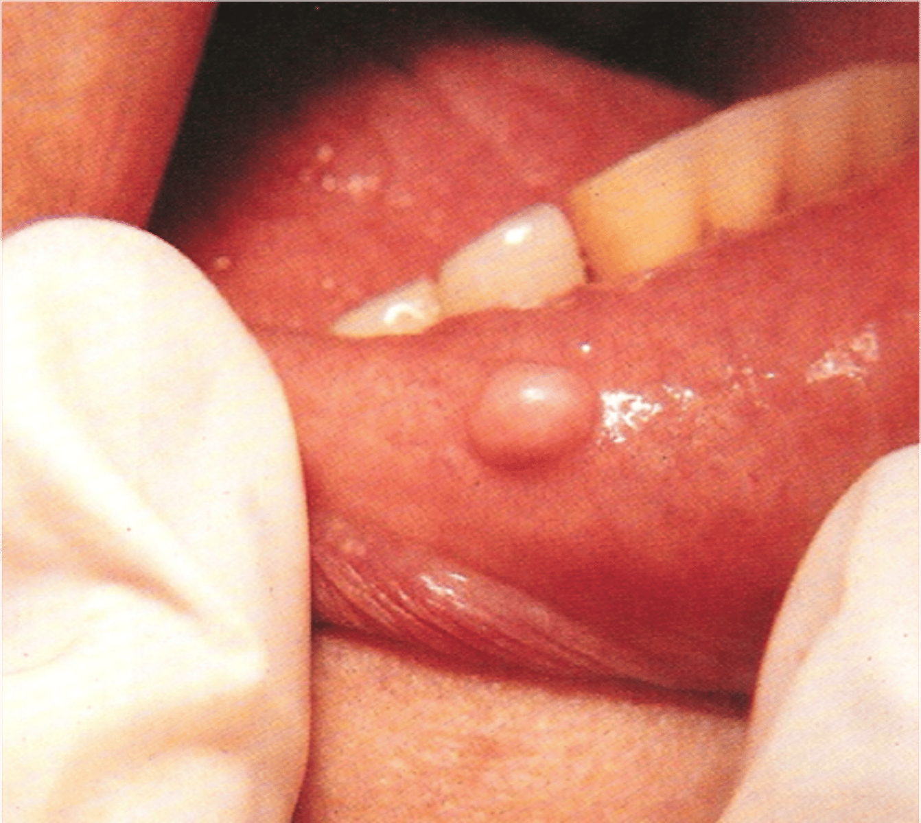
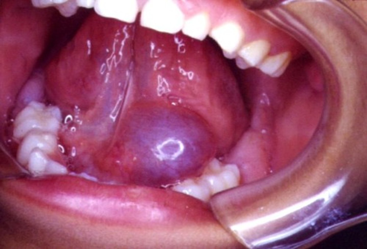
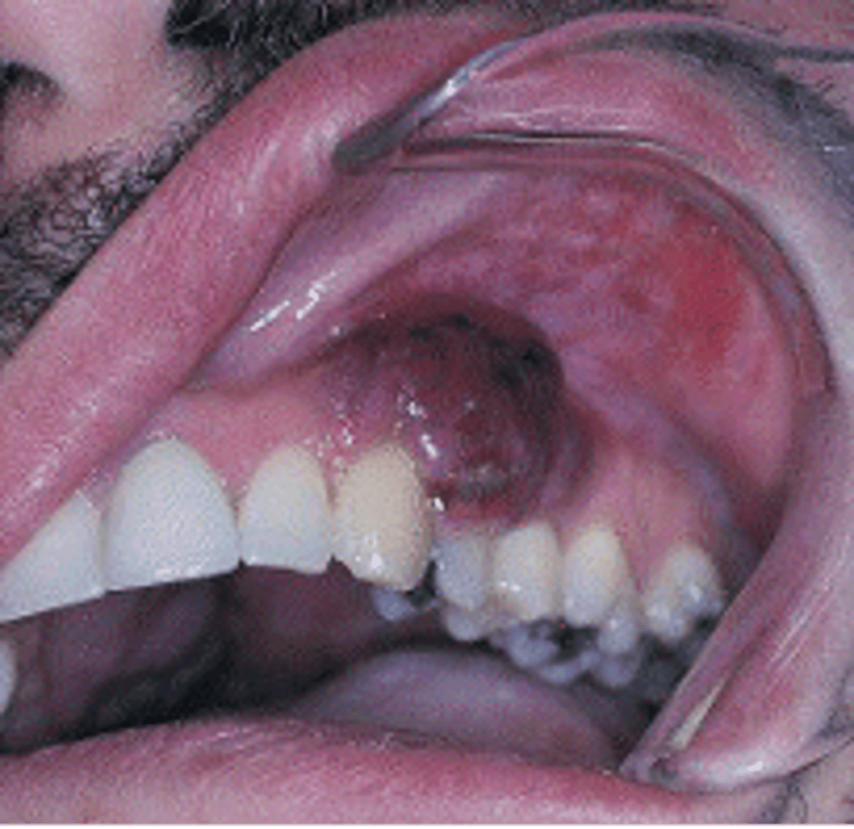
The major cause of a mucocele is
Ranula
is a floor of mouth submucosal swelling caused by trauma to the sublingual gland.
Necrotizing sialometaplasia
results from ischemia and infarction of minor salivary glands and typically involves the palate.
A lesion formed when a salivary gland duct is severed and the mucous salivary gland secretion spills into the adjacent CT
Mucous Retention Lesion: Mucocele
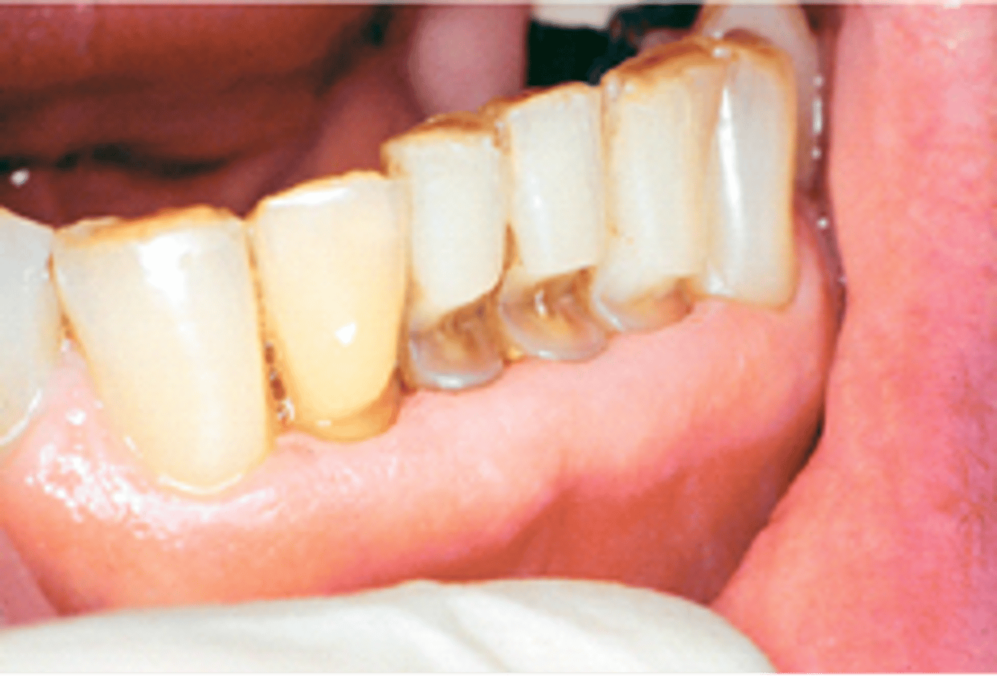
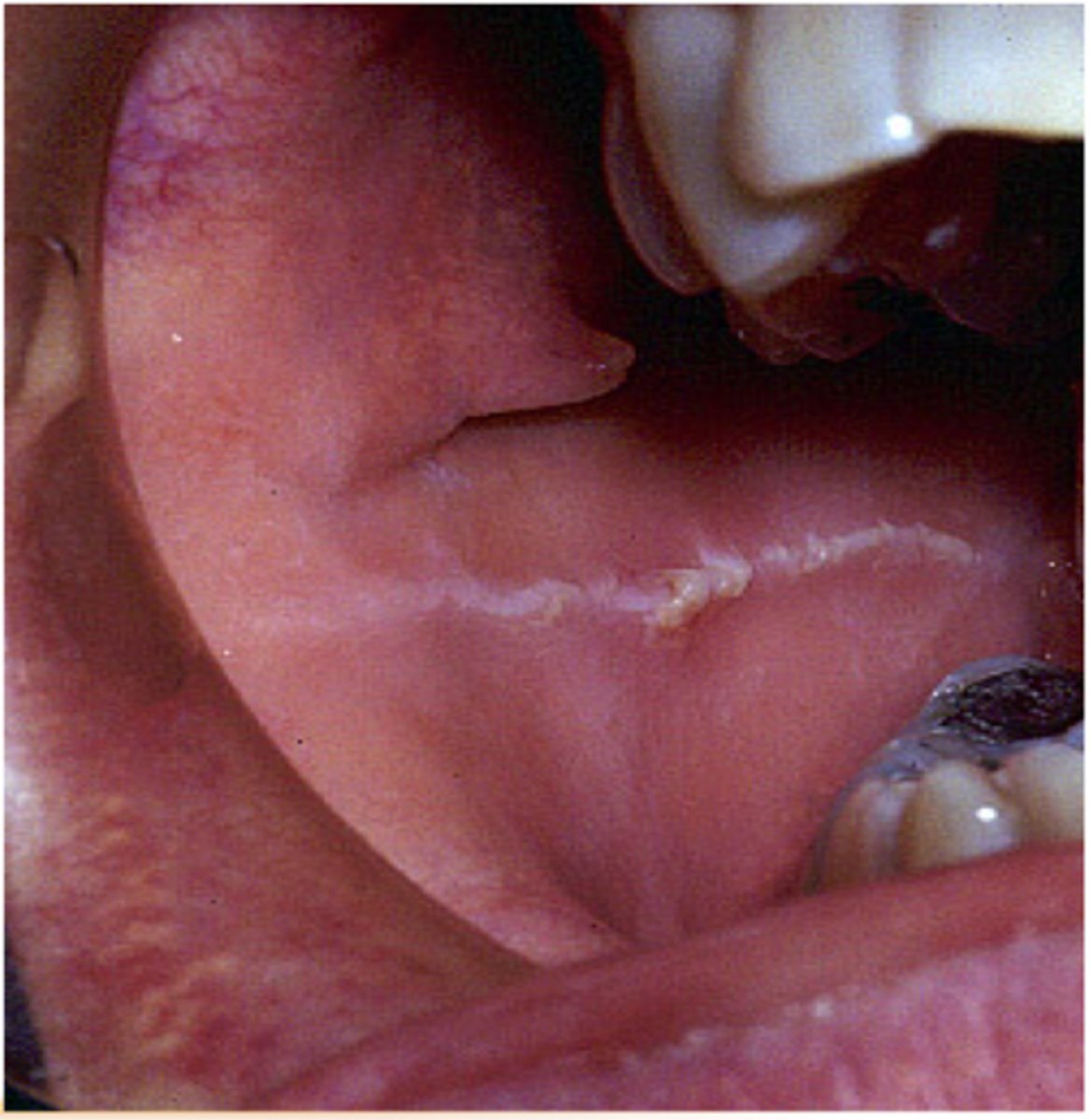
Linea alba
refers to a white line seen along the occlusal plane and is the result of clenching
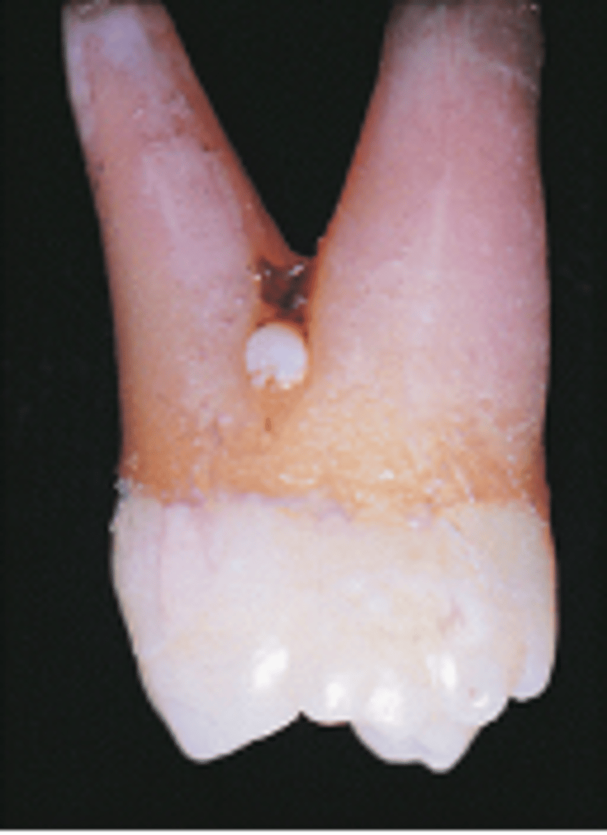
-Ectopic enamel
-Tooth enamel on the root surface (usually in the furcation)
Enamel Pearl
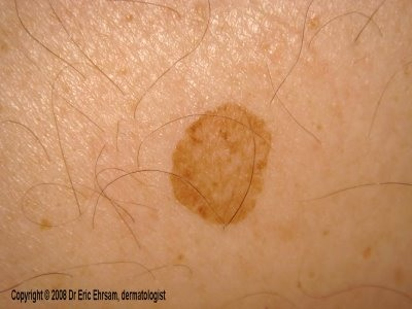
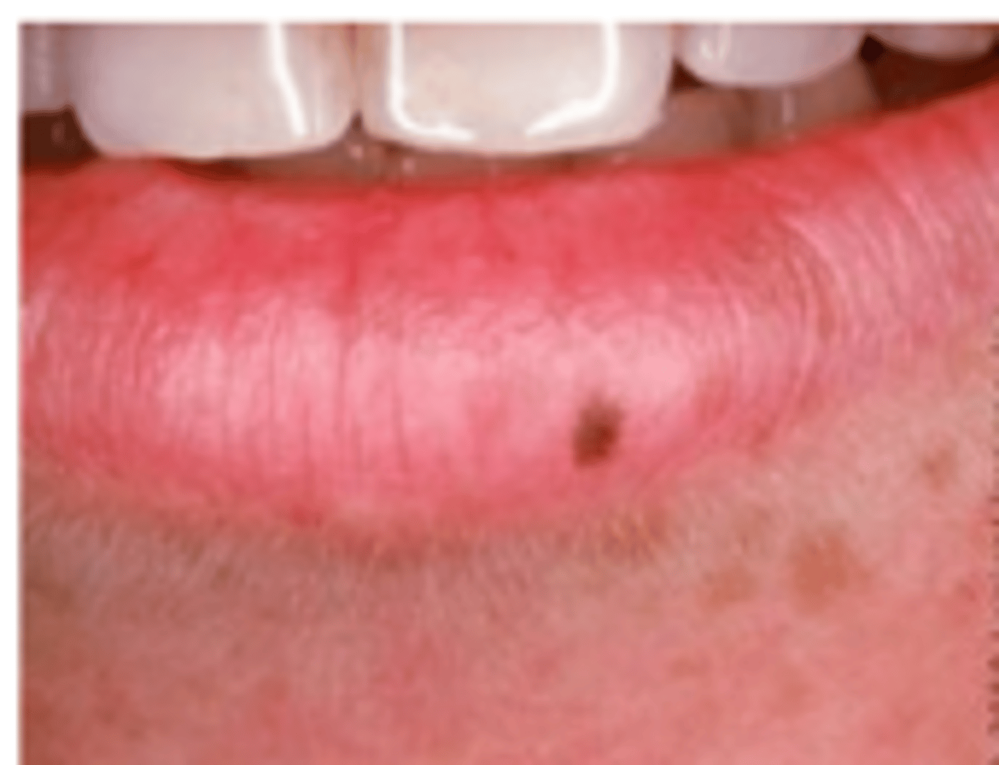
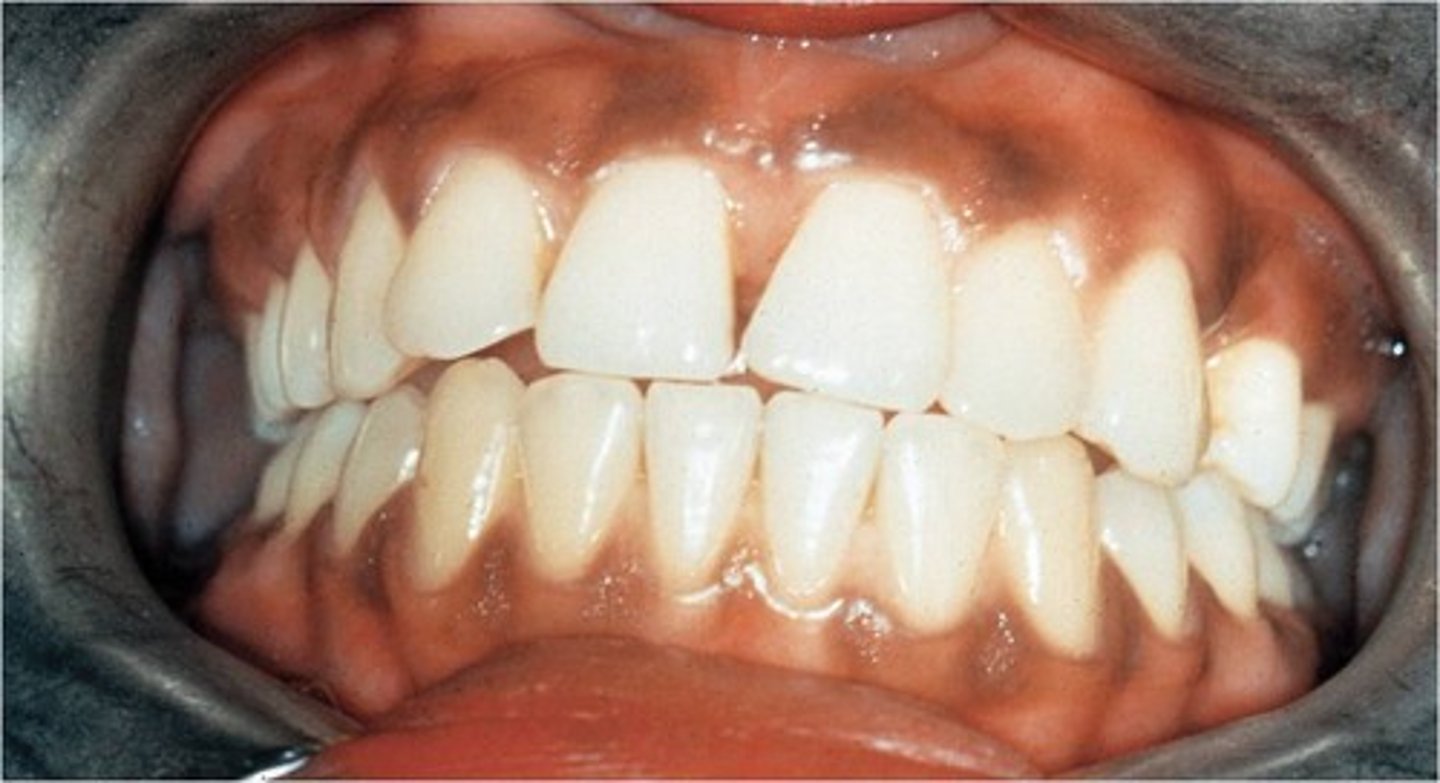
Localized macules of pigmentation caused by presence of melanin
Melanin pigmentation
-Pigment that gives colour to skin, eyes, hair, mucosa and gingiva
-Most common in dark skinned individuals
Melanin Pigmentation
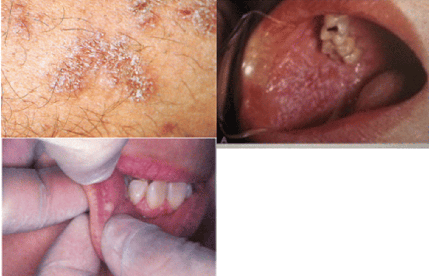
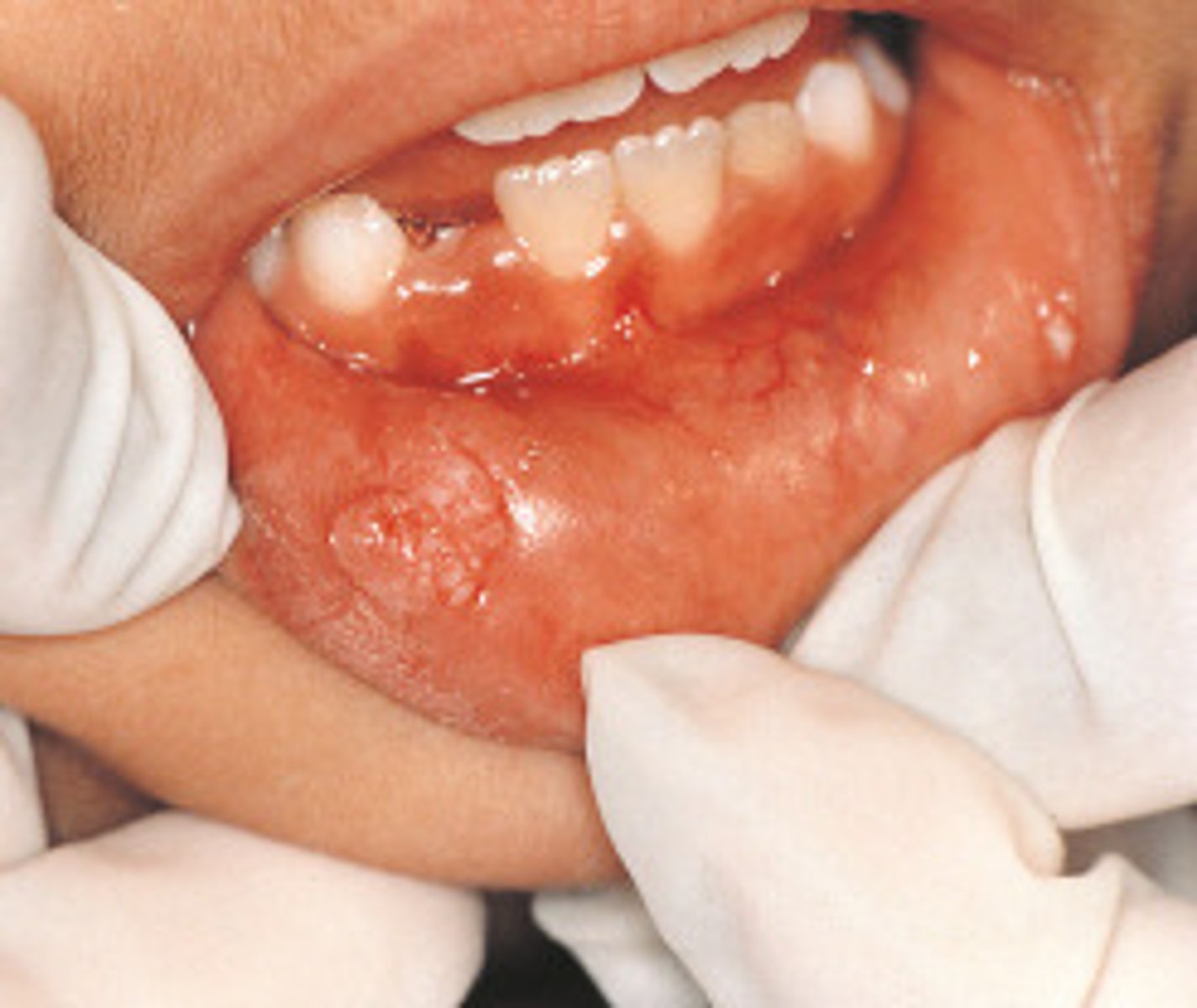
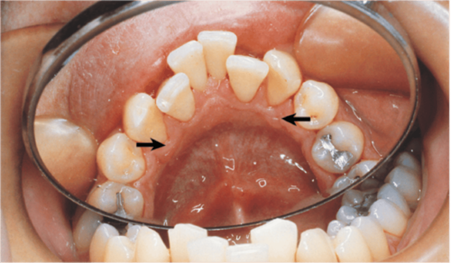
Sessile nodule on gingival margin of the lingual aspects of the mand cuspids
Retrocuspid Papillia
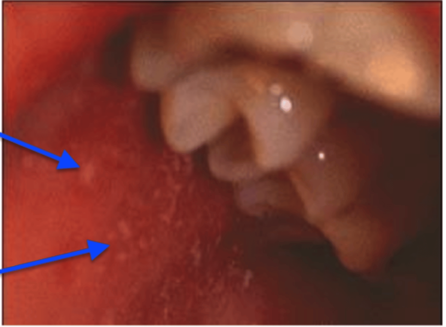
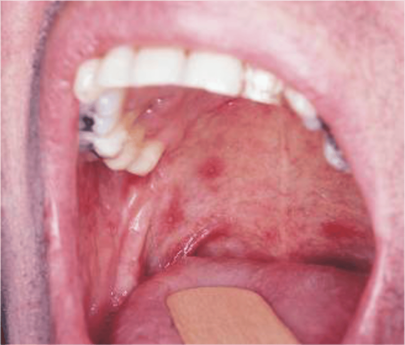
Koplik spots
are erythematous macules with white necrotic centers that may occur in the oral cavity in patients with measles
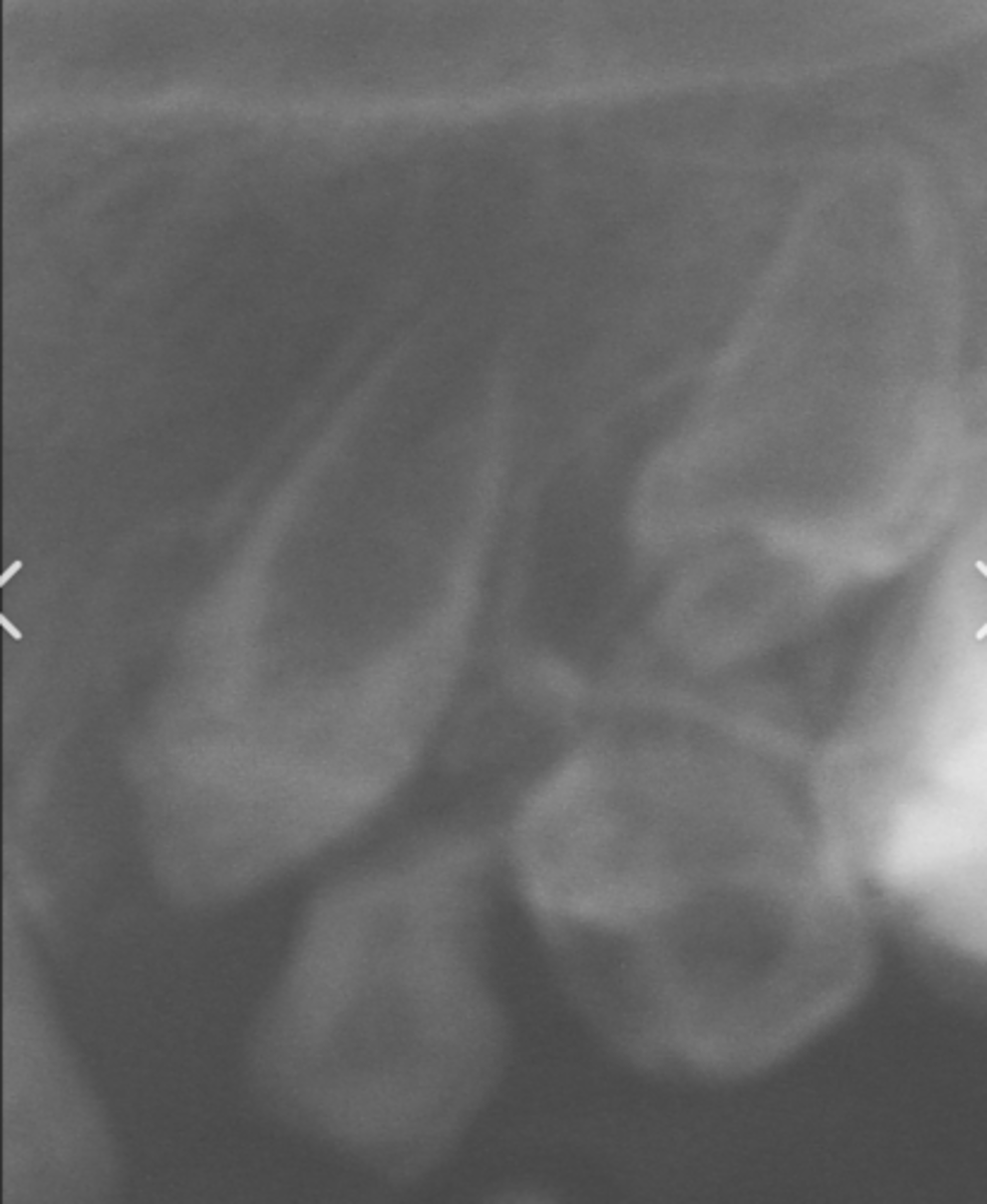
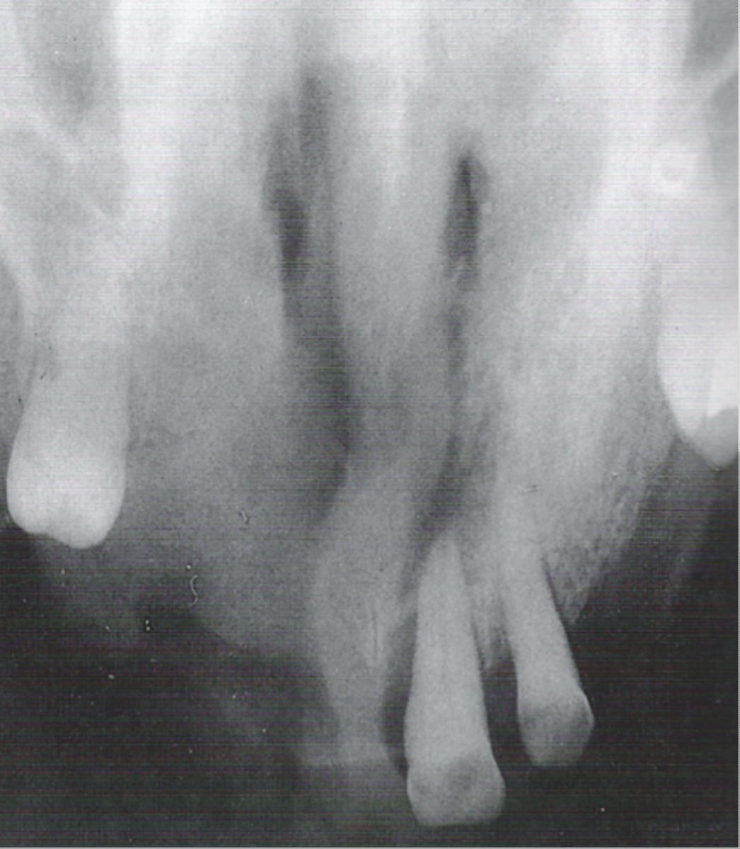
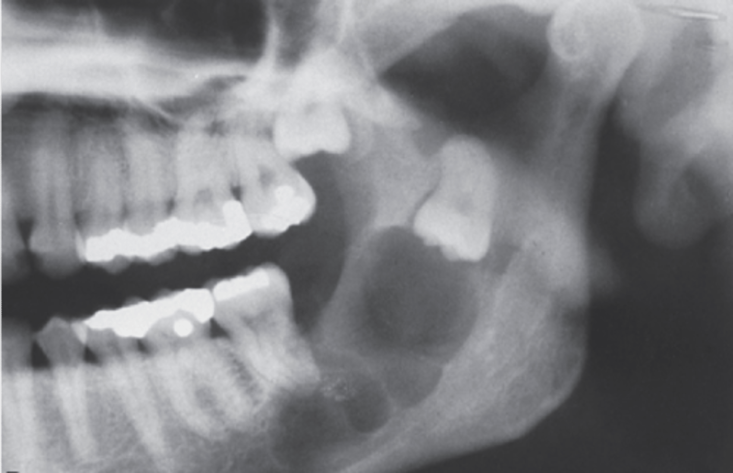
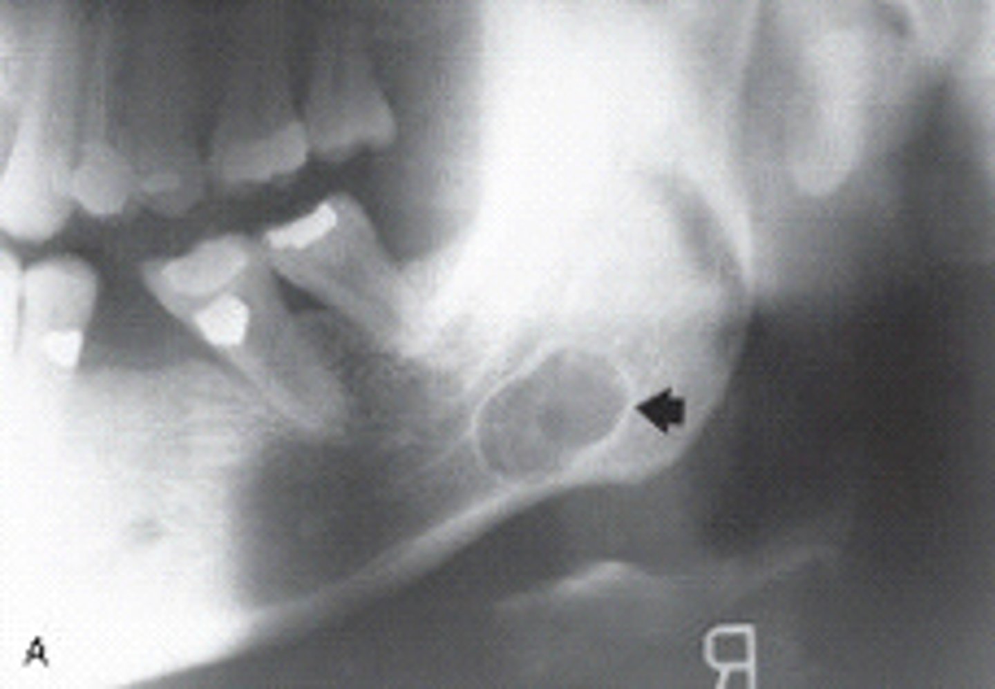
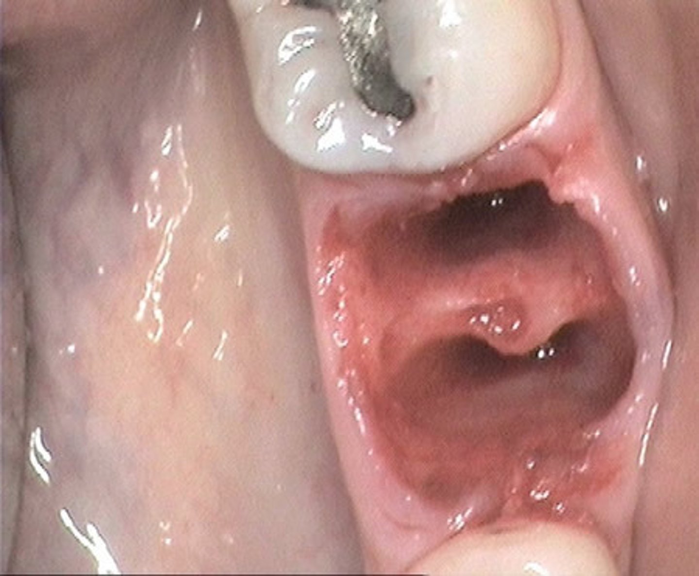
Radiolucency in posterior mandible below mandibular canal due to lingual concavity of jaw
A _____usually presents as an oval radiolucency anterior to the angle of the ramus and inferior to the mandibular canal.
Stafne bone defect
Stafne bone cyst
a pseudocyst filled with salivary gland tissue and may be an extension of the sublingual gland. ct.
trauma
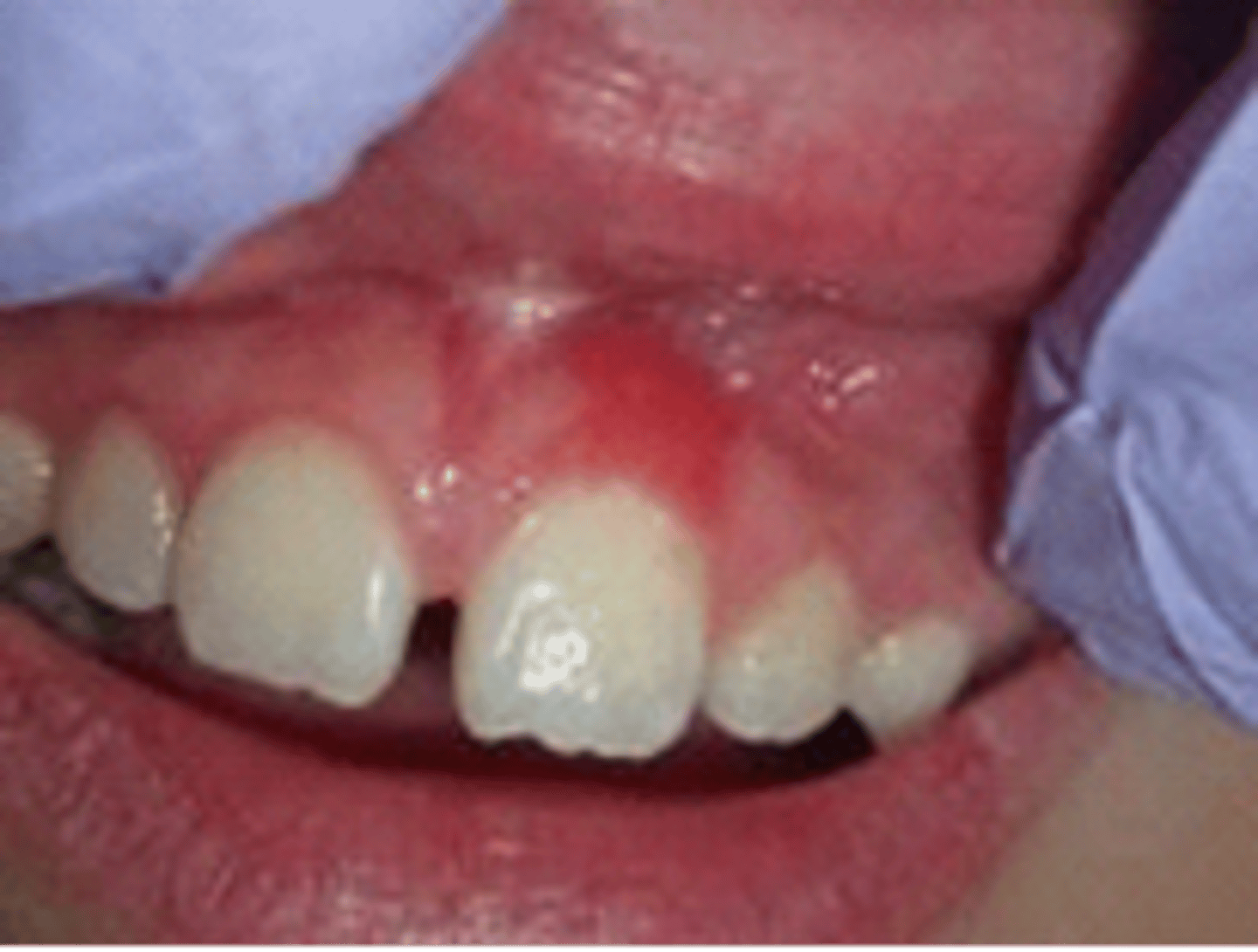
is one of the precipitating factors associated with minor aphthous ulcers and these ulcers do heal in 7 to 10 days.
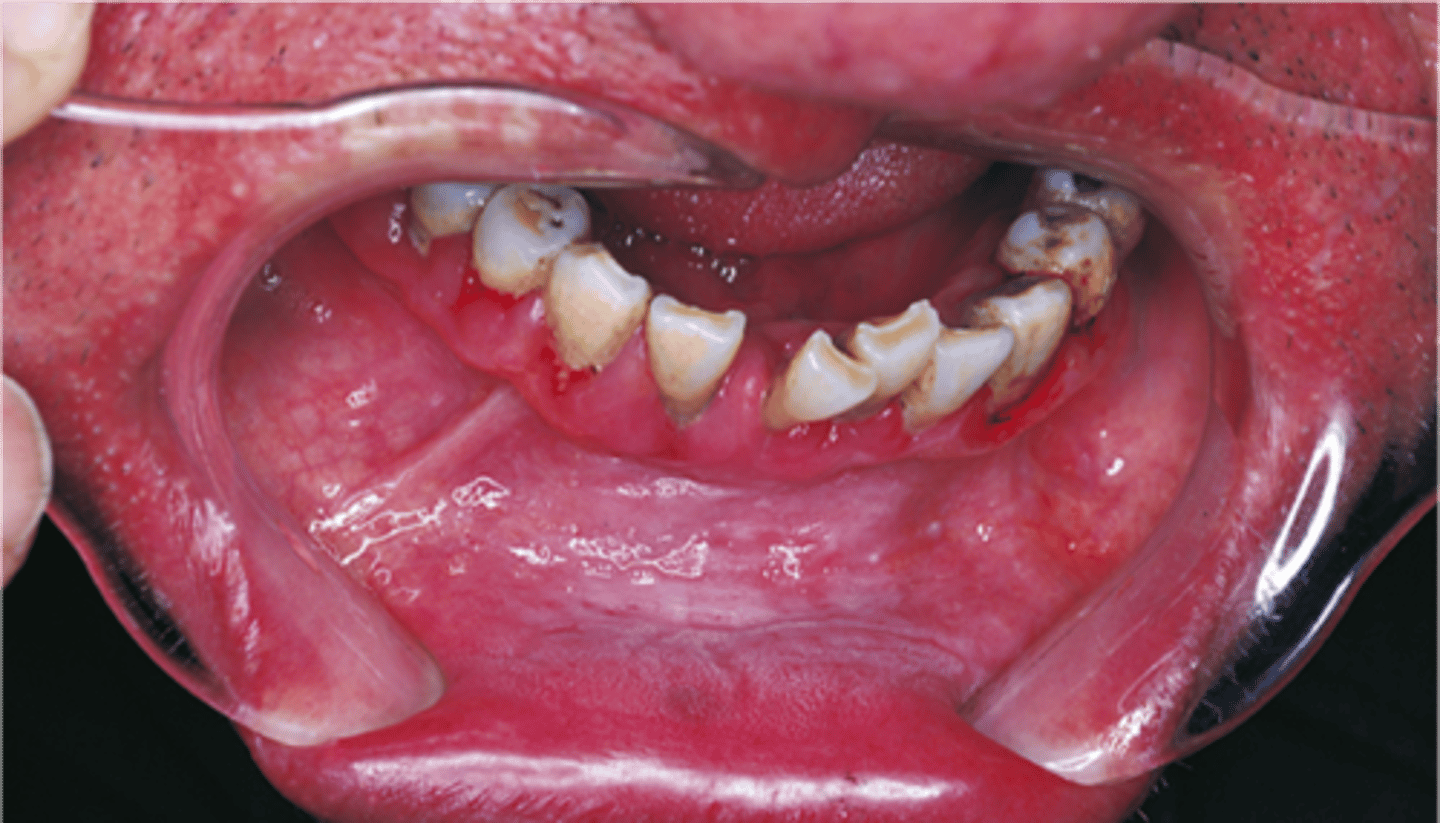
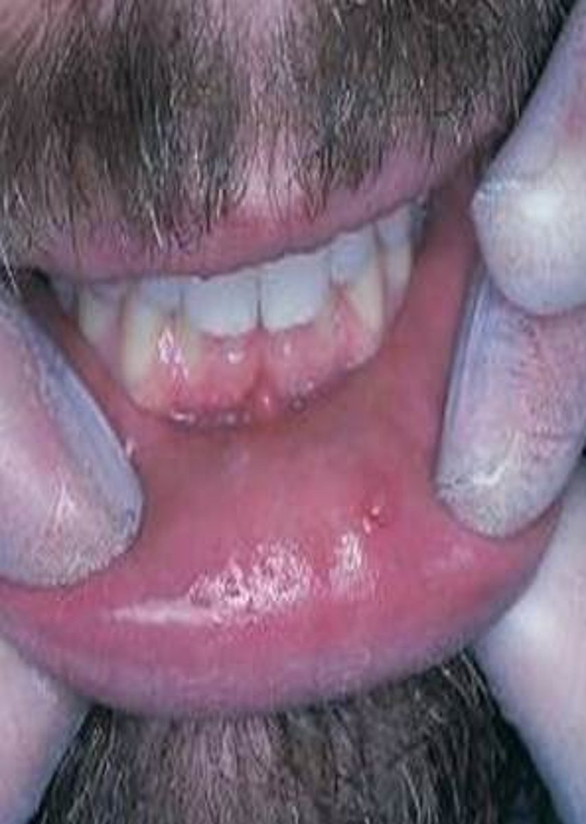
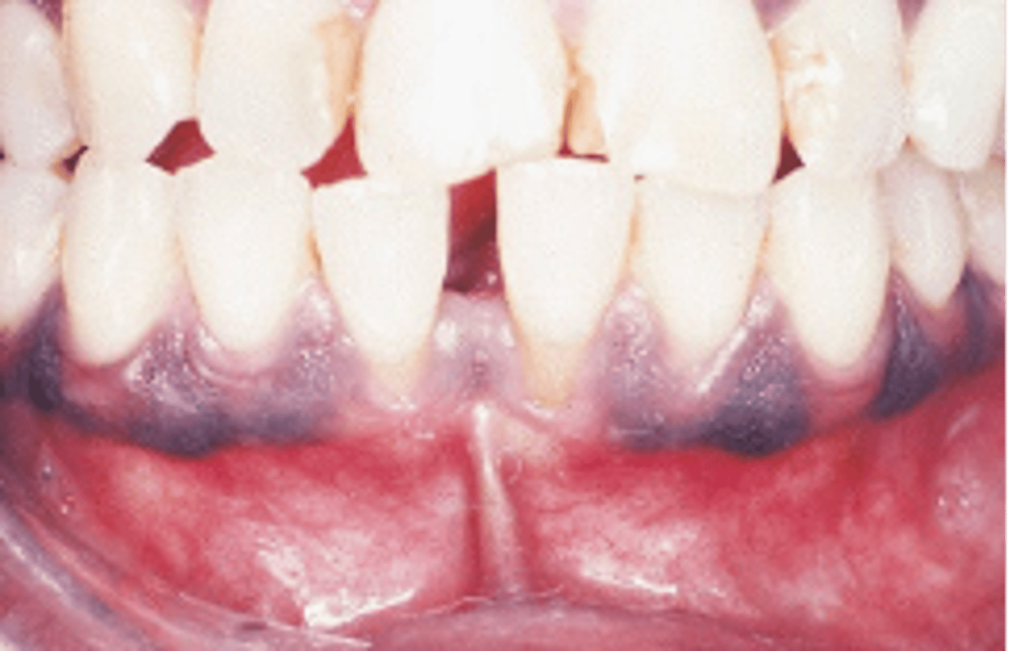
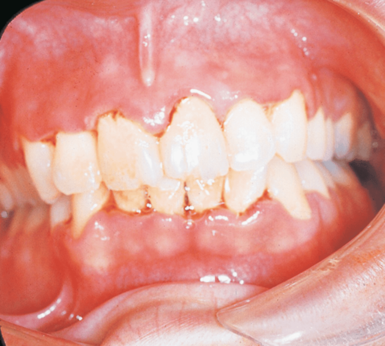
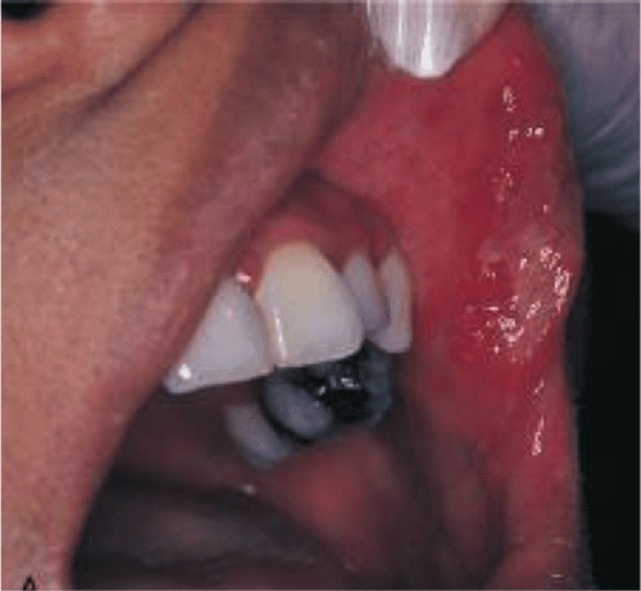
The gingival enlargement in this patient is caused by a calcium channel blocker. Which one of the following medications is the cause?
Nifedipine
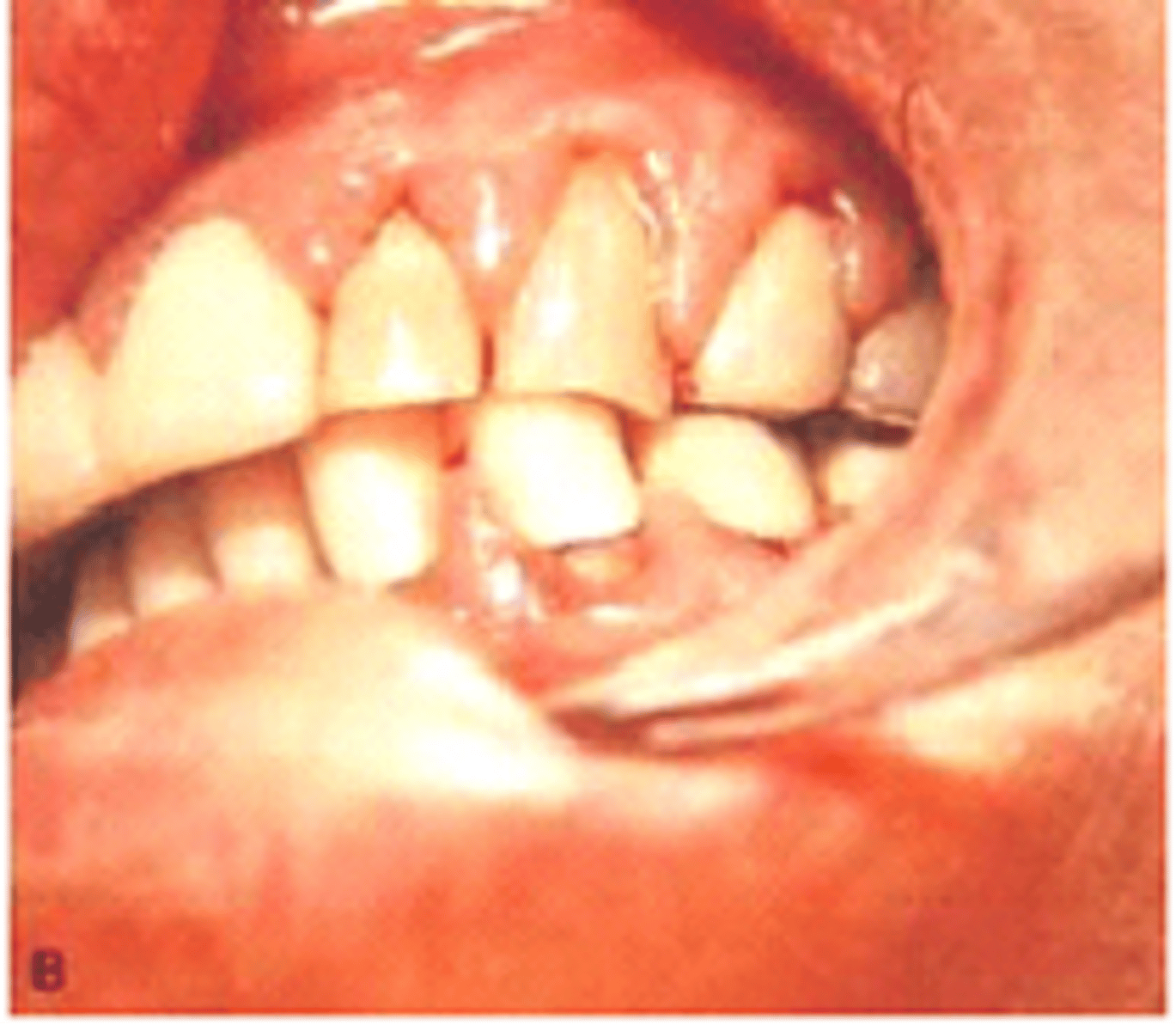
Seen by a patient taking nifedipine (Ca Channel blocker)
Gingival Enlargement by a calcium channel Blocker

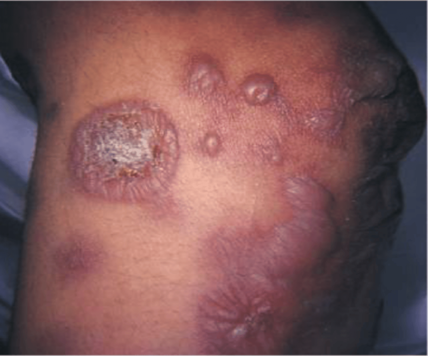
Cancer caused by the human herpes virus 8 (HHV-8) that mainly affects the skin and mucous membranes but may also cause extensive visceral organ involvement; also called malignant neoplasm of soft tissue
Kaposi sarcoma (KS)
Dilantin
is an anticonvulsant medication used to control seizures and other neurologic conditions that does cause gingival enlargement
Tetracycline
is an antibiotic responsible for discoloring teeth
Cyclosporine
is an immunosuppressant drug that also causes gingival enlargement, and is used to prevent rejection of organ transplants
Gingival enlargement
A medical history of a patient prescribed a calcium channel blocker may reveal which condition?
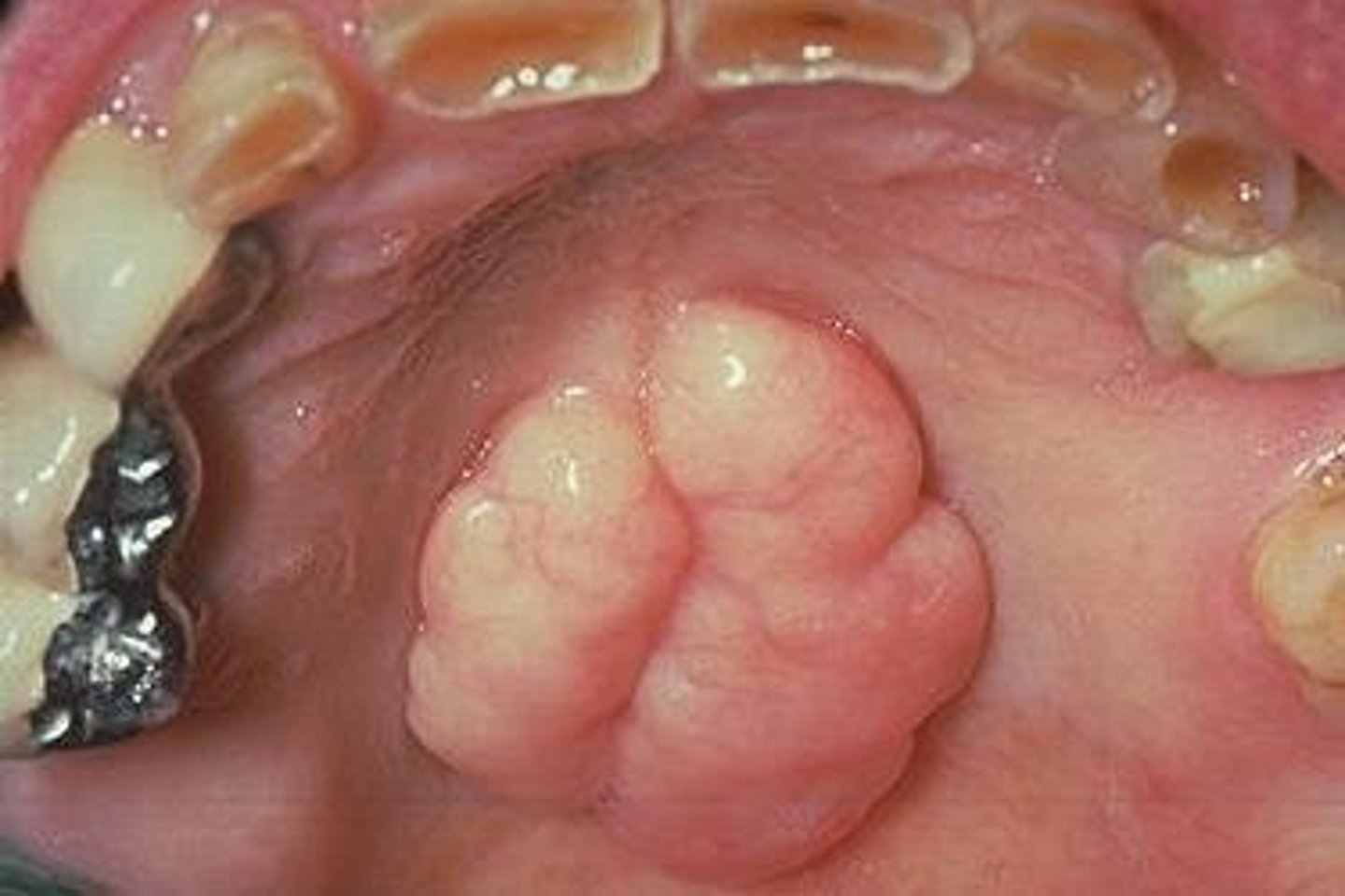
Outgrowths of dense bone found on lingual aspect of mandible in areas of the premolars above mylohyoid ridge
Mandibular Tori
-Exophytic growth of normal compact bone
-Observed clinically in midline of the hard palate
-Inherited, gradual formation
-More common in women
-Takes on various shapes and sizes, covered by normal soft tissue, can be lobulated
-No treatment, unless it interferes with speech, swallowing, or prosthetic appliance
Torus Palatinus
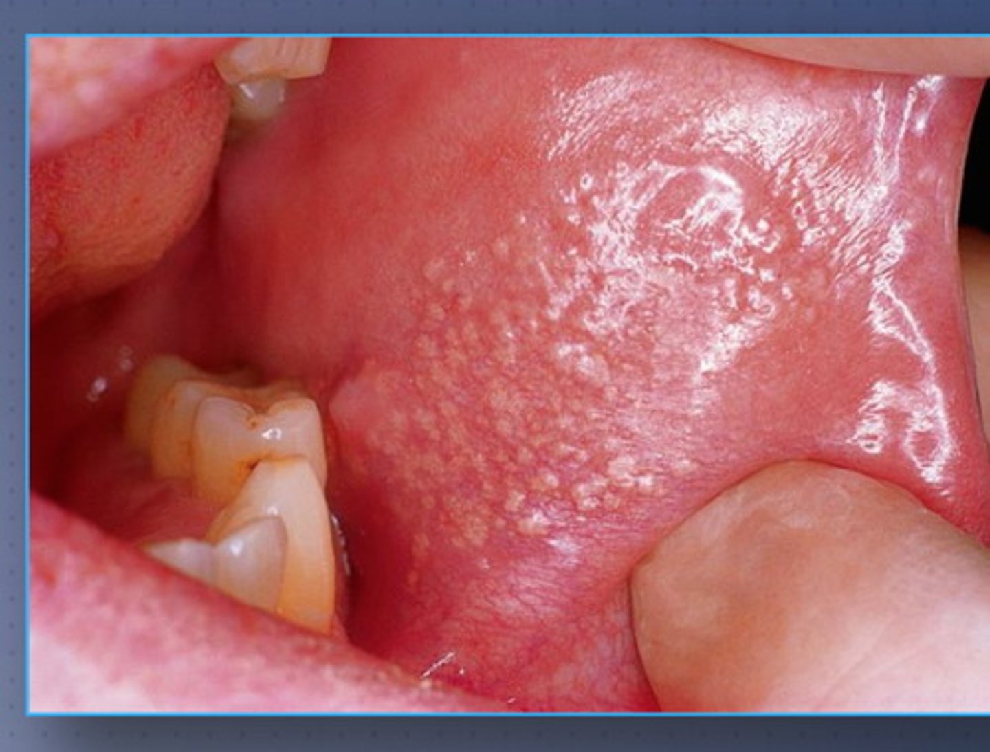
-Clusters of ectopic sebaceous glands
-Yellow lobules in clusters
-Common on vermillion border and buccal mucosa
-No treatment
Fordyce Granules
Nitroglycerin
Which does not cause gingival enlargement?
Nifedipine
The gingival enlargement in this patient is caused by a calcium channel blocker. Which one of the following medications is the cause?
Acute osteomyelitis
Acute osteomyelitis of the jaws commonly results from extension of a periapical abscess. Acute osteomyelitis can result from surgery, but this is not a common reaction. Acute osteomyelitis can result from fracture of the jaw, but again this is not a common reaction. Paget disease has been associated with chronic osteomyelitis.
Erythematous Candidiasis
is an erythematous, often painful mucosa that may be localized or generalized.
Thick, white, raised patches in the mouth
Candidiasis (Thrush)
Chronic atrophic candidiasis or denture stomatitis
presents as erythematous mucosa with lesions that vary from petechiae-like to more generalized and granular.
This granular, erythematous papillary surface of the palatal vault was caused by an ill-fitting suction area of a maxillary denture. Inflammatory soft tissue lesion; Papillary lesions under the MAXILLARY DENTURE, especially if denture is NEVER REMOVED; Excise tissue and remove denture
Papillary hyperplasia of the palate
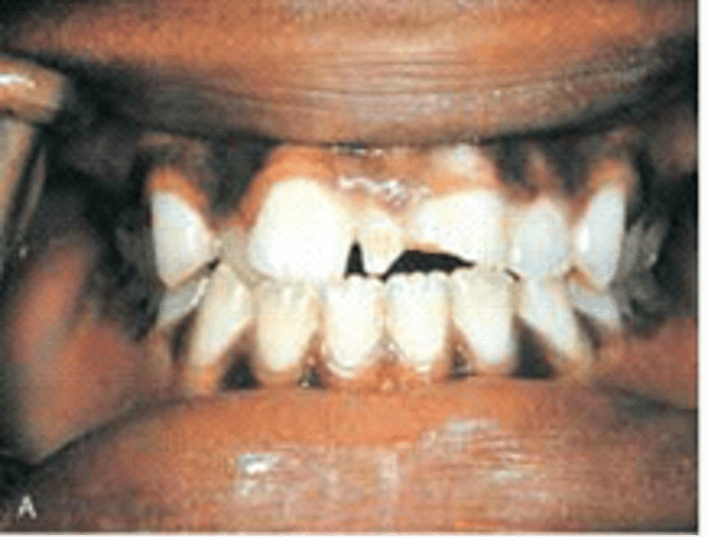
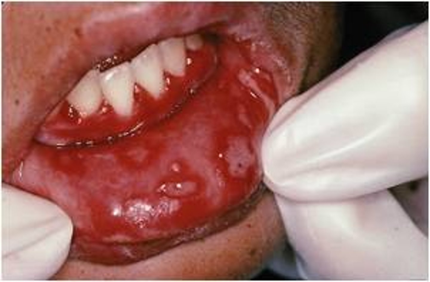
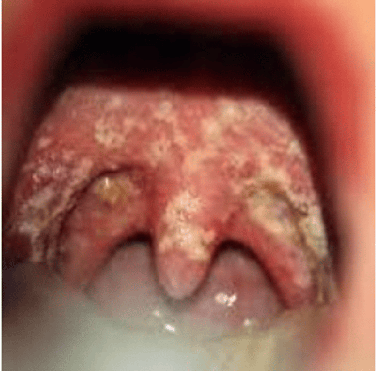
NUG (Necrotizing Ulcerative Gingivits)
The gingivae shown are painful and erythematous. The interdental papillae appear as punched-out, necrotic, cratering areas. The overall sloughing of the necrotic tissue appears as a pseudomembrane over the tissues. The patient experiences a foul odor and metallic taste. On the basis of these features, which condition is suspected?
Fusiform bacillus and Spirochete cause this, cratered papilla, and white tissue because of cell death
Pseudomembranous
A white curdlike material is present on the oral mucosa and can be wiped off. The underlying mucosa is erythematous and the patient experiences a burning sensation. Which type of candidiasis do you suspect?
tiny (1-2 mm)
resemble herpes simple ulcers
painful
occurs in groups
herpetiform aphthous ulcers
larger that 1 cm
deeper and longer lasting that minor
very painful
occurs in the posterior of the mouth more ofter
Major aphthous ulcers
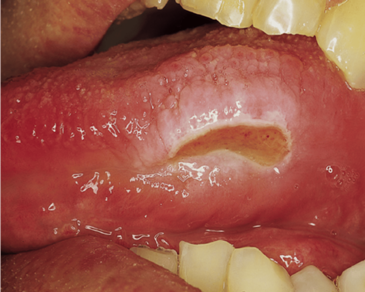
Ulcerative lesion due to trauma- Healing usually lasts 7-14 days
Traumatic Ulcer
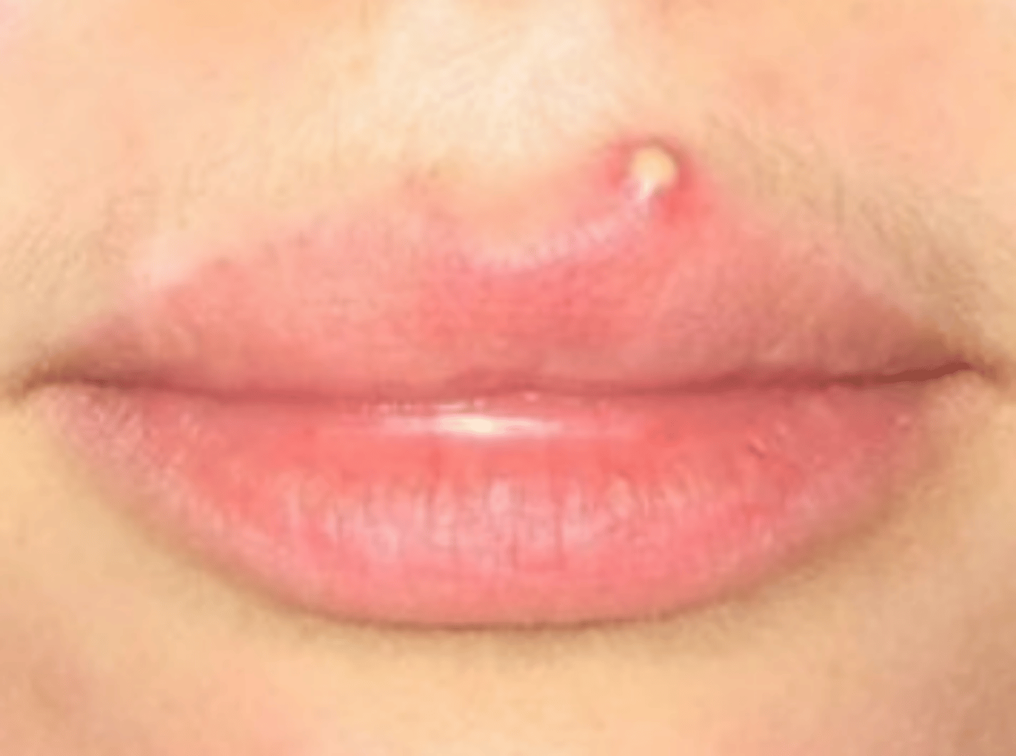
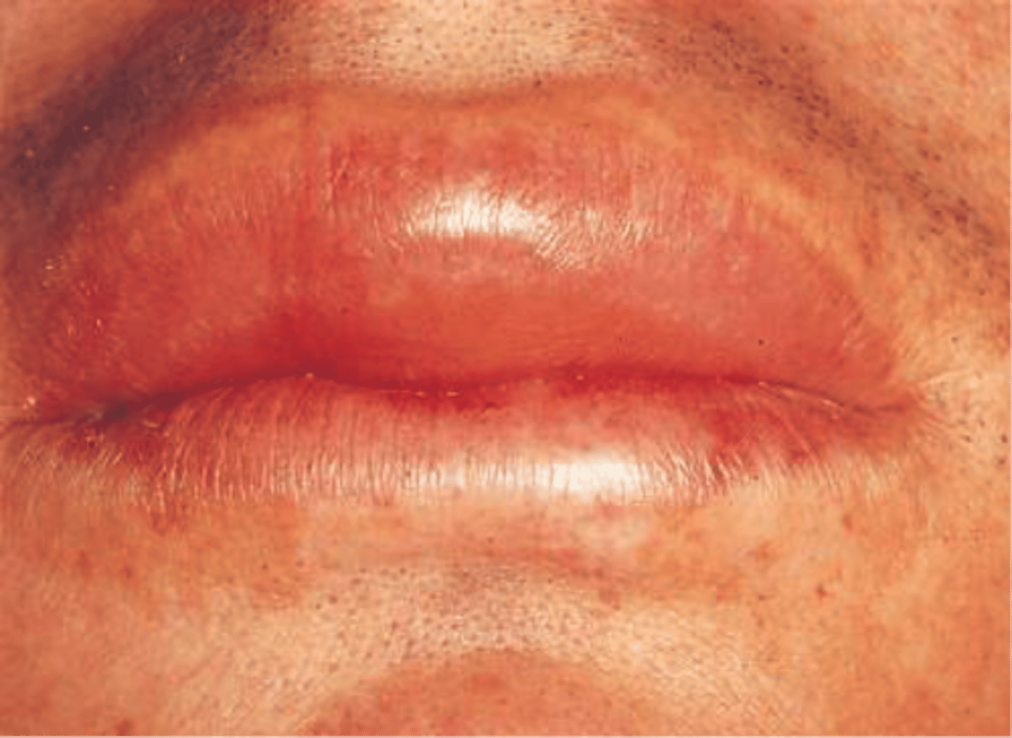
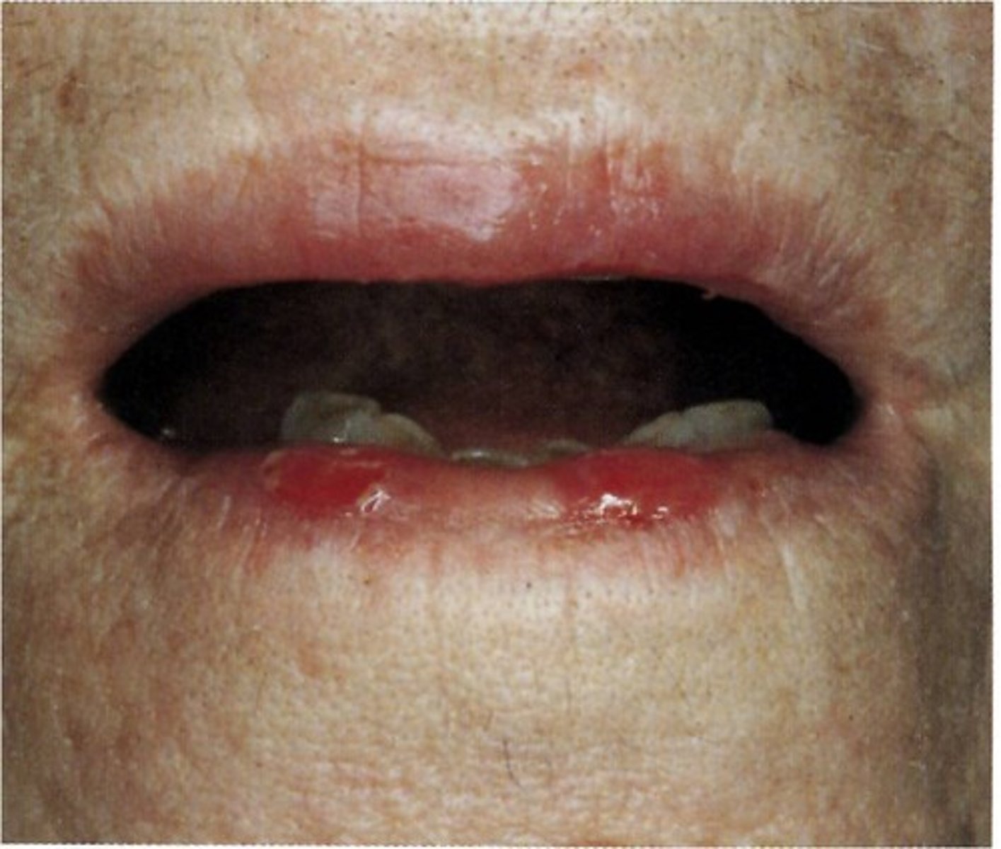
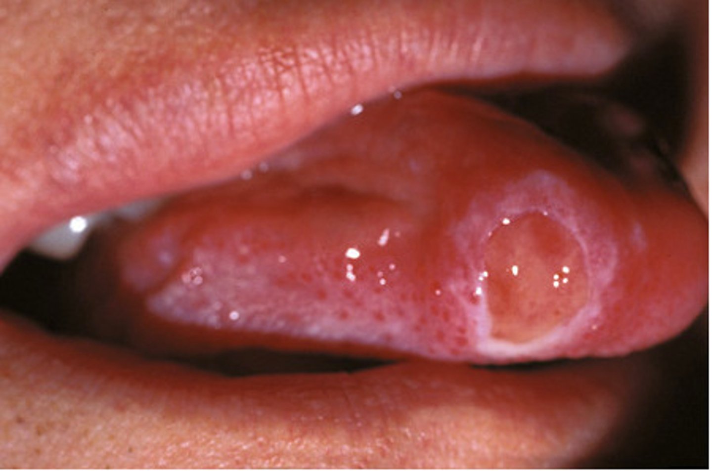
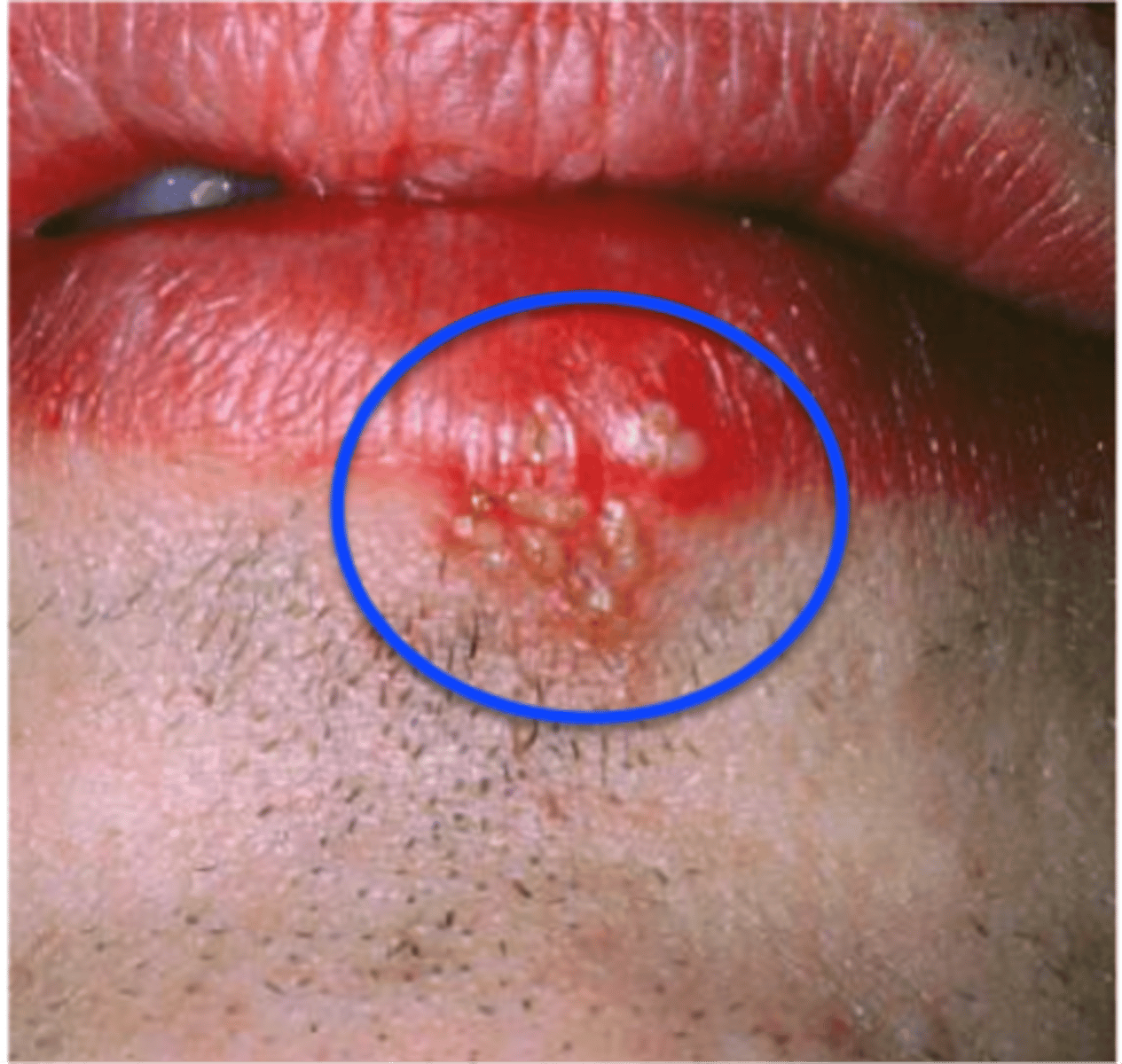
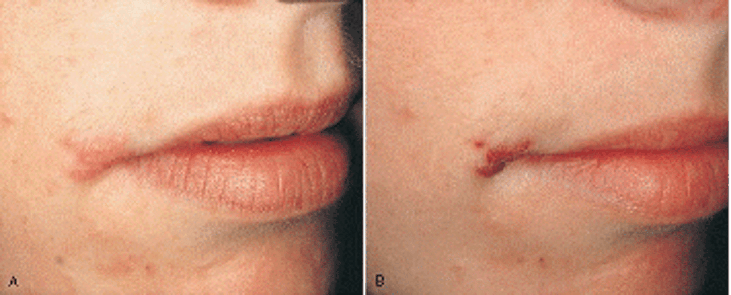
The most common form of recurrent herpes simplex infection is termed
Herpes Labialis
gingival lesions have been called desquamative gingivitis
cicatricial pemphigoid
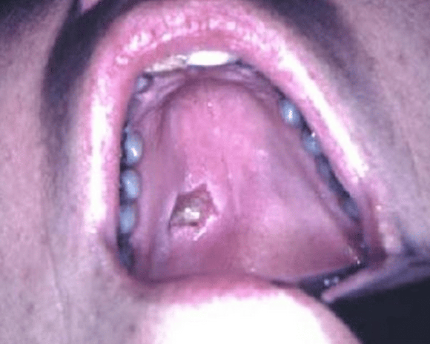
A postoperative complication following tooth removal in which the blood clot is lost before healing can take place, leaving raw, exposed nerve endings
Alveolar Osteitis ("Dry Socket")
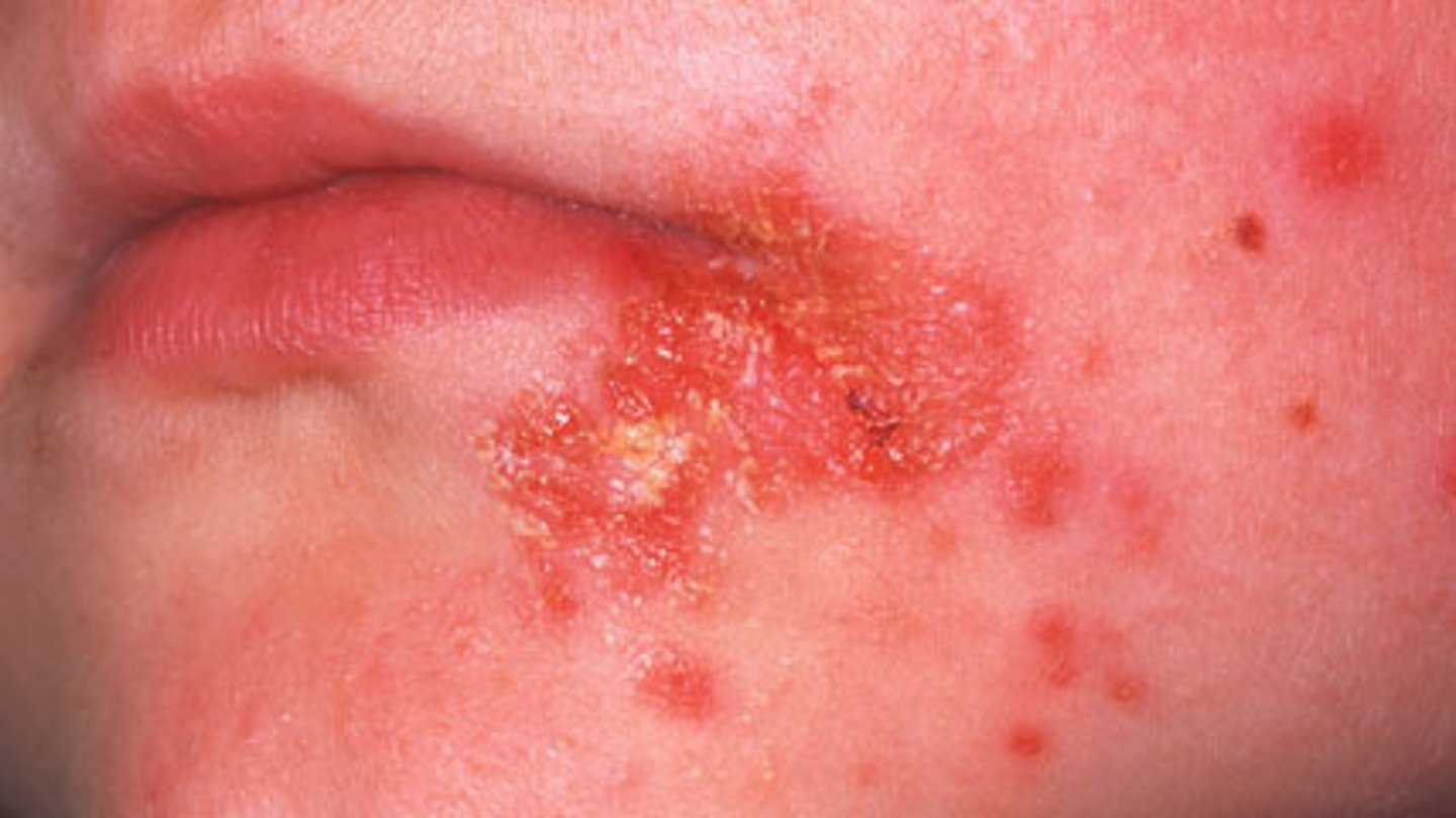
blister-like sores on the lips and adjacent facial tissue that are caused by the oral herpes simplex virus type 1 (HSV-1); also known as cold sores or fever blisters
Herpes Labialis
Type 2 herpes simplex
is most commonly associated with genital infections.
Herpes simplex virus
causes primary herpetic gingivostomatitis and recurrent herpes simplex virus infections.