Chapter 22: Heart
1/150
There's no tags or description
Looks like no tags are added yet.
Name | Mastery | Learn | Test | Matching | Spaced | Call with Kai |
|---|
No analytics yet
Send a link to your students to track their progress
151 Terms
Heart
________ connects to blood vessels that transport blood between this and body tissues
Arteries
send blood away from heart
Veins
Bring blood back to heart
Capillaries
serve as the sites of exchange (between blood and alveoli, or between blood and systemic cells)
Great vessels
The arteries and veins entering and leaving the heart are called
unidirectional
Heart valves ensure its blood flow is
lungs; body tissues
The heart is two side-by-side pumps that work at the same rate and pump the same volume of blood
One pump directs blood to the lungs
One pump directs blood to body tissues
blood pressure
The heart generates ______________ through alternate cycles of contraction and relaxation
This is the force of the blood pushing against the inside walls of blood vessels
It is essential to circulate blood throughout the body
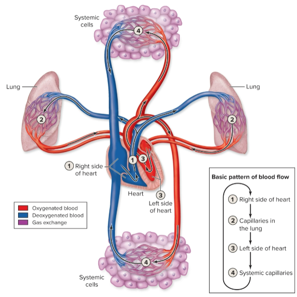
Pulmonary circulation
Starts with right side of heart pumping deoxygenated blood through pulmonary arteries to capillaries in lungs
After oxygen pickup and carbon dioxide release, pulmonary veins carry blood to left side of heart
Systemic circulation
Left side of the heart pumps oxygenated blood through systemic arteries to systemic capillaries
Nutrients, respiratory gases, and wastes are exchanged, and systemic veins carry the blood back to the right side of the heart
Cardiovascular system
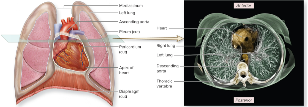
Mediastinum
Heart located slightly left of midline, deep to the sternum, in space called the
Right; Left
Heart is slightly rotated so _______ border is located more anteriorly while the _______ border is located more posteriorly
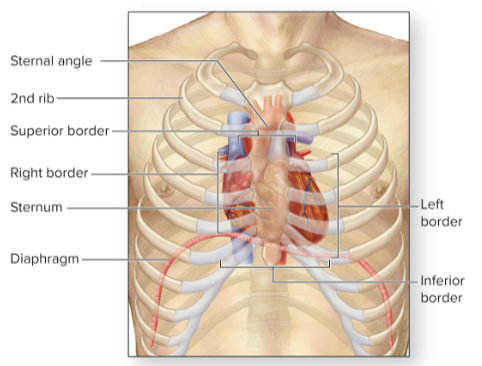
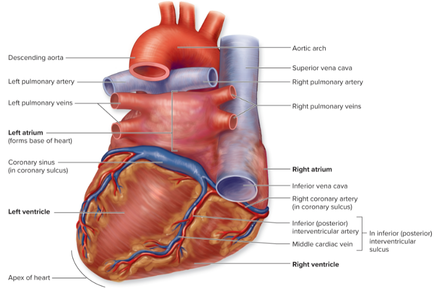
Base
_____ of heart: its posterosuperior surface—mainly left atrium
Superior border
___________ of the heart is formed by the great arterial vessels and the superior vena cava
Apex
inferior conical end of the heart
Inferior border
formed by the right ventricle
Borders of the heart
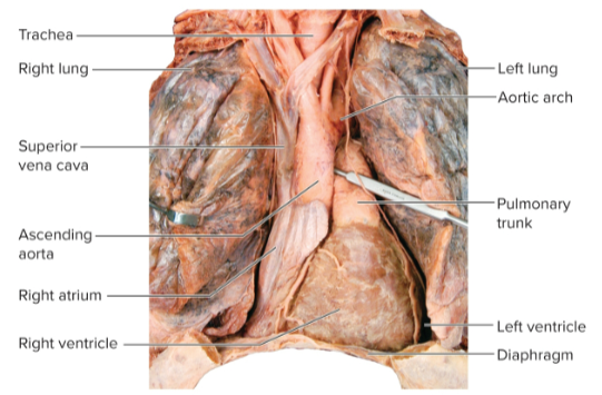
Heart and lungs, anterior view
Serous membranes of the heart + cross-sectional view in CT scan
pericardium
Heart is enclosed within a tough sac called the __________ (pericardial sac)
Restricts heart movements so that it moves only slightly within the thorax; prevents heart from overfilling with blood
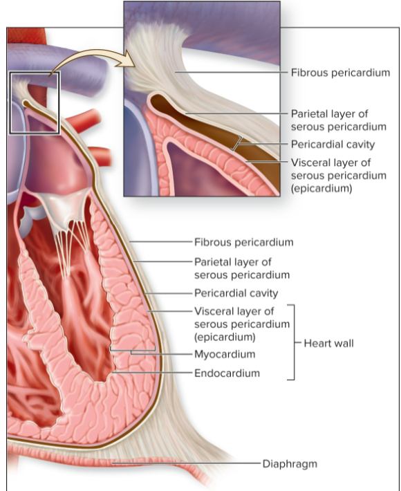
Fibrous pericardium
tough outer sac
Serous pericardium
composed of parietal and visceral layers
Pericardial cavity
thin potential space between layers of serous pericardium containing serous fluid
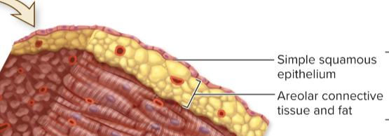
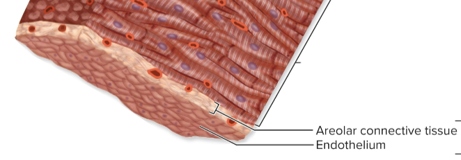
Epicardium
Myocardium
Endocardium
Three layers in heart wall (from superficial to deep):
Epicardium
visceral layer of serous pericardium and areolar connective tissue
Myocardium
cardiac muscle; thickest of the three layers
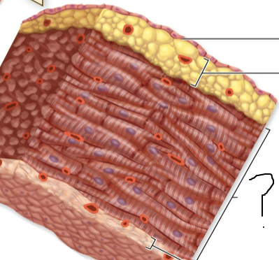
Endocardium
internal surface of heart chambers; simple squamous epithelium and areolar connective tissue
2 atria and 2 ventricles
Heart is composed of four chambers
Atria
Two superior, smaller chambers of the heart
Ventricles
two inferior, larger chambers of the heart
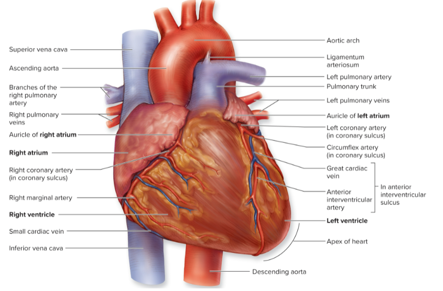
Auricle
Anterior part of each atrium forms a flaplike extension called an _______
Coronary sulcus
groove separating atria and ventricles
Anterior interventricular sulcus and inferior interventricular sulcus
Sulci located between the ventricles
External anatomy and features of the heart- Anterior
External anatomy and features of the heart- Posterior
Right atrium, right ventricle, left atrium, left ventricle
Name the 4 hollow chambers that make up the heart.
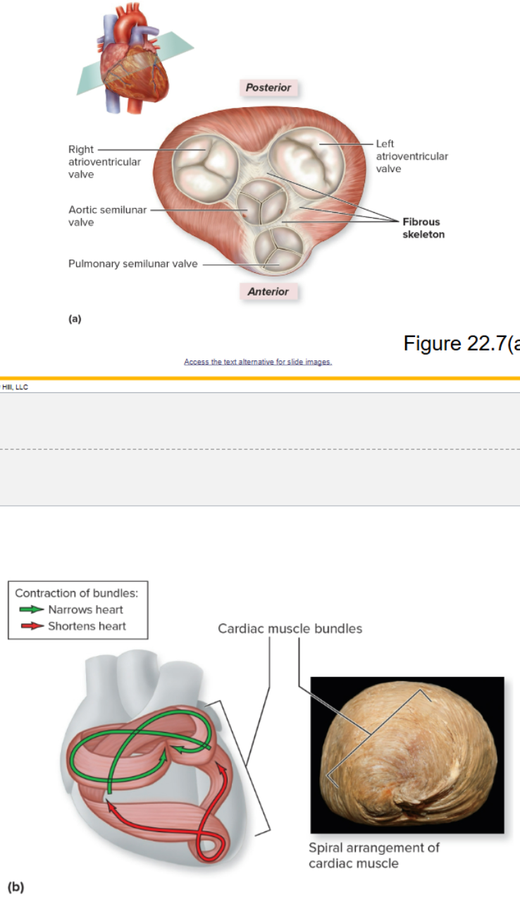
2 Atrioventricular (AV) Valves
Their closure causes the first heart sound “lubb”
semilunar valves
Two ___________ at base of great arteries
Their closure causes the second heart sound “dupp”
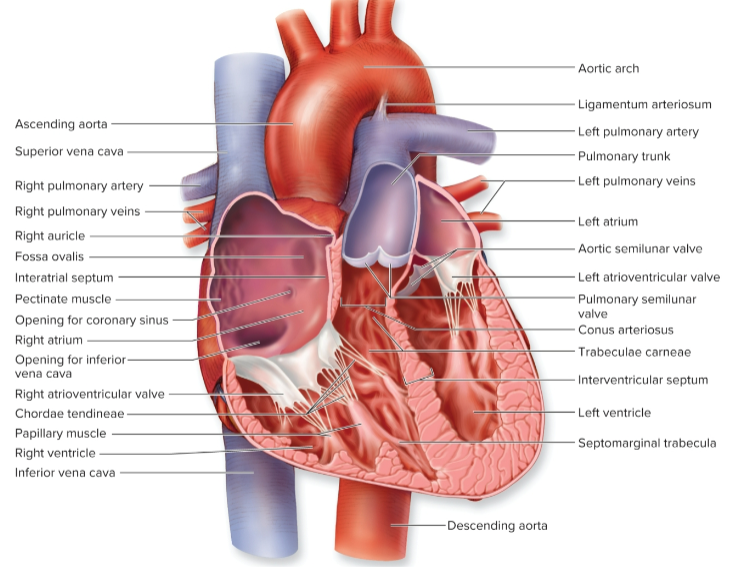
Internal anatomy of the heart
Fibrous skeleton of the heart
Formed from dense regular connective tissue between the atria and ventricles
Provides structural support at boundary between atria and ventricles
Forms supportive fibrous rings to anchor heart valves
Provides framework for attachment of cardiac muscle
Acts as an electrical insulator to prevent action potentials in atria from passing to ventricles, so that ventricles don’t contract at the same time as the atria
Right atrium
receives venous blood from heart and systemic circulation through the superior vena cava, inferior vena cava, and coronary sinus
superior vena cava, inferior vena cava, and coronary sinus
Right atrium receives venous blood from heart and systemic circulation through the __________________
Interatrial septum
divides right atrium from left; fossa ovalis present in interatrial septum
Fossa ovalis
present in interatrial septum
Pectinate muscles
ridges on internal surface of atrial wall
Right Atrioventricular (AV) Valve
ensures one-way blood flow from right atrium to right ventricle through right atrioventricular opening
Right ventricle
receives deoxygenated blood from right atrium
Interventricular septum
thick wall between right and left ventricles
trabeculae carneae
Inner wall of each ventricle displays irregular muscular ridges called ____________
Papillary muscles
cone-shaped muscle projections anchoring chordae tendineae
chordae tendineae
attach muscle to atrioventricular valve and prevent cusps from flipping into atrium when ventricle contracts
Septomarginal trabecula
connects anterior papillary muscle of right ventricle to atrioventricular septum
Conus arteriosus
smooth funnel-shaped region at superior end of right ventricle leading to pulmonary semilunar valve
Semilunar valve
ensures one-way flow from ventricle to pulmonary trunk
pulmonary arteries
Pulmonary trunk divides into right and left _______________
Left atrium
Oxygenated blood from the lungs travels through the pulmonary veins to the
Left atrioventricular valve
controls flow through opening between left atrium and ventricle
Also referred to as the bicuspid valve (two cusps) or mitral valve
Valve is forced shut when the left ventricle contracts
Left ventricle
pumps blood through entire systemic circulation, Generates very high pressure
Aortic semilunar valve
controls flow from left ventricle to aorta; located at superior end of left ventricle
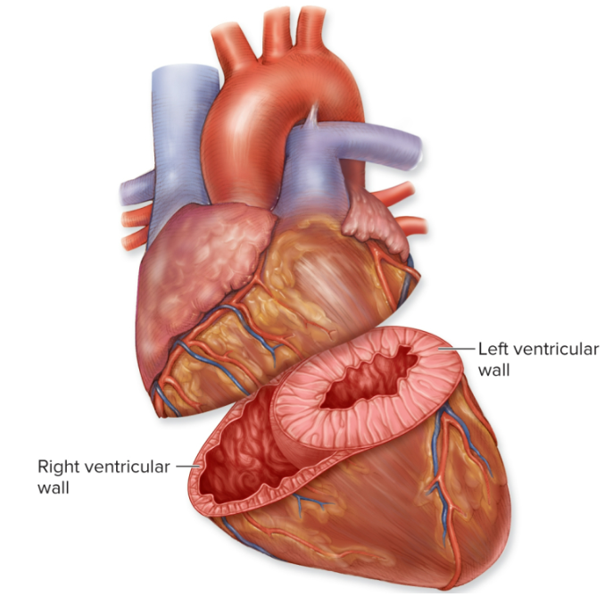
Wall of left ventricle
_______________________ is about three times thicker than right ventricle
Must generate enough pressure to force blood through entire systemic circulation
Right ventricle only needs to pump blood to the nearby lungs
Ventricular walls
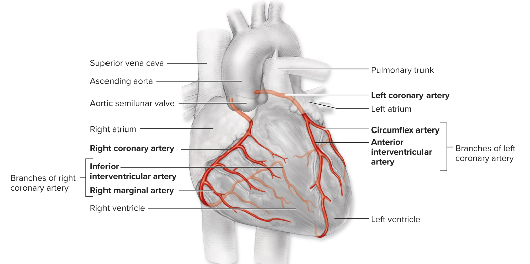
Right and left coronary artery
travel within coronary sulcus and supply heart wall with oxygen and nutrients and branch off ascending aorta just superior to aortic valve
Right marginal artery and inferior interventricular artery
Right coronary artery branches into 2 branches, name them
Right marginal artery
supplies right border of heart
Inferior interventricular artery
supplies posterior surfaces of the left and right ventricles
Anterior interventricular artery, Circumflex artery
Left coronary artery branches into 2, name them
Anterior interventricular artery
also called left anterior descending artery; supplies anterior surface of both ventricles and most of the interventricular septum
Circumflex artery
supplies the left atrium and ventricle
Coronary arteries
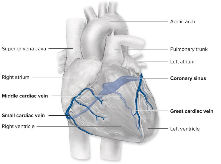
coronary sinus
The three major cardiac veins drain into the ____________, which drains into the right atrium
Great cardiac vein, middle cardiac vein, small cardiac vein
Name the 3 major cardiac veins
Great cardiac vein
runs alongside anterior interventricular artery
Middle cardiac vein
runs alongside inferior interventricular artery
Small cardiac vein
travels close to the right marginal artery
Coronary veins
conduction system
For the heart to be an effective pump, contraction of chambers needs to be coordinated
This is possible due to the heart’s ____________
capillary networks
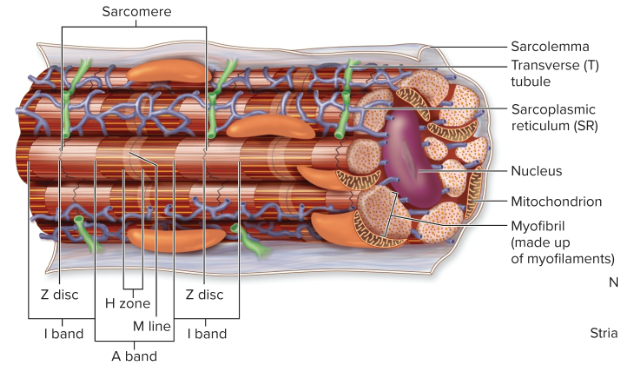
Cardiac muscle is striated with extensive ________________ (similar to skeletal muscle)
Characteristics of Cardiac Muscle Tissue
Less SR quantity and organization
No terminal cisternae
Less contact between SR and T-tubules
Organization and Histology of Cardiac Muscle
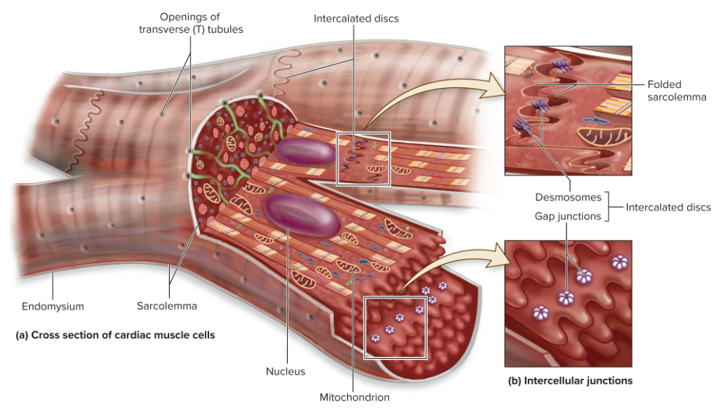
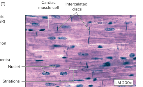
Longitudinal view of cardiac muscle cell
Longitudinal section of cardiac muscle
gap junctions
Cardiac muscle fibers contract as a single unit because they are all connected with _______________
intercalated discs
Gap junctions are parts of _______________ between adjacent fibers
electrical impulse
Each _______________ is distributed immediately and spontaneously throughout the myocardium
Desmosomes
_____________ within intercalated discs prevent cardiac muscle cells from pulling apart
autorhythmicity
Heart exhibits _______________—it initiates its own heartbeats
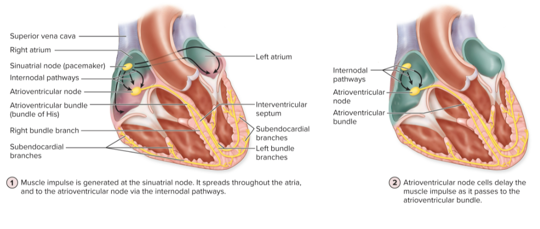
Conducting system
consists of specialized cells that start and propagate electrical impulses to contractile cells
Sinoatrial node, Atrioventricular node, Atrioventricular bundle, Left and right bundle branches, Subendocardial branches (Purkinje fibers)
Passage of signals through the conducting system:
sinoatrial (SA) node
Electrical impulse begins here (heart’s pacemaker)
located on posterior wall of right atrium adjacent to the opening of the superior vena cava
atrioventricular (AV) node
Impulse from SA node travels via gap junctions to left atrium and ________________ on the floor of right atrium
Impulse is paused at AV node delaying activation of ventricles as they fill with blood
atrioventricular (AV) bundle (bundle of His)
Impulse leaves AV node, enters the ________________, extending into interventricular septum
left and right bundle branches
Once within the septum, the AV bundle divides into ______________________
subendocardial branches (Purkinje fibers)
The left and right bundle branches pass the impulse to ___________________ that begin at the heart apex
spread the impulse superiorly from the apex to all of the ventricular myocardium
Conducting system of the heart
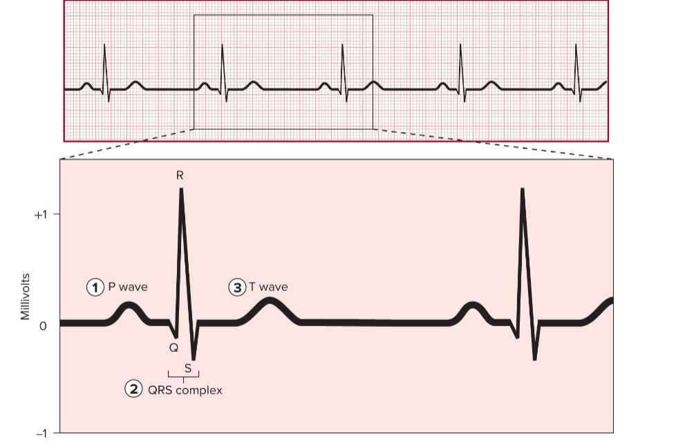
Electrocardiogram (ECG)
a composite tracing of all electrical impulses in heart; can be used diagnostically
P wave
atrial depolarization
QRS Wave
ventricular depolarization (and atrial repolarization)
T wave
ventricular repolarization
Electrocardiogram example