Male reproductive biology, spermatogenesis, and endocrinology
1/97
There's no tags or description
Looks like no tags are added yet.
Name | Mastery | Learn | Test | Matching | Spaced | Call with Kai |
|---|
No analytics yet
Send a link to your students to track their progress
98 Terms
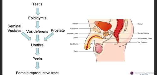
Journey of sperm
Seminiferous tubules > lumen > rete testis > ductuli efferentes > epididymis > vas deferens . penis

Why sperm leaving testis cannot fertilise
They lack motility and fertilisation capacity until epididymal maturation
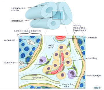
Two compartments of testis and functions
Seminiferous tubules perform spermatogenesis
Interstitium performs steroidogenesis
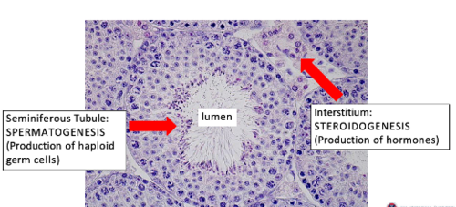
Structure function of seminiferous tubules
Highly organised epithelium supports germ cell development from basal to lumen
Components of interstitium
Leydig cells - produce testosterone
Capillaries - carry released testosterone to circulation
Fibroblasts: produce ECM of interstitium and testis
Macrophages: activated in inflammation
Lymphatics: lymph drainage
Peritubular myoid cells: surround seminiferous tubules and are contractile
Mechanism of testosterone synthesis
LH binds Leydig receptors: stimulates conversion of cholesterol into testosterone and testosterone > 5-alphaDHT by 5-alpha reductase
Why DHT is important
DHT has higher androgen receptor affinity producing stronger effects
Experimental evidence for Leydig function
3beta HSD staining localises steroidogenesis to Leydig cells
EDS destroys Leydig cells leading to reduced testosterone and degeneration of spermatogenesis - Stem Leydig cells repopulate interstitium after EDS treatment
Structure of peritubular myoid cells
Smooth muscle like cells surrounding seminiferous tubules
Contract to move sperm toward the rete testis
Structure of epididymis
Highly coiled tubule with tall columnar epithelium and stereocilia
Highly coiled to provide long tubule length for sperm maturation within the space
Concentrates sperm and induces motility and fertilisation competence
Acidic to maintain sperm in inactive state for maturation
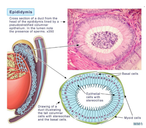
Structure function of stereocilia
Increase surface area for absorption of fluid concentrating sperm
C ros knockout experiment
Loss of C ros causes alkaline epididymal pH tail defects and infertility
the acidic environment is essential for sperm maturation
Effect of androgen receptor knockout in epididymis
Epithelial involution loss of stereocilia and impaired function
androgens are therefore essential for epididymal maintenance
Changes to sperm in epididymis
Membrane protein lipid modification and acquisition of motility
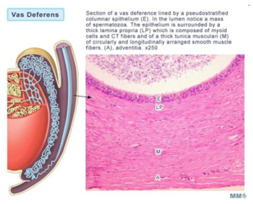
Structure of vas deferens
Low cuboidal epithelium and thick smooth muscle tube which contracts to move sperm along narrow lumen and strong peristaltic contractions propel them during ejaculation
Stores sperm
Vasectomy mechanism
Vas deferens is cut preventing sperm entry into semen sperm are phagocytosed
Contraline ADAM mechanism
Injectable hydrogel forms physical barrier in vas deferens and can be reversed
undergoing clinical trials as a male contraceptive
Composition of semen
Sperm: 2-5%
seminal vesicles secretions: 60-75 %
prostate secretions: 25-30 %
Function of seminal vesicles
Provide fructose for sperm energy and semenogelin proteins for coagulation after ejaculation
Structure function of seminal vesicles
Secretory glands adapted to produce large volume nutrient rich fluid
dependent on androgens - as castration reduces size, while testosterone restores structure
Prostate gland function
Produces alkaline fluid with zinc and PSA
prostatic fluid is alkaline to neutralise the female reproductive tract to improve sperm survival
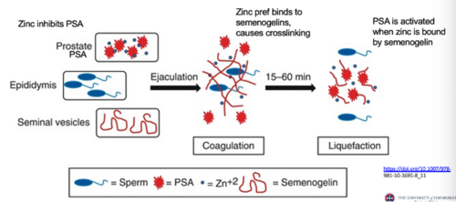
Function of PSA
Protease that cleaves semenogelin to liquefy semen
used as a diagnostic tool as PSA is elevated in prostate disease like BPH and cancer
Zinc mechanism in semen
Initially inhibits PSA allowing coagulation then binds semenogelin freeing PSA for liquefaction
present in semen
Importance of coagulation then liquefaction
Coagulation retains semen then liquefaction allows sperm motility
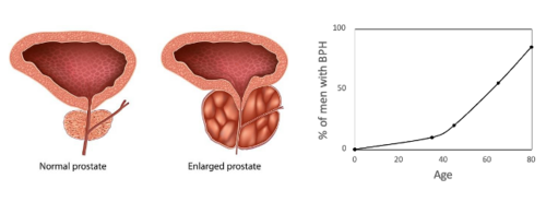
BPH mechanism
Enlargement of prostate gland due to cell proliferation
Age related
non-cancerous
Structure of penis
Contains corpus cavernosum with vascular spaces and smooth muscle
delivers sperm to reproductive path
Mechanism of erection
Anatomical: blood entering the corpus cavernosum due to smooth muscle relaxation
Sexual stimulation activates the cavernous nerve > releases Nitric oxide > stimulates guanylate cyclase to convert GT > cGMP > protein kinase decreases intracellular calcium causing smooth muscle relaxation and blood filling to the corpus cavernosum
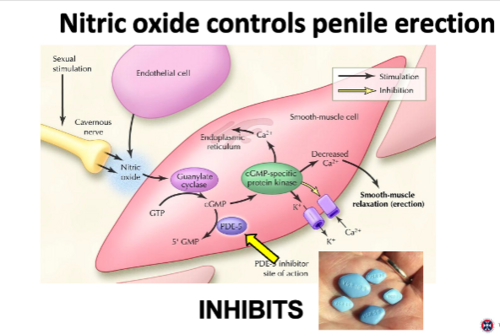
Viagra mechanism
Inhibits PDE5, which prevents cGMP breakdown prolonging erection
Semen coagulation and liquefaction
Zinc in prostatic fluid inhibits PSA, preventing cleavage of semenogelin - this allows semenogelin to cross link and causes semen coagulation
Later, zinc binds to semenogelin instead, freeing PSA to cleabe semenogelin for liquefaction
Overall male reproduction
The seminiferous tubules produce haploid germ cells, and the interstiitum produces tesosterone for male reproduction. PTM cells move sperm out of testes by peristalsis. In the epididymis, sperm are concentrated and undergo maturation for motility and fertilisation. The vas deferens stores and transports sperm. while prostate and seminal vesicles add most of seminal fluid for alkalinity energy and coagulation of sperm for reproduction. the penis then delivers sperm to the female.
Purpose of spermatogenesis
Produce haploid and genetically diverse, motile sperm
occurs in the seminiferous epithelium from basal surface to luminal surface
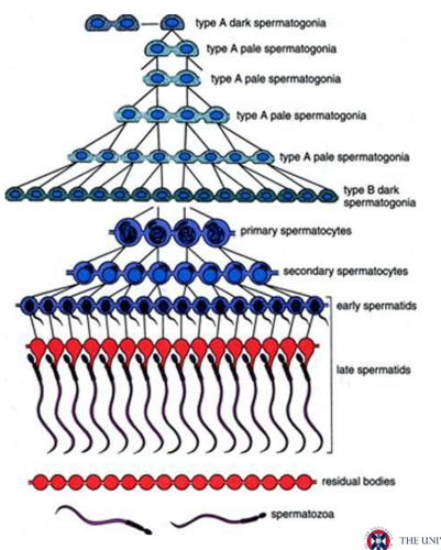
Cell sequence
Spermatogonia > spermatocytes > spermatids to>spermatozoa

Phase 1 proliferation
Spermatogonia undergo mitosis at basement membrane
PCNA staining in mice marks these cells here
Stem cell niche definition
Local environment providing signals regulating self renewal and differentiation
Key factors are GDNF, FGF2, CXCL12
Key niche factors
GDNF FGF2 CXCL12
GDNF mechanism
Binds receptors on spermatogonia promoting self renewal
KO - depletion of spermatagonial stem cells and infertility
Role of retinoic acid
Triggers differentiation and entry into meiosis
occurs at puberty in males - initiates spermatogenesis
Reduced RA leads to impaired spermatogonial differentiation
thus RA is essential for differentiation
RA mechanism
genomioc RA receptor: acts as transcription factor inducing Stra8 expression
Non-genomia RA receptor: prmotes translation of KIT from existing mRNA
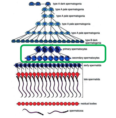
Phase 2: meiosis
Reduces chromosome number and generates genetic diversity
produces haploid cells from diploids
Germ cells > spermatocytes
what is diploid vs haploid
diploid: 2 homologous chromosomes in each homologous pair
Meiosis I mechanism
Homologous chromosome pairs (mother and father) separate, crossing over to exchange genetic information, and independent assortment occur
chromosome number is reduced from diploid (2n) to haploid (n)
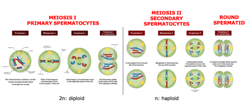
Meiosis II mechanism
Sister chromatids separate and convert secondary spermatocytes to round spermatids - maintaining haploid chromosome
Cell progression through meiosis
Primary spermatocyte > secondary spermatocyte > spermatid
Blood testis barrier structure
Formed by Sertoli cell tight junctions that separate the basal from the apical side of the seminiferous epithelium
protects haploid germ cells from the immune system
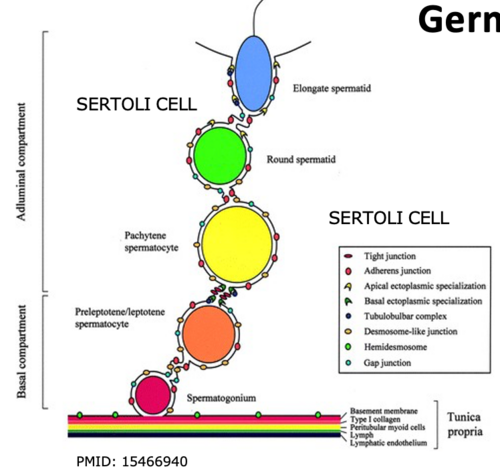
Why barrier must open
to allow germ cells to move across the epithelium and spermatocytes to move to adluminal compartment
the tight junctions dynamically reorganise during germ cell development
Role of androgens in meiosis
Required for progression beyond meiosis into spermiogenesis
as loss of testosterone prevents formation of post meiotic cells
Phase 3: spermiogenesis
Transformation of spermatids into spermatozoa
produces structural changes needed to ensure sperm is fast, motile, and able to fertilise
phases: golgi phase > cap > acrosomal > maturation phase
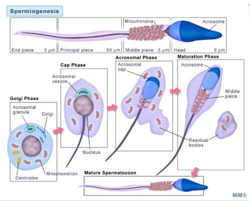
Golgi phase
Round cell becomes polar - the golgi moves to the head end and centrioles move to the tail end
Cap phase
Acrosome vesicle forms a cap over nucleus and primitive flagellum develops
Acrosomal phase
Acrosome spreads around he nucleus as nucleus elongates
Maturation phase
Mitochondria arrange in midpiece and excess cytoplasm removed - the cell becomes a mature spermatazoon
DNA packaging mechanism
Histones replaced by specialised histones then by transition proteins then protamines for greater chromatin condensation
creates compact hydrodynamic sperm head
Transcription in spermiogenesis
Stops in early spermatid nucleus so proteins must be pre synthesised
Loss of BrUTP incorporation shows transcription stops in early spermatids
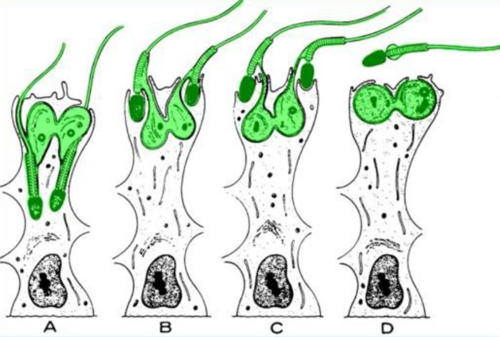
Phase 4: spermiation
Release of sperm from Sertoli cells into seminiferous lumen
Sertoli germ cell junctions remodel allowing release
the residual body is phagocytosed by sertoli cells
Organisation of spermatogenesis
Occurs in waves along seminiferous tubules
controlled by Pulses of retinoic acid coordinate timing
Human arrangement
Helical distribution of stages
Human spermatogenic cycle
Approximately 16 days per cycle
so total duration of spermatogenesis is 64 days
Integrated function of system
Testis produces sperm, epididymis matures, vas deferens transports, glands support, penis delivers
What is the male HPG axis?
GnRH > LH and FSH > act on testes (Leydig + Sertoli cells) to regulate testosterone production and spermatogenesis.
What is the mechanism of GnRH action?
GnRH is released in a pulsatile manner → binds GnRH receptors on pituitary gonadotrophs → stimulates LH and FSH synthesis and secretion.
What are LH and FSH?
Gonadotrophins from anterior pituitary: LH acts on Leydig cells (testosterone synthesis); FSH acts on Sertoli cells (spermatogenesis support).
Nehative feedback: testosterone inhibits GnRH and LH secretion,
inhibin B from sertoli cells selectively inhibits FSH secretion
How does negative feedback regulate the HPG axis?
Testosterone inhibits GnRH and LH secretion; Inhibin B from Sertoli cells selectively inhibits FSH secretion at pituitary level.
What experimental evidence shows testosterone negative feedback?
Castration studies: removal of testes → ↑ LH and FSH → demonstrates loss of testosterone-mediated inhibition.
Which neurons mediate steroid negative feedback?
Kisspeptin neurons in hypothalamus (especially arcuate nucleus) regulate GnRH pulse frequency.
Kisspeptin binds GPR54 receptors on GnRH neurons → modulates (reduces) GnRH pulse frequency → alters LH/FSH output.
What evidence supports role of kisspeptin neurons?
They express androgen/oestrogen receptors and mediate feedback rather than direct action on GnRH neurons.
What does LH do in the testis?
LH binds LHCGR on Leydig cells → activates cAMP pathway → stimulates testosterone synthesis.
LHCGR expression is restricted to Leydig cells (receptor localisation studies).
What happens if LH is absent?
Normal male external genitalia but delayed puberty and low testosterone → shows role in postnatal development.
What happens in LHCGR mutations?
Female external phenotype
Why can fetal development occur without LH?
hCG from placenta can activate LHCGR → substitutes for LH during fetal life.
Describe LH signalling mechanism in Leydig cells.
LH → LHCGR (GPCR) which activates adenyl cyclase to convert ATP → cAMP → PKA activation →
pKA activates StAR activation and transcription of steroidogenic enzymes which = cholesterol → pregnenolone → testosterone.
What is the role of StAR?
Transfers cholesterol from outer to inner mitochondrial membrane → rate-limiting step of steroidogenesis.
What experimental evidence supports steroidogenesis pathway?
Activation of enzymes like 3β-HSD observed in Leydig cells during testosterone synthesis.
What are Δ5 vs Δ4 pathways?
Δ5 pathway predominant in humans; Δ4 pathway predominant in rodents for testosterone synthesis.
Why is testosterone converted to DHT?
Peripheral tissues have lower testosterone → conversion to more potent androgen DHT ensures effective masculinisation.
Which enzyme converts testosterone to DHT?
5α-reductase found in skin
What is the function of DHT?
Drives external genital masculinisation and secondary sex characteristics (e.g. pubic hair, aggressiveness etc)
What is the androgen receptor mechanism?
Ligand (testosterone/DHT) binds → receptor conformational change → acts as transcription factor → regulates gene expression.
What are structural domains of androgen receptor?
DNA-binding domain + ligand-binding domain.
What is CAIS?
Complete androgen insensitivity syndrome: AR mutation → XY genotype but female phenotype.
Why are Wolffian structures absent in CAIS?
Testosterone present but no functional receptor → no maintenance of epididymis
Do germ cells express androgen receptor?
No → androgen effects on spermatogenesis are indirect.
Androgens act via Sertoli and other somatic cells
Mechanism of PTM androgen action?
Stimulates GDNF production → supports spermatogonial mitosis.
What happens in LCARKO mice?
Leydig cells undergo apoptosis in adulthood but fertility maintained.
KO mice in male androgen receptors
ARKO mice: no spermatogenesis beyond spermatogonia and failed Wolffian development. - supports androgen receptor
Sertoli-cell AR knockout → meiosis arrest + no haploid sperm + blood-testis barrier disruption - supports that androgen signalling in sertoli cells is essential for meiosis and barrier integrity
PTM-ARKO mice: reduced sperm production at all stages
LCARKO mice: leydig cells undergo apoptosis in adulthood, but fertility maintained - indicates that androgens are important for leydig cell survival but no for fertility
What are the phases of testosterone production?
Embryonic surge > neonatal surge > puberty surge > post-pubertal surge
Role of embryonic testosterone surge?
Development of Wolffian ducts + testicular descent.
What is neonatal testosterone surge?
Transient rise after birth; function unclear but may influence brain development.
What happens post-puberty?
Continuous testosterone production → spermatogenesis + secondary sexual characteristics.
Why is intratesticular testosterone important?
Levels are much higher than circulating → required for spermatogenesis.
What happens with exogenous testosterone (contraception)?
Suppresses LH → reduces intratesticular testosterone → inhibits spermatogenesis despite normal systemic androgen effects.
What does FSH do?
Stimulates Sertoli cells → supports spermatogenesis and Sertoli function.
peptide protein, so must use membrane receptors - stimulates mitosis in sertoli cells
What is ABP?
Androgen-binding protein → concentrates testosterone in seminiferous tubules.
What is inhibin B?
Hormone from Sertoli cells that selectively inhibits FSH secretion.
Heterodimer: alpha subunti (sertoli) + Beta subunit (germ cells)
What evidence suggests alternative FSH signalling?
FSH receptor KO mice still produce some sperm
What is AMH?
Anti-Müllerian hormone from Sertoli cells during embryogenesis.
regresses mullerian duct in females, keeps wollfian in males
What is 5α-reductase type II deficiency?
Mutation in Srd5a2 → impaired DHT production.
XY individuals appear female at birth bc of lack of external masculinisation
at puberty: partial masculinisation due to type I enzyme compensation
How does inhibin B regulate spermatogenesis?
Normal spermatogenesis → ↑ inhibin B → ↓ FSH → maintains homeostasis.
Overall integration of HPG axis?
GnRH → LH/FSH → Leydig (testosterone) + Sertoli (spermatogenesis + inhibin B); testosterone and inhibin B provide negative feedback to maintain balance.