Nano-Optics
1/53
There's no tags or description
Looks like no tags are added yet.
Name | Mastery | Learn | Test | Matching | Spaced | Call with Kai |
|---|
No analytics yet
Send a link to your students to track their progress
54 Terms
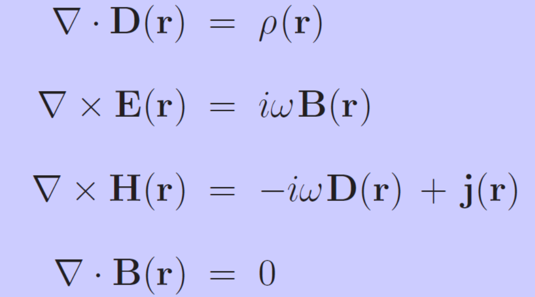
What are the Maxwell equations for complex fields?
Gauss law
Faraday’s law
Ampères law
Gauss law for magnetism
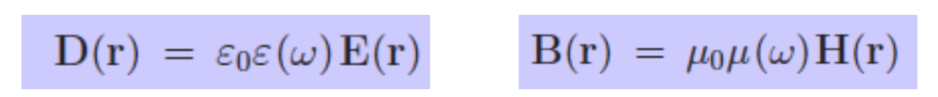
What are the constitutive equations to the Maxwell equations?
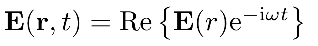
How is the monochromatic field defined?
With E(r) being the phasor.
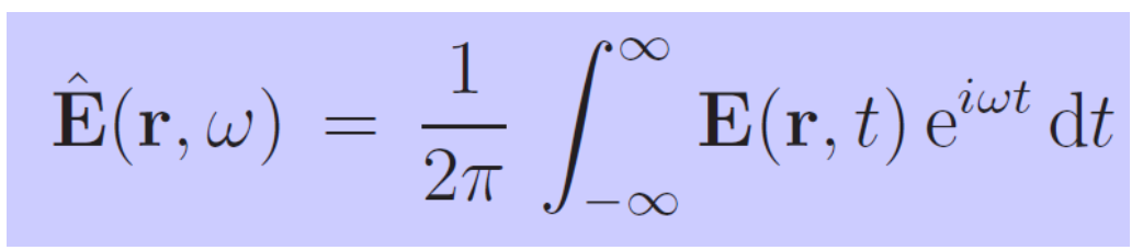
What is the spectral (Fourier transform) of the complex E-field?
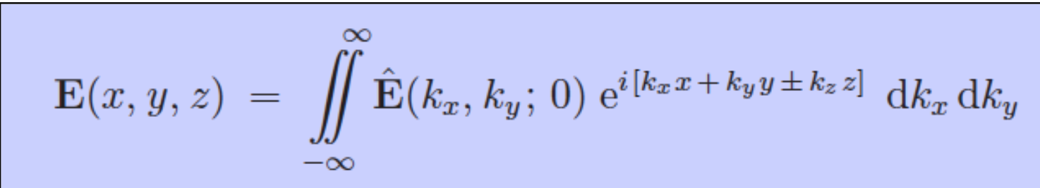
Explain what the angular spectrum is. What does the Helmholtz equation say?
The idea of the angular spectrum is:
If I know the E field at a location e.g z=0. I now it everywhere else. It decomposes the field into plane waves at z=0 that all have different directions (kx and ky). The Fourier coefficient Ê is saying how much of each direction is present.
Monochromatic waves need to satisfy the Helmholtz equation. This will yield the result Ê = Ê(kx, ky;0)e+-i*k_z*z.
Together they form the final result.
Remember that kz can be evanescent! That’s why we can’t image arbitrarily fine structures from far away!
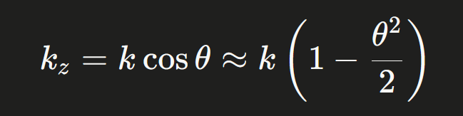
How is the paraxial approximation used? When is it valid'?
If fields propagate mainly in z-direction or they only deviate with a small angle from z.
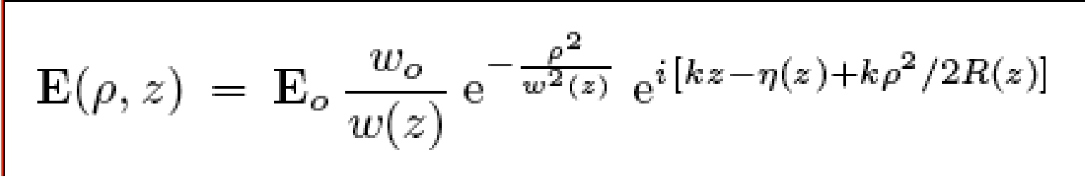
This is the formula for the Gaussian beam:
Describe the following variables:
w(z)
R(z)
n(z)
z_0
theta
w(z) = w0 * (1+z²/z0²)1/2 → Is the beam waist
R(z) = z * (1+z0²/z²) → Wavefront radius
n(z) = arctan(z/z0) → Gouy phase (Phase correction)
z0 = kw0/2 → Rayleigh length
At the rayleigh length the beam radius increases by the factor sqrt(2)thata = 2/(k*w0) → total angular spread
Questions about the Gaussian beam:
Which paramter of the Gaussian beam is free?
When is it a good approximation?
Does it contain evanescent components?
w0, the beam waist. It’s dependent on the optical system.
When it’s paraxial.
Under the paraxial condition it shouldn’t.
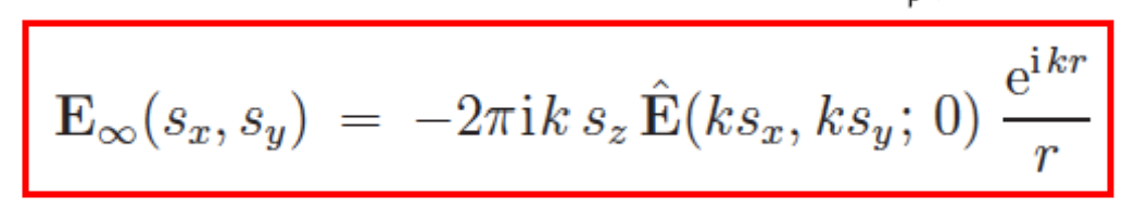
What is the angular spectrum very far from the source? Hence in the far-field?
In the far field, each direction s corresponds to exactly one plane wave component from the angular spectrum.
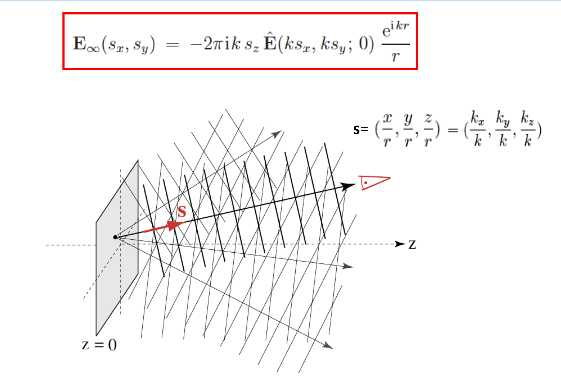
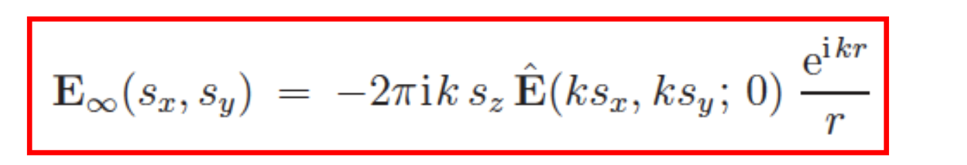
The formula for the angular spectrum is as follows:
What does it contain?
Ê(ks_x,ks_y,0) = Angular spectrum of the source field, evaluated at the spatial frequencies selected by direction s
e^(ikr) / r = Spherical wave: oscillates at rate k, amplitude decays as 1/r (energy conservation)
s_z = k_z/k = cos(theta) = Obliquity/projection factor — suppresses plane waves traveling nearly parallel to the source plane (grazing angles)
−2πik = Prefactor from the stationary phase evaluation of the integral
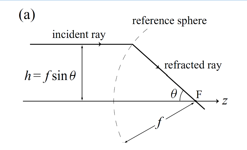
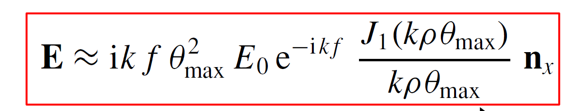
How can we treat strongly focused fields?
Use far field approximation in “reverse“
The angular spectrum in the focal region is (proportional to) the real-
space field distribution far away (i.e., on the reference sphere)
So, the crucial step is finding the real field on the reference sphere!
Since every point on the reference sphere is the far field of the focus we can directly apply our formula:
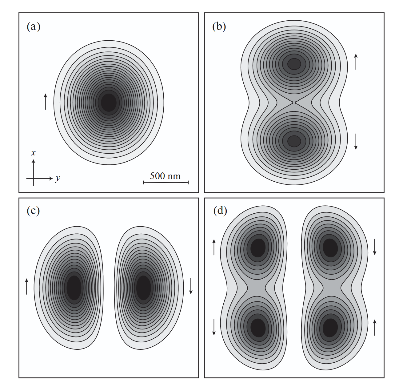
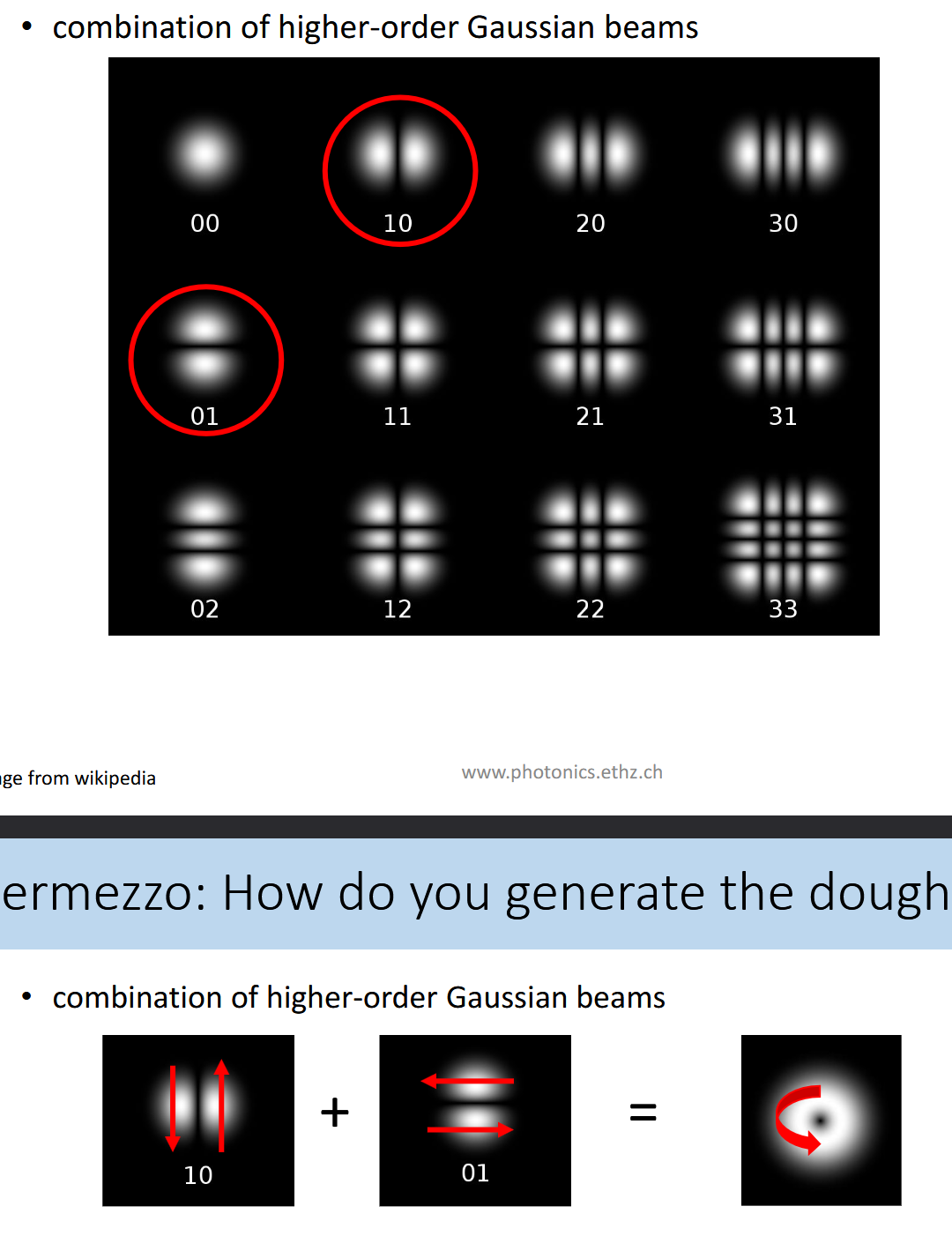
How do the first four Hermite-Gaussian modes look like?
a) (00) mode (Gaussian
mode), (b) (10) mode, (c) (01) mode, and (d) (11) mode.
How can weakly focused beams be treated?
Assume:
Strongly overfilled back-aperture
Assume small NA
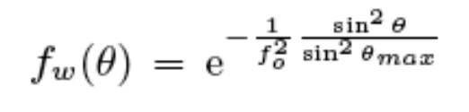
Explain under and overfilling of the aperture.
It can be explained via the apodization function:
f_0 yields information about how filled the aperture is. Is it small → Gaussian beam is narrow.
Is it underfilled → beam is smaller than the aperture, is it overfilled → the beam is larger than the aperture
What is the implicatons if you have a over or an underfilled aperture?
Underfilled:
Not the full NA of the lens is used. Only shallow angles theta are contributing to the Debye integral. The high-theta angles that create the tightest focus are missing.
Overfilled:
The aperture clips the beam BUT all angles up to theta_max are used. Full NA is used.
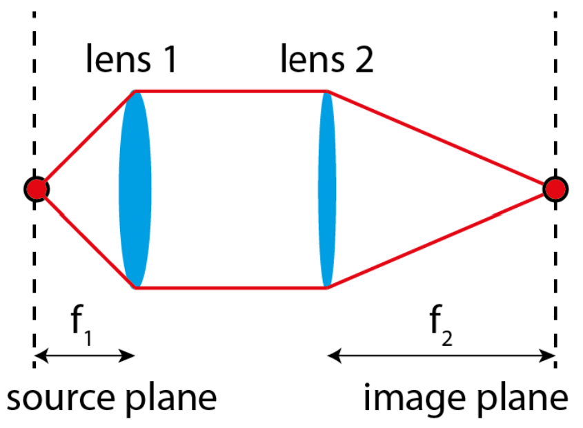
Which element determines resolution of this imaging system?
What is the magnification of this imaging system?
Resolution:
NA = n*sin(theta_max) = n*sin(arctan(D/2*f_1)
Magnification:
M = -f_2/f_1
f2 > f1 → magnification
f2 < f1 → demagnification
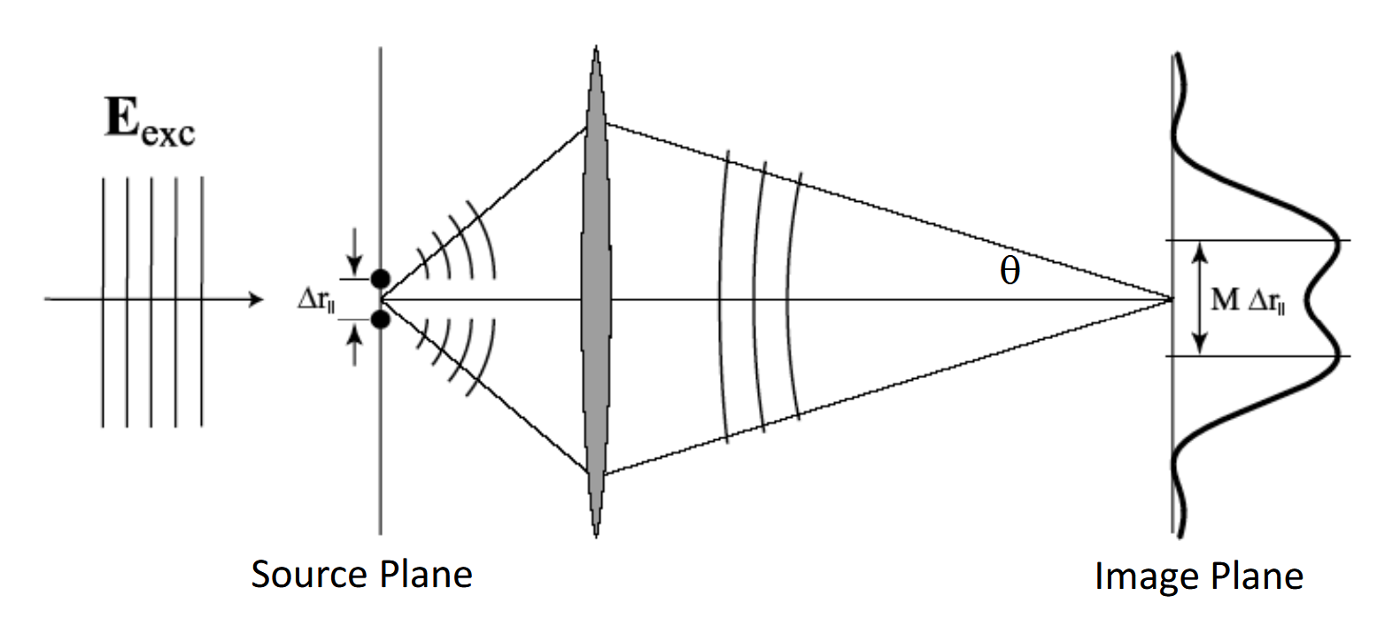
What is the PSF?
The Point-Spread-Function is the image of a (mathematical) point source.
What is the classical resolution limit?
It is stated by Abbe (1873!):
min(delta_R) = 0.6098* lambda/NA
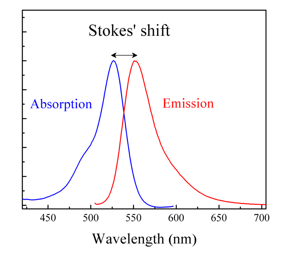
What is the role of Stokes shift in fluorensence microscopy?
Stokes shift of fluorescence allows to spectrally separate (intense) pump
light from (weak) fluorescence
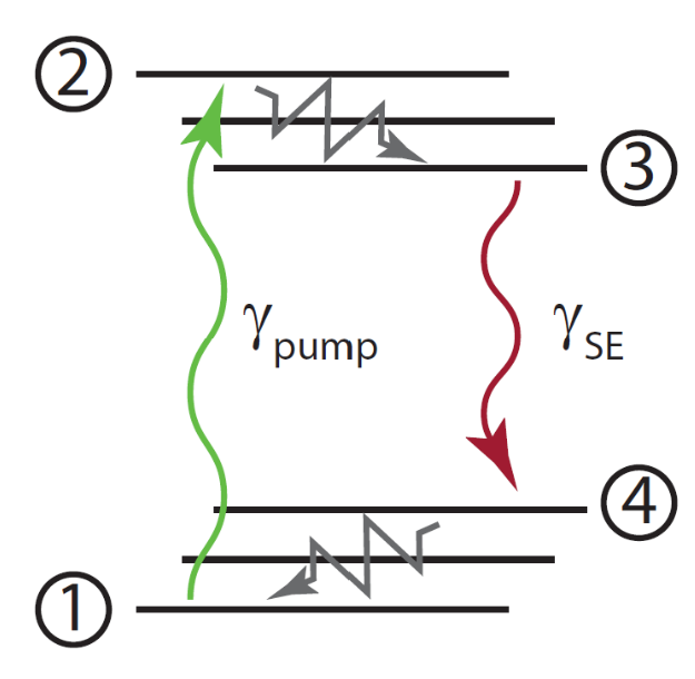
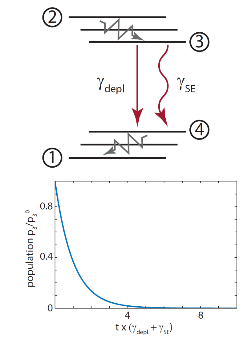
Explain this diagram:
It is a 4-level system used to describe fluorescent molecules.
1-2: Absorption of pump photon, the molecule is excited from the ground state to a high level of the excited state.
2-3: Fast vibrational relaxation, settling into the lowest vibrational level of the excited state. (100fs)
3-4: Stimulated emission (or spontaneous fluorescence),the molecule emits a photon and drops to a vibrational level of the ground state
4-1: Fast vibrational relaxation back to the true ground state
emitted photon is always lower energy (longer wavelength) than the absorbed photon.
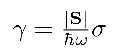
How is the interaction rate between a fluorophore in a light field defined?
|S|: Poynting vector magnitude — the intensity of the light field (energy per area per time)
hw: Energy per photon
|S|/hw: Photon flux — number of photons arriving per area per time
sigma: Absorption cross section — effective area the molecule presents to the incoming photons
What is a fluorophore?
A fluorophore is simply a molecule (or part of a molecule) that can absorb a photon and re-emit a photon at a longer wavelength — i.e. it fluoresces.
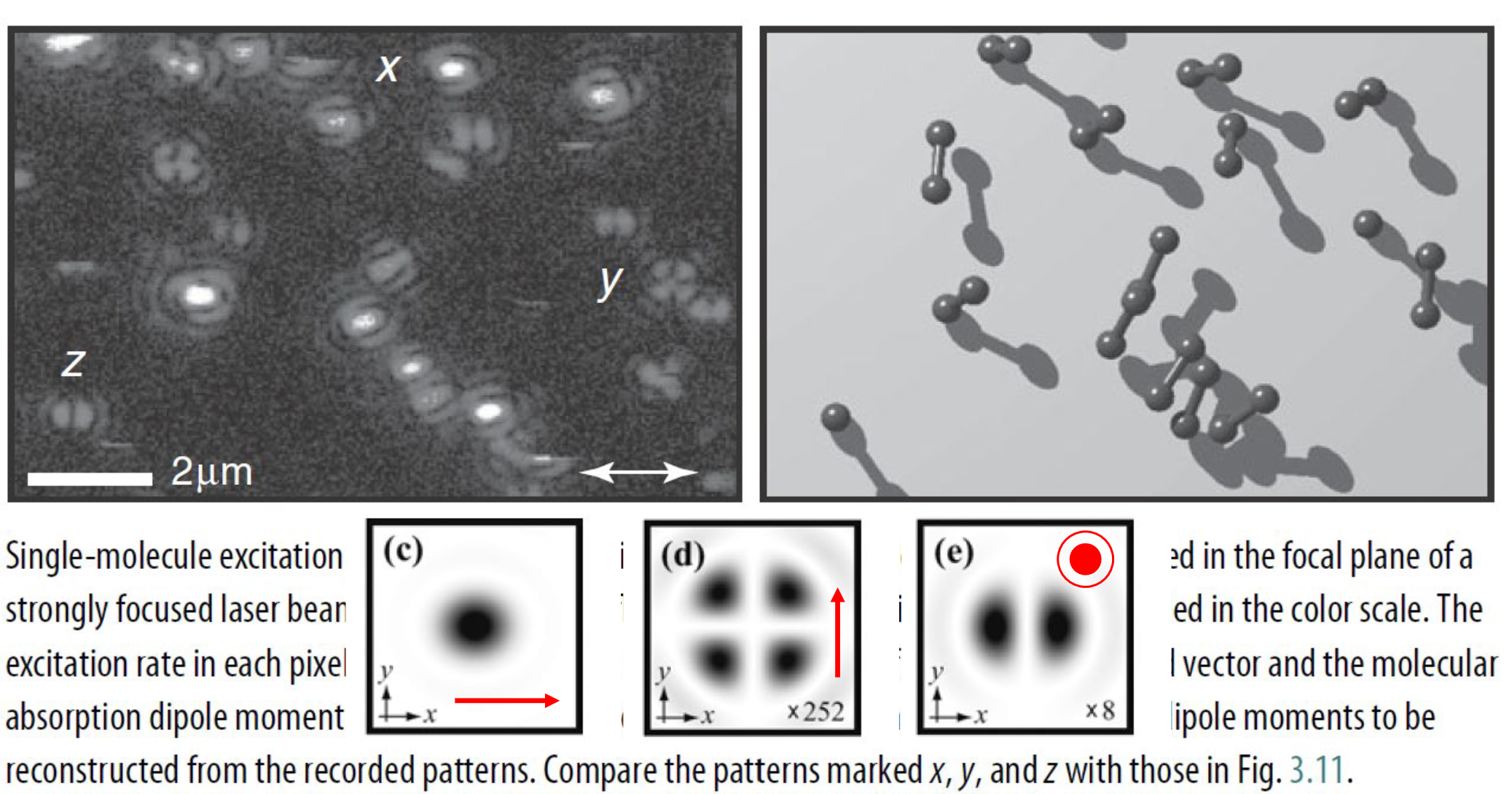
Explain the term:
Excitation rate∼∣μ⋅E(x,y,z_0)∣²
This is the quantum mechanical expression for how strongly the molecule couples to the field. Two things determine it:
The dot product μ·E means only the component of the electric field aligned with the transition dipole moment μ drives the transition. If the field is perpendicular to μ, the excitation rate is zero — the molecule is "blind" to that polarization. This is why polarization matters in fluorescence microscopy and why you can use it to measure molecular orientation.
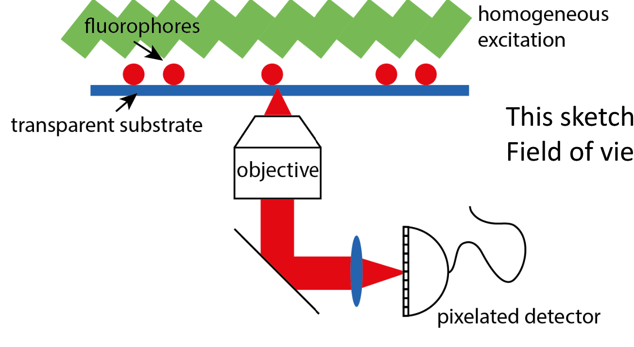
Explain the Epi-Illumination set up
Epi greek for above:
Illuminate entire sample homogeniously
Image sample plane on pixelated detector
Each fluorophore generates a signal according to the PSF
Resolution is:
x0=lambda/2*NA
How does the Intensitiy look like on the pixelated detector with Epi-Illummination?
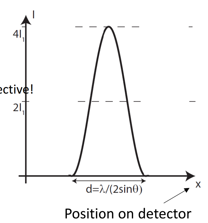
In fluorescence microscopy what’s the difference between scanning and wide field?
Both are limited by diffraction!
Scanning technique. Resolution set by size of pump spot on sample
Wide-field imaging. Resolution limited by PSF of imaging system

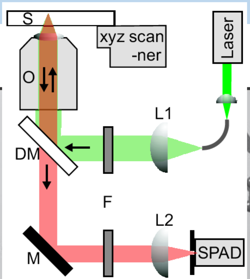
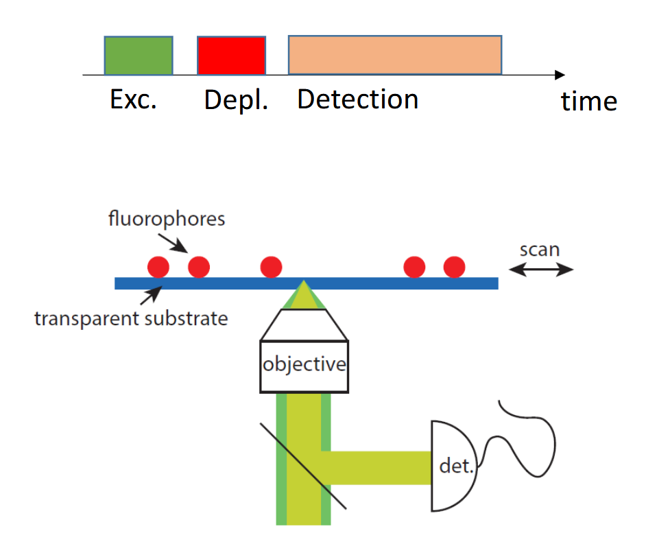
Explain the set up for single molecule detection
Laser pumps the molecules into a higher energy state. When they relax back into the ground state they emit fluorescence which is sampled on the SPAD → Single Photon Avalanche Diode
The dichroic mirror is crucial — it separates excitation from emission by wavelength, which is what makes the epi geometry work cleanly.
How are the orientation of molecules imaged?
By the mode which is sampled.
What is STED microscopy and what are the “ingredients“?
STED = stimulated emission depletion
It allows fluorescence microscopy beyond the diffraction limit
Ingredients:
(at least) 4-level system
pump laser
depletion laser
What are the assumptions and key points of explaining the population of the excited state in absence of the STED beam?
4-level system created by two electronic states (of a fluorophore) and vibrational relaxation
Vibrational relaxation is infinitely fast
Start in ground state, turn on pump
population of excited state as a function of time follows “charging“ curve of capacitor
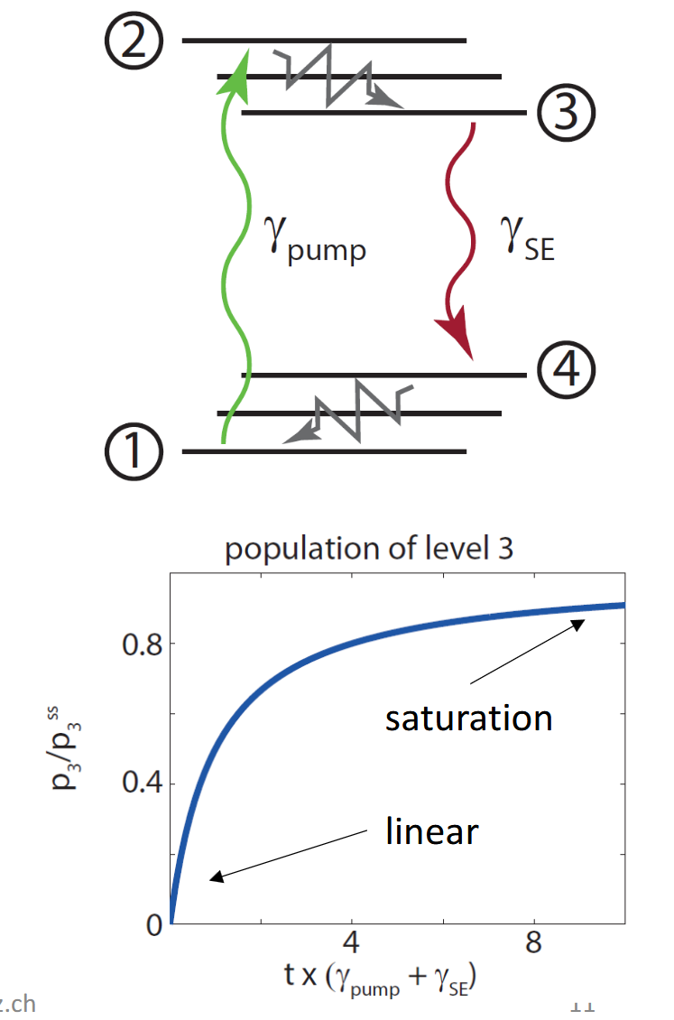
In STED how is the charging curve formulated and what do the variables mean?
Charging is calculated with:
1-exp(-t/tau)
So its exponantial growth which saturates. tau is the rate or time constant. After 4*tau the state is virtually full.
tau = rate_pump + rate_SE
What are the assumptions and key points of explaining the population of the excited state in presence of the STED beam?
Start in excited state (with certain probability), turn on depletion laser
Exponential decrease of population as function of time
Depletion field “helps“ spontaneous emission
depletion = exp(-t/tau)
tau = rate_depl + rate_SE
STED - How it works
Set up overlapping excitation and depletion lasers (both can naturally only be focused to the diffraction limit!)
We discuss a “pulsed“ scheme: sample us hit by different laser pulses and detection is only “on“ during specific time
STED - How it works. How do each pump pulses look like in a formula? What do terms mean? And how does the detected field look like?
Pump pulse:
p_ex(x) Probability of a fluorophore at position x being excited
σ_ex Absorption cross-section at the excitation wavelength — how likely the molecule is to absorb a photon
τ_ex Duration of the excitation pulse
I_ex(x) Intensity of the excitation beam at position xx x (Gaussian profile)
ℏω_ex Energy of one excitation photon ( ℏ = reduced Planck constant,ω_ex = angular frequency)
So the whole expression I_ex(x) / (ℏω_ex) is simply the photon flux (photons per area per time), and multiplying by σ_ex *τ_ex gives a dimensionless probability.
The same is for the depletion layer with p_ex is simply the starting condition — it's the population of molecules that were excited by the pump pulse in the first place.
The exponential describes stimulated emission depletion — the more STED photons arrive, the more molecules are forced back to the ground state before they can fluoresce. Where I_STED is large, pSTED→0
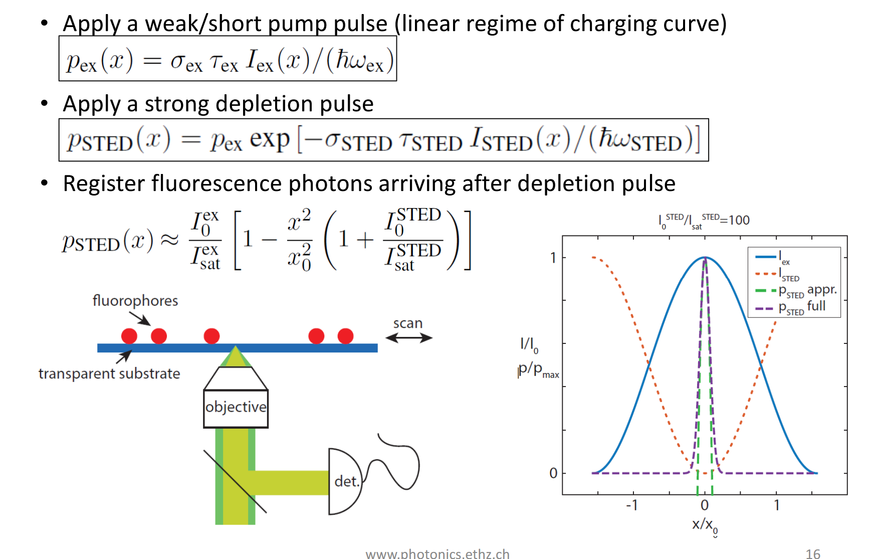
STED - how it works. What is now the whole magic? Why is the resolution beyond the diffraction limit?
Resolution before: x_0 = lambda/2NA
It’s the donut shape of the STED beam!
At the center (x=0): I_STED=0, so no depletion — molecules fluoresce freely
At the periphery: I_STED is large, exponential →0, fluorescence is suppressed
This is exactly what the graph shows — only a very narrow region around x=0 survives, far narrower than the diffraction-limited excitation spot x_0.
The zero in the donut remains at the center, but increasing STED power makes the intensity climb faster away from that zero, so the region where fluorophores are not depleted becomes smaller than the diffraction-limited excitation spot.
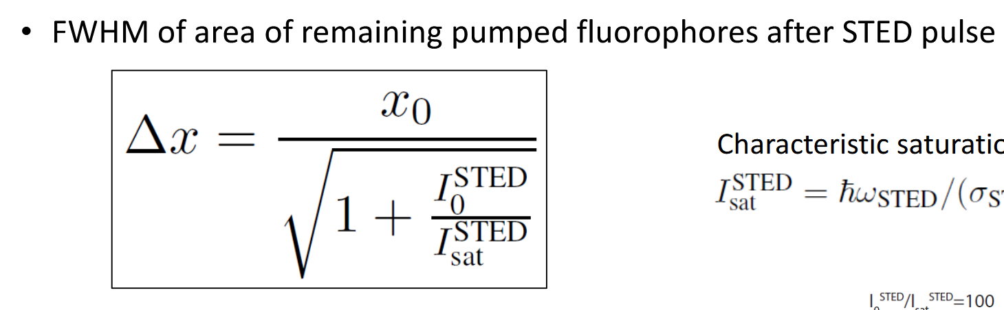
For STED:
Why do I need pulses?
Could I also do this with CW (continuous wave) lasers?
If yes, how?
The key is temporal ordering.
Excitation pulse fires → molecules go to excited state
STED pulse fires immediately after → depletes the periphery before fluorescence occurs
Then you collect fluorescence from the surviving central molecules
Pulses let you precisely control this sequence. The STED pulse must arrive within the fluorescence lifetime (~nanoseconds), so timing matters enormously.
Yes, CW can be used:
Excitation and depletion happen at the same time
There's no clean temporal separation
How can it work:
Keep excitation power very low so only few molecules are excited at any moment
Keep STED power very high so depletion is effective at all times
Gives worse signal to noise because sted beam can excite molecules as well (Anti stokes)
How can you generate the donut for STED?
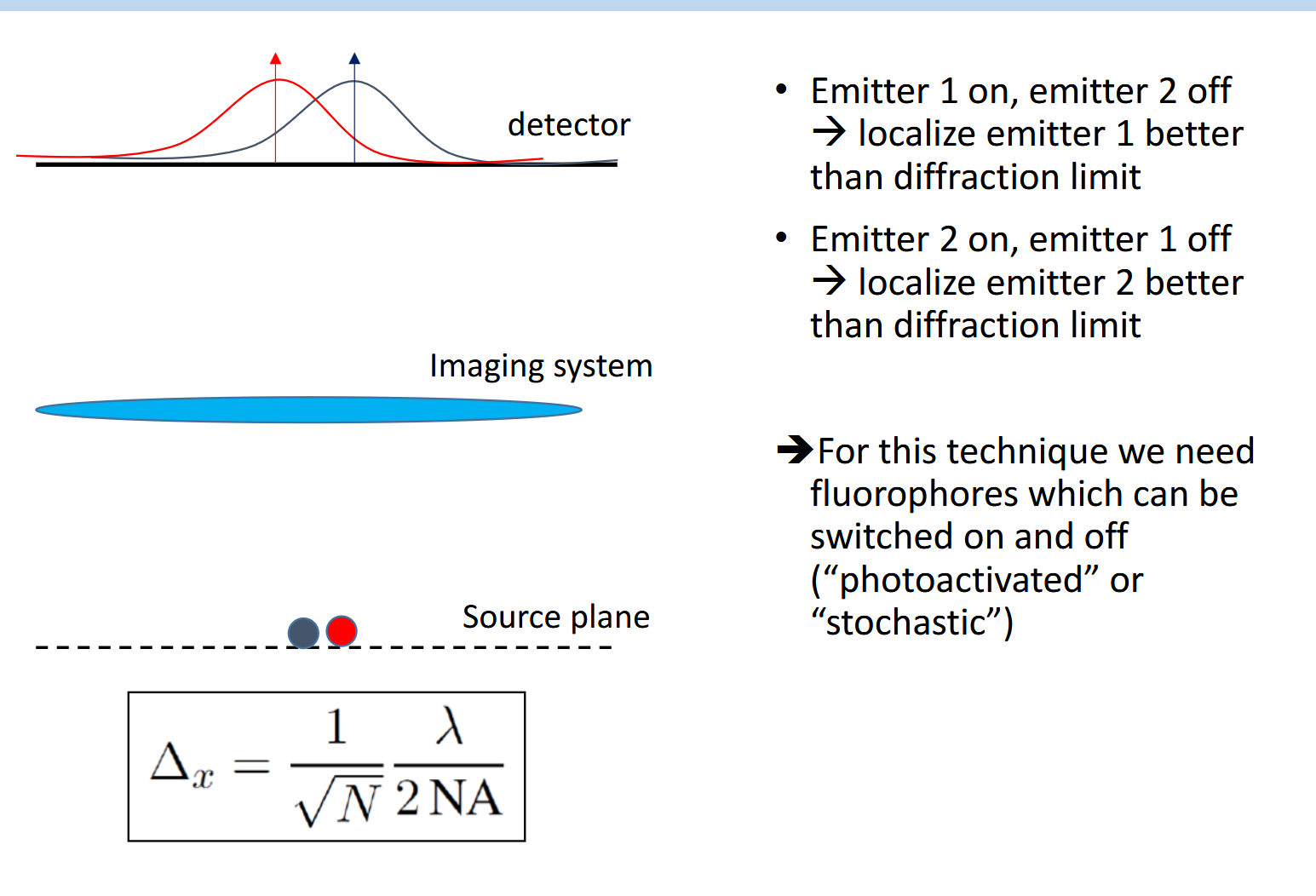
Two kind of same techniques for localization microscopy
PALM: Photoactivated localization microscopy
STORM: Stochastic optical reconstruction microscopy
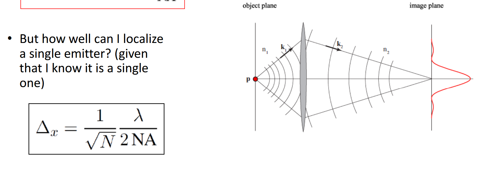
Given that you know there is a single emitter. How well can you localize it?
With N being the number of detected photons.
This is pure statistics. Each detected photon gives you a noisy estimate of the emitter's position (spread over the PSF). But by averaging NN N independent measurements, the uncertainty on the mean shrinks as 1/sqrt(N) — this is just the standard error of the mean.
Describe the STORM process
You need special fluorophores that can be switched between a dark (off) state and a fluorescent (on) state — called photoactivatable or photoswitchable fluorophores.
The randomness is controlled — you tune the activation laser power to ensure only a sparse subset is on at any moment. Too much power → too many on → PSFs overlap. Too little → acquisition takes forever.
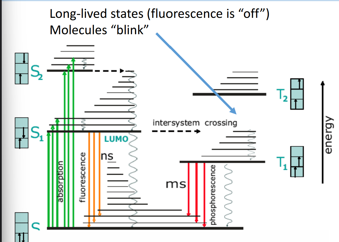
Think about the Jablonski diagram. What is the difference between fluorescnece and phosphorescence?
Fluorescence:
Absorption → singlet excited state S_1
Electron returns directly to GS S_0
Spin is conserved → transition allowed
Emission in ns
Stops immediately after pumping is off
Phosphorescence:
Absorption → S_1 → intersystem crossing to triplet state T_1
Spin flip makes return to GS quantum mechanically forbidden
Electron is stuck - Can only return slowly via a rare spin-flip event
Emission is very slow, ms to s to h. During this time “dark state“
Hence if one fluorophore is continuously pumped, sometimes it get’s stuck in Triplet state and is in the dark state. So it’s blinking.
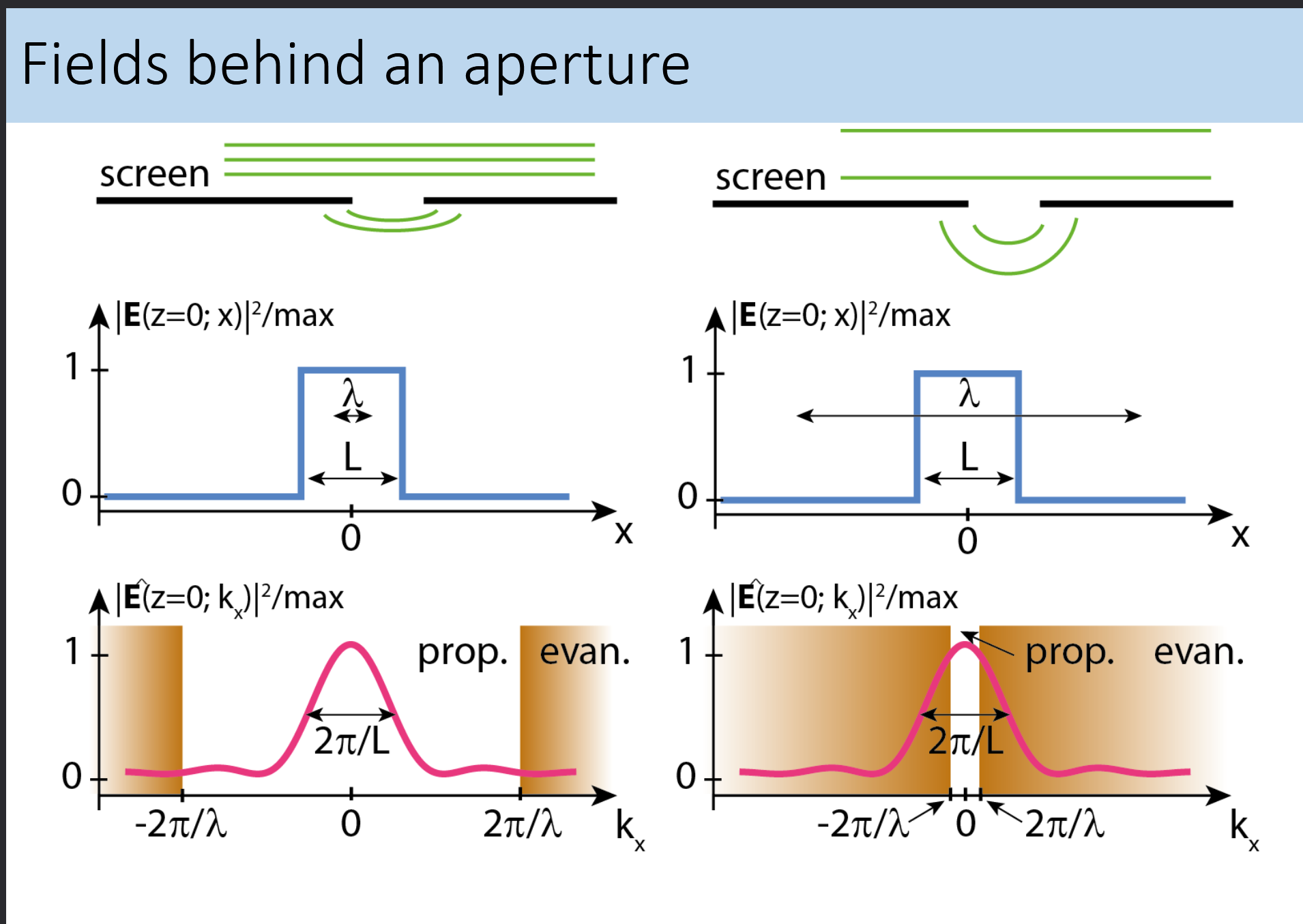
How do fields behind an aperture look like? And what is the reason for evanescent waves?
This example is 2D and relates kx to kz.
The real space looks like a top hat function which has the width of the aperture. Purely geometrical.
The k space jinc is from the fourier transform of a top hat. With the FWHM of 2pi/L.
The interesting part is when the wave propagating in z direction becomes evanescent. The shaded region is given by 2pi/lambda. Which is the value of k. k is given by 2pi/lambda.
sqrt(kx² + kz²) = k = 2pi/lambda. If kx becomes larger than 2pi/lamda, kz must be imaginary and hence the wave becomes evanescent in z direction.
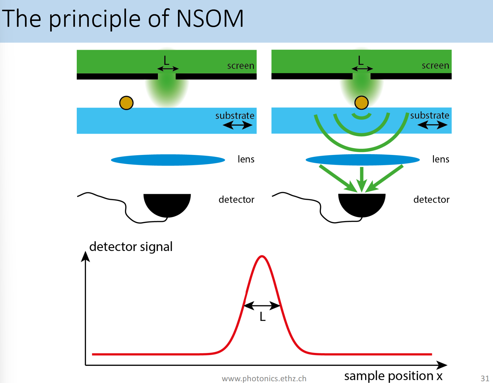
What is the principle of NSOM?
NSOM = Near-field scanning optical microscope.
Light passes through a tiny aperture L < < Lambda. Fields are evanescent and decay rapidly. If a particle is in the region of evanescent fields it acts as a dipole scatterer and scatters (our probably more in line with th ecourse → fluorescence) propagating photons which than can be collected. With this method the diffraction limit can be beat.
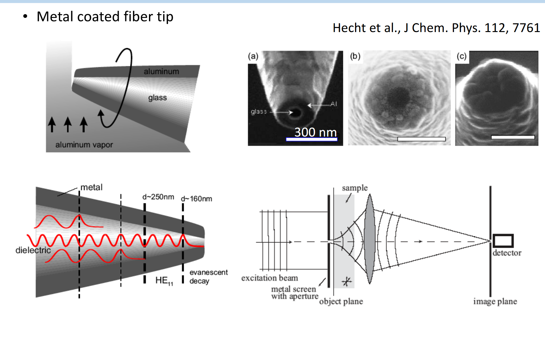
How does an NSOM aperture look like and why?
Glass acts as a waveguide and propagates with total internal reflection. The aluminum is here to prevent leaks which could disturb the measurement.
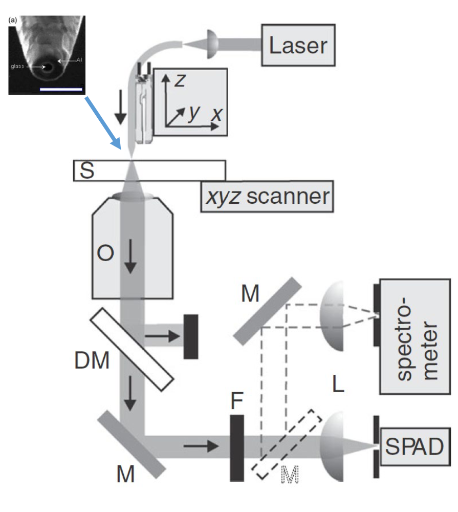
Describe the set up used in NSOM.
Laser for the monochromatic light source
Aperture made of glass and aluminium connected to stage
sample with fluorophores
objective to collect the light
dichroic mirror to seperate pump laser from stimullated emission (stokes shift)
Filter to filter out remaining laser light
SPAD → single photon avelanche diode
If we look at radiating sources, what are the characterstics of classical radiation sources and quantum emmitters?
Classical radiation sources:
Are driven by voltage/current source with tunable frequency
May have resonances given by their geometry
Quantum emitters:
Have a discrete level scheme
Emit one photon at a time
have a fluorescence lifetime (= inverse of decay rate gamma_SE)

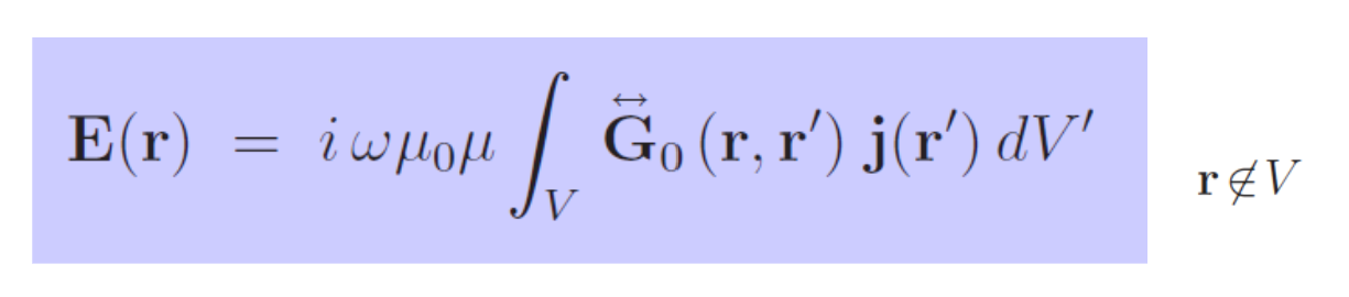
What is this formula for? What does it say and what are the different variables?
The formula incoorporates the Green’s function it’s a tool to solve linear differential equations with a source. If you know the field produced by a point source, you can find the field from any source by superposition on different locations.
E(r): electric field at observation point r (outside the source volume)
j(r’): current density at source point r’ inside the volume. It’s whats driving the field
G(r,r’): The dyadic Greens function, it acts as the propagator. How much field is produced at r by a current at r’
Integral over V: Sums contributions from all source points r’ in the volume, weighted by the Green’s function
iwu0u: Prefactor converting current density units into field units
What can you tell about the nature of the Green’s function in that formula?
The dyadic (tensor, indicated by double arrow) nature of G tells that it’s a 3×3 matrix. Because a current in one direction can produce field components in multiple directions!
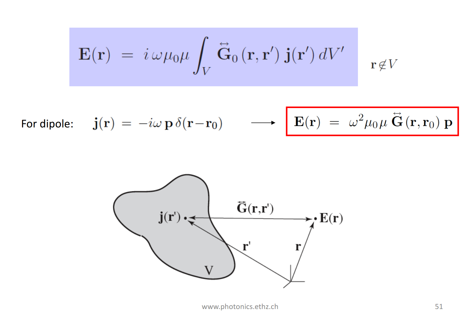
We use a simplification here. What is it and what’s the implication?
We know the current density of a dipole!
j(r) = -iwpδ(r-r0)
p: dipole moment vector
δ(r-r0): Dirac delta - it’s 0 everywhere, except at r0, making it a point source!
-iw: Comes from the time derivative relating current to dipole moment
Plugging this into the integral, the delta function collapses it! Hence the formula simplifies to:
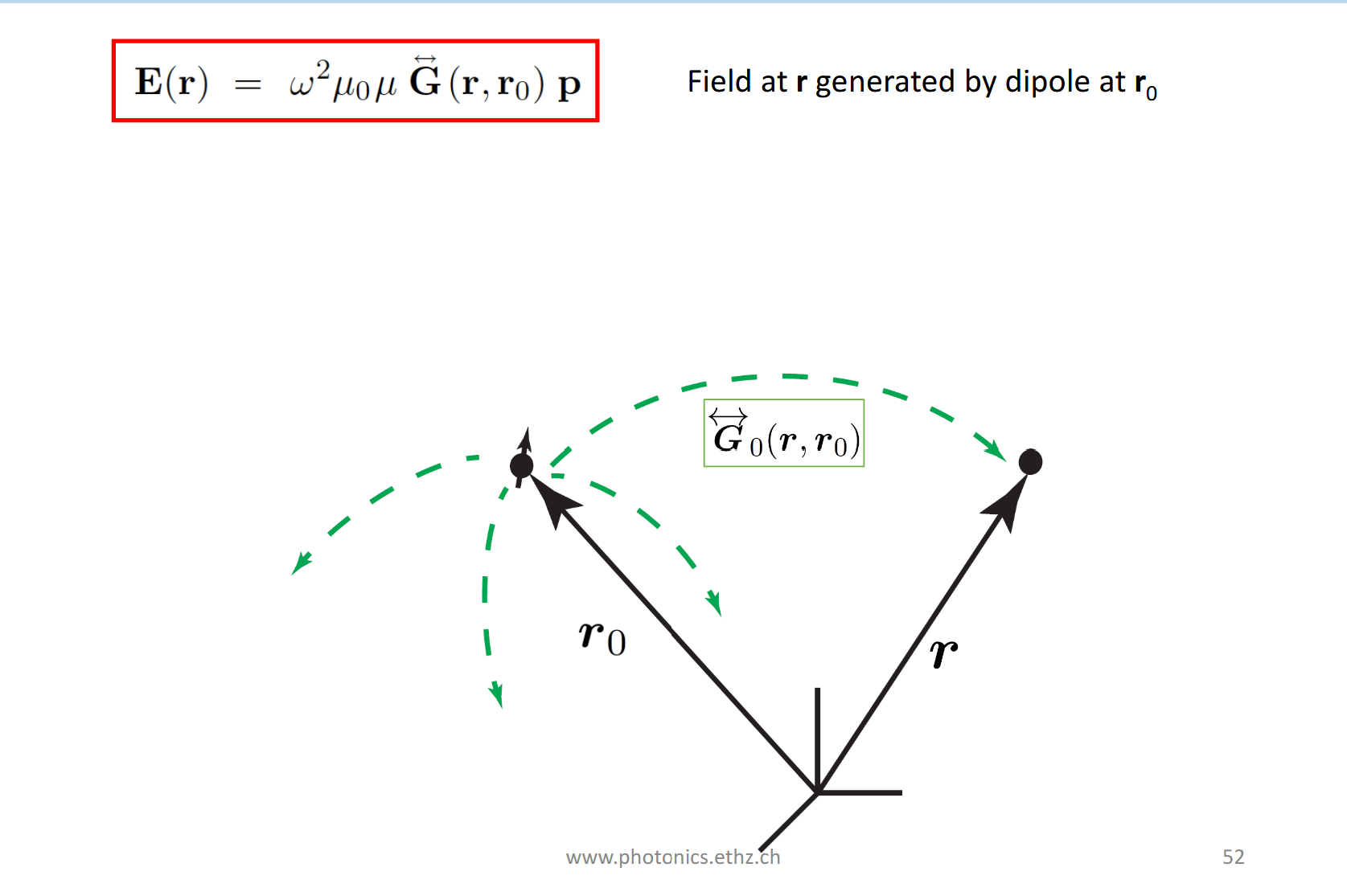
Result:
No integral needed
The field anywhere is just G acting on the dipole moment p
G encodes all physics of how the field propagates from r0 to r
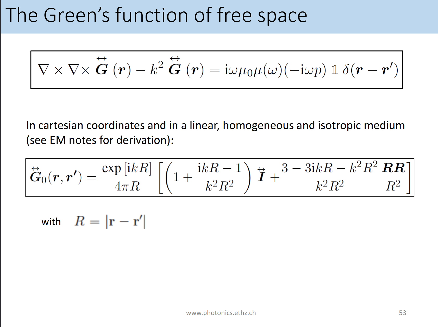
What can you tell about the Green’s function in free space?
Above equation is the wave equation with a point source.The Green’s function is defined as the solution to this equation.
RHS: Prefactor from dipole w²pu0uδ(r-r’) is recovered
𝟙 being the identity tensor, meaning the point source radiates equally into all polarization components
This all is the generalization of the scalar Helmholtz Green’s function. The extra tensor (𝟙) is needed because ligth is a vector field with polarization
CONTUNUE WITH BOTTOM SAVED IN CLAUDE