Clin Lab III: Urine Sediment ID + Urolithiasis Info.
 Call Kai
 Learn
Learn Practice Test
 Spaced Repetition
 Match
Match Flashcards
 Knowt Play
1/34

There's no tags or description
Looks like no tags are added yet.
Name | Mastery | Learn | Test | Matching | Spaced | Call with Kai |
|---|
No analytics yet
Send a link to your students to track their progress
35 Terms
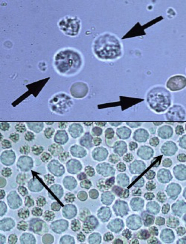
Spherical in shape, can appear dull gray or greenish-yellow in color.
ID’d by characteristic granules or lobulations of the nucleus.
In concentrated urine, they shrink.
In diluted urine, they swell.
White Blood Cells.
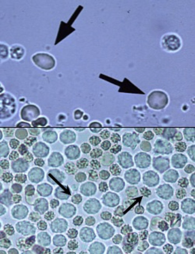
In fresh urine, they’re small, round, usually smooth edged, biconcave disc shaped.
Somewhat refractile.
Orange or yellow in color, some may be colorless.
In concentrated urine, they shrink and crenate.
In dilute or alkaline urine, they swell and may lyse.
Red Blood Cells.
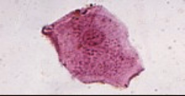
Largest cells observed in urine sediments.
Flat, thin cells with a homogeneous appearance and often have straight edges with distinct corners that sometimes curl or fold.
Squamous epithelial cells.
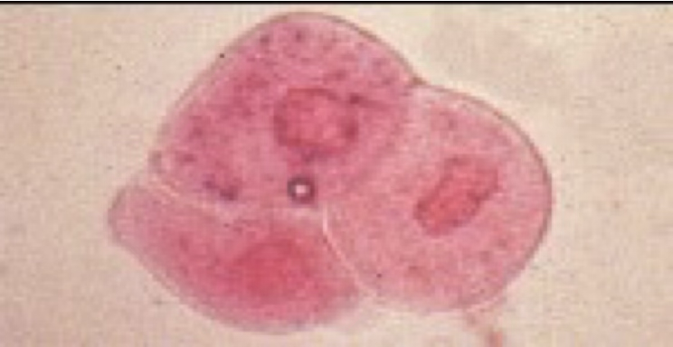
Usually round, but may appear pear-shaped or caudate (have a tail)
A low number may be seen due to sloughing of old cells.
An increased number seen can indicate cystitis or pyelonephritis
Transitional epithelial cells.
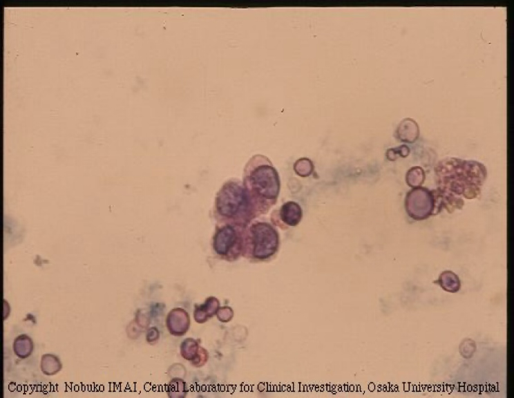
The smallest epithelial cell observed in urine sediment.
Rarely found, but am increased number seen can indicate renal tubular disease or degeneration/diseases of the kidney parenchyma.
Renal epithelial cells.
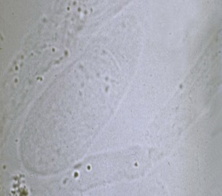
Clear, colorless and somewhat transparent.
A few seem is normal in urine sediment.
An increased number seen can indicate the mildest form of renal irritation, as well as fever, poor renal perfusion, strenuous exercise or general anesthesia.
Hyaline Cast.
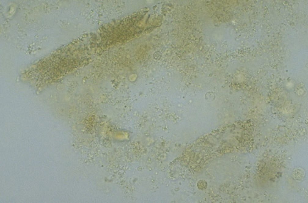
The most common type of cast seen in animals.
A few can be seen in normal urine sediment.
An increased number seen can indicate acute nephritis, more sever kidney damage than the hyaline cast.
Granular Cast.
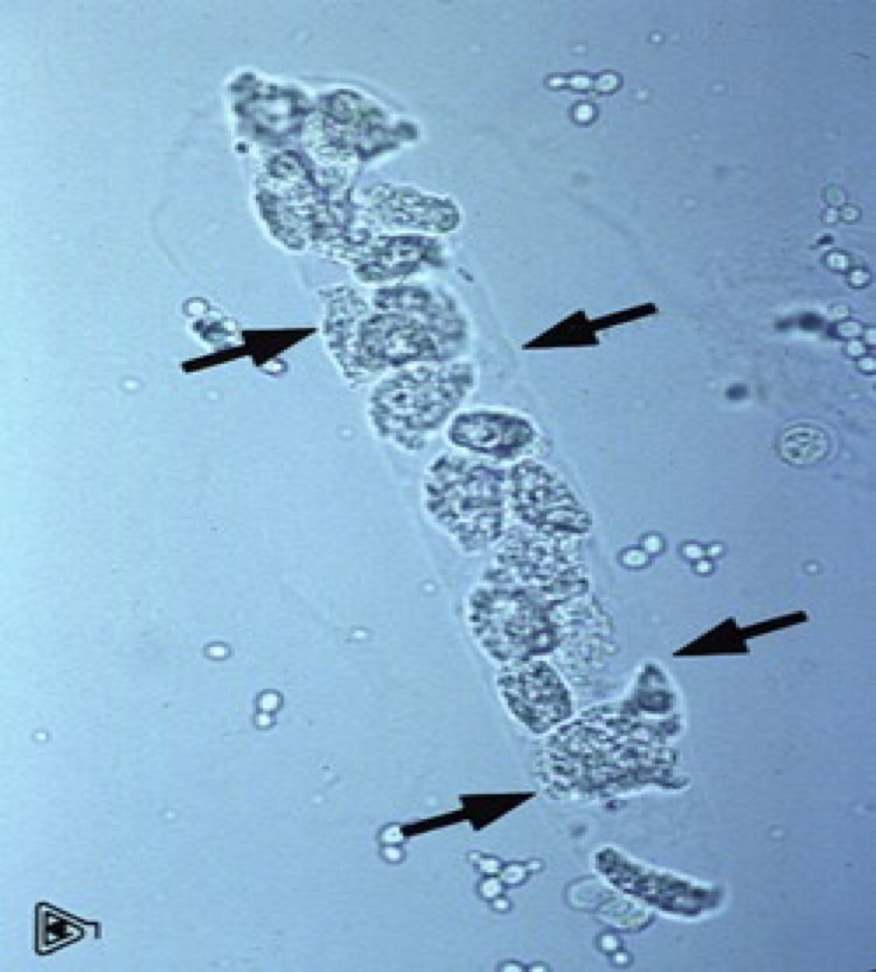
Consist of epithelial cells from the renal tubules imbedded in a hyaline matrix.
Formed by epithelium sloughing in the tubules.
Epithelial cast.
Contain WBC’s, predominately neutrophils.
Their presence indicate inflammation in the renal tubules or pyelonephritis.
WBC cast.
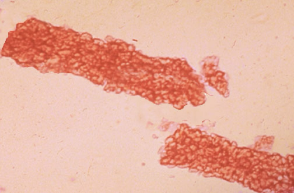
Form when RBC’s aggregate/clump within the lumen of the tubule.
Presence indicates renal bleeding.
RBC cast.
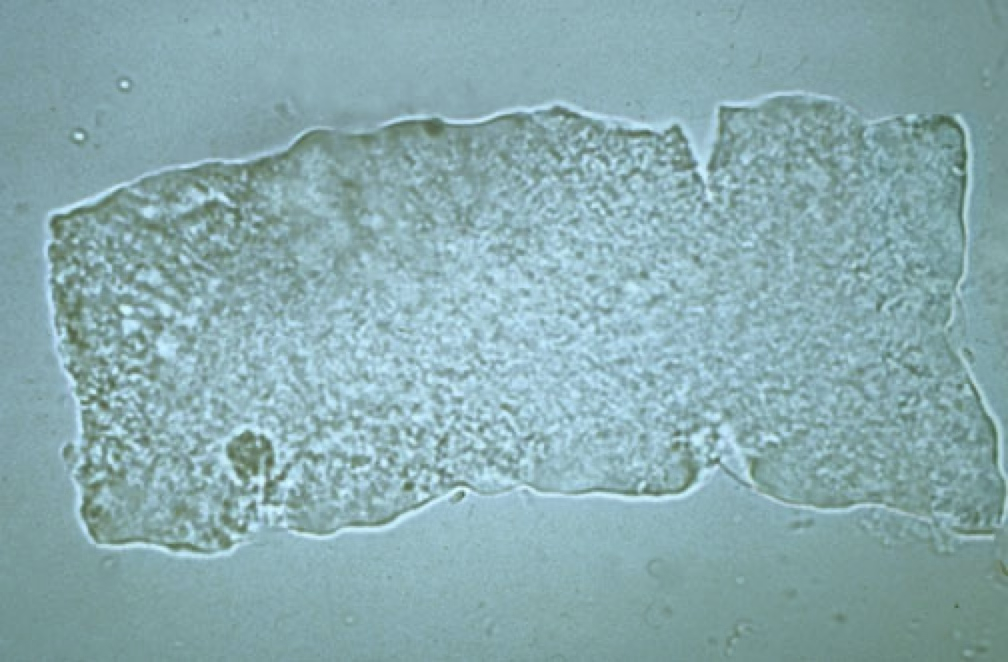
Resemble hyaline casts, but typically wider with square ends.
Their presence indicates chronic, severe degeneration of the renal tubules.
Waxy casts.
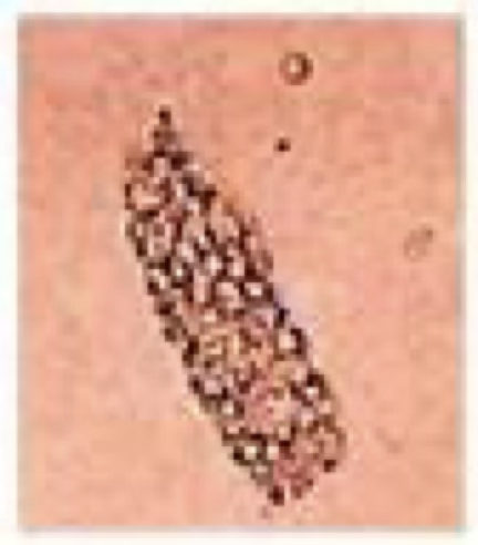
Contain small droplets of fat.
More commonly seen in cats due to high lipid content of their tubular epithelium.
Fatty cell cast.
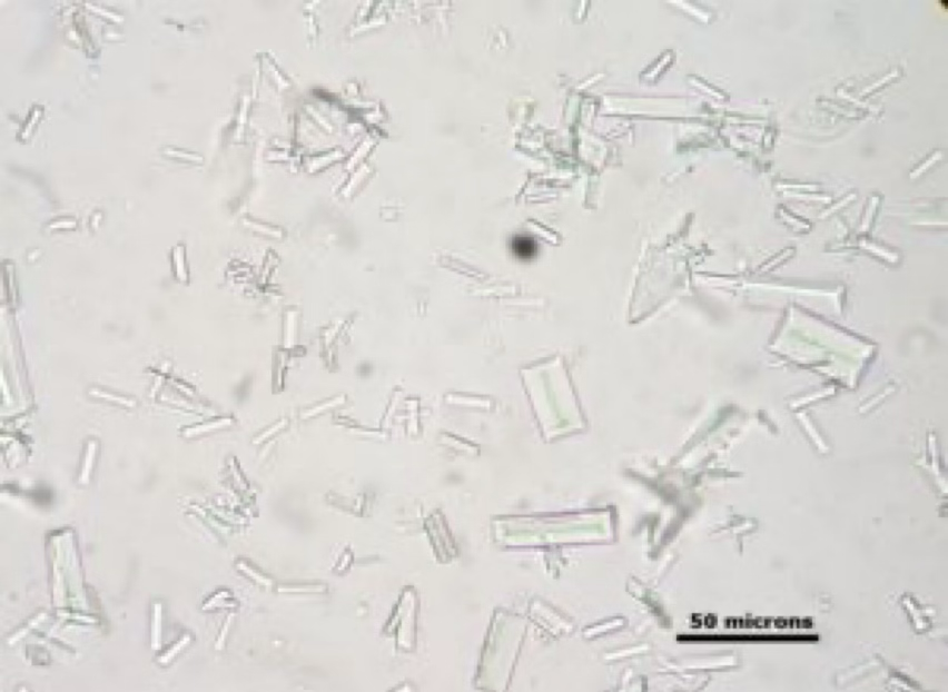
Typically seen in alkaline urine.
Resemble “coffin lids”
Very common urine crystal found in dogs and some cats.
Struvite crystals.

Seen in alkaline urine.
Appear as granular precipitate.
Amorphous phosphate crystals.
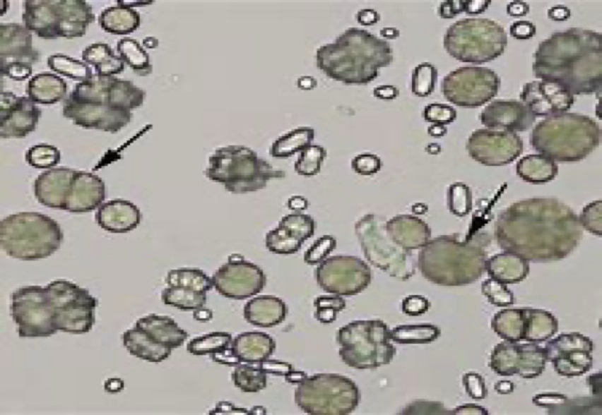
Typically seen in alkaline or neutral urine.
Commonly seen in the urine of horses and rabbits.
“Dumbbell shape”
No clinical significance.
Calcium Carbonate crystals.
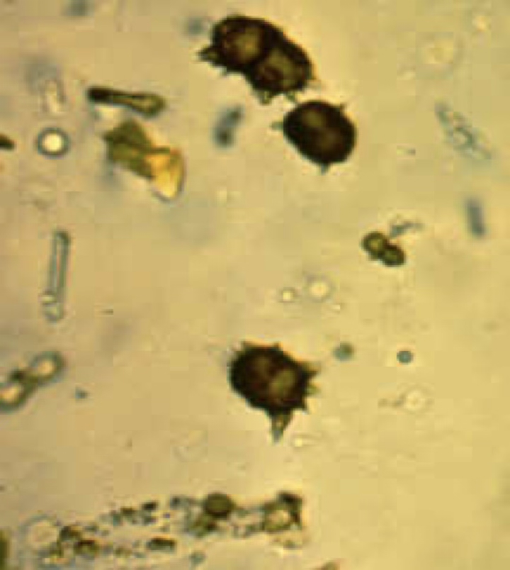
Typically seen in slightly acidic, alkaline, or neutral urine.
Resemble “thorn apples”.
Commonly seen in animals with severe disease, such as portacaval shunts.
Ammonium biurate crystals.
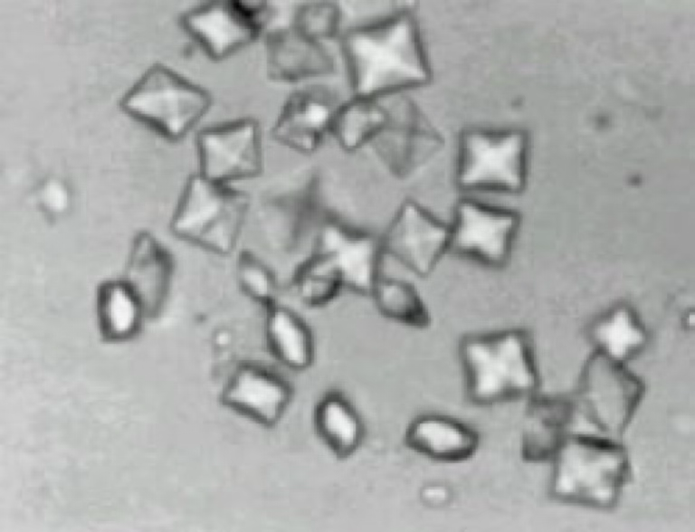
Resemble the back of an envelope, or the “xbox” logo.
Small number is commonly seen in dogs and horses.
Calcium oxalate crystals.
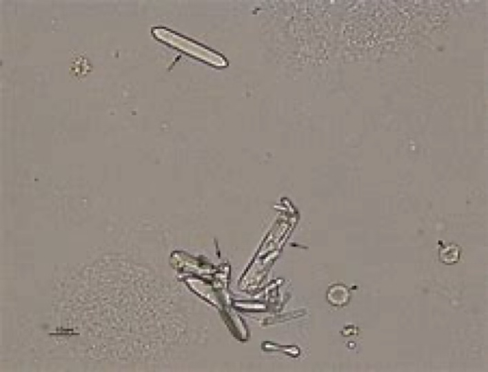
Small and dumbbell shaped.
Resemble a slat from a picket fence.
A large number is seen in animals with ethylene glycol (antifreeze) toxicity.
Calcium Oxalate Monohydrate crystals.
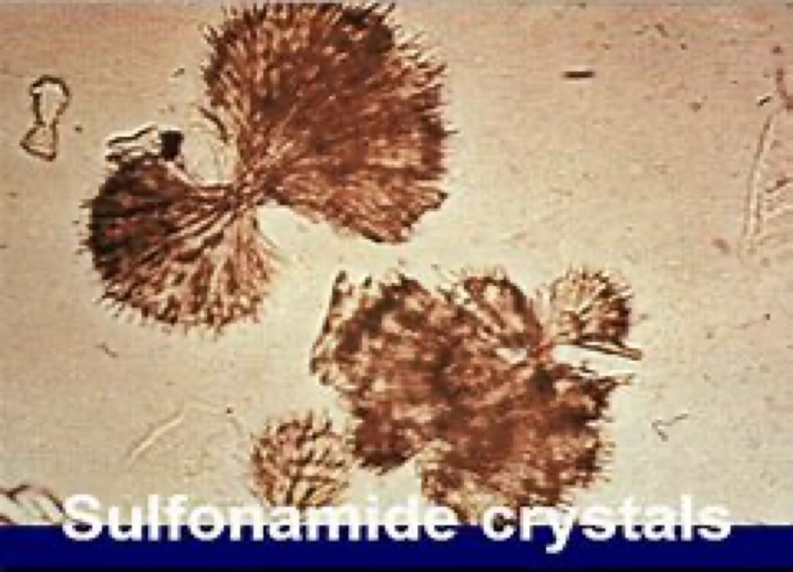
Seen in animals treated with sulfonamides.
Less likely to be observed in alkaline urine because they are more soluble in alkaline urine.
Sulfonamide crystals.
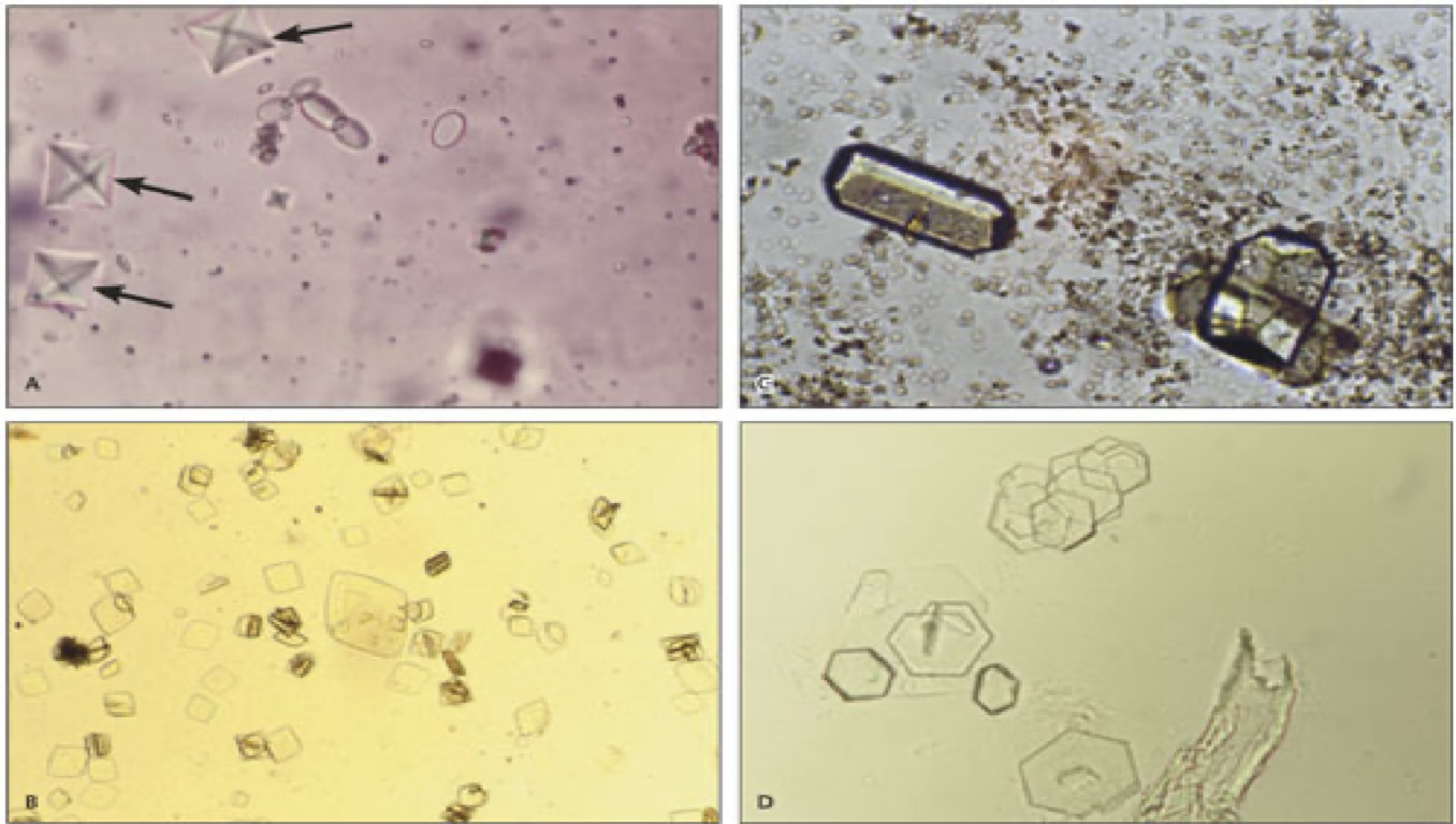
Typically seen in acidic urine.
Diamond or rhomboid in shape.
Yellow or yellow-brown.
Commonly found in dalmations.
Uric acid crystals.
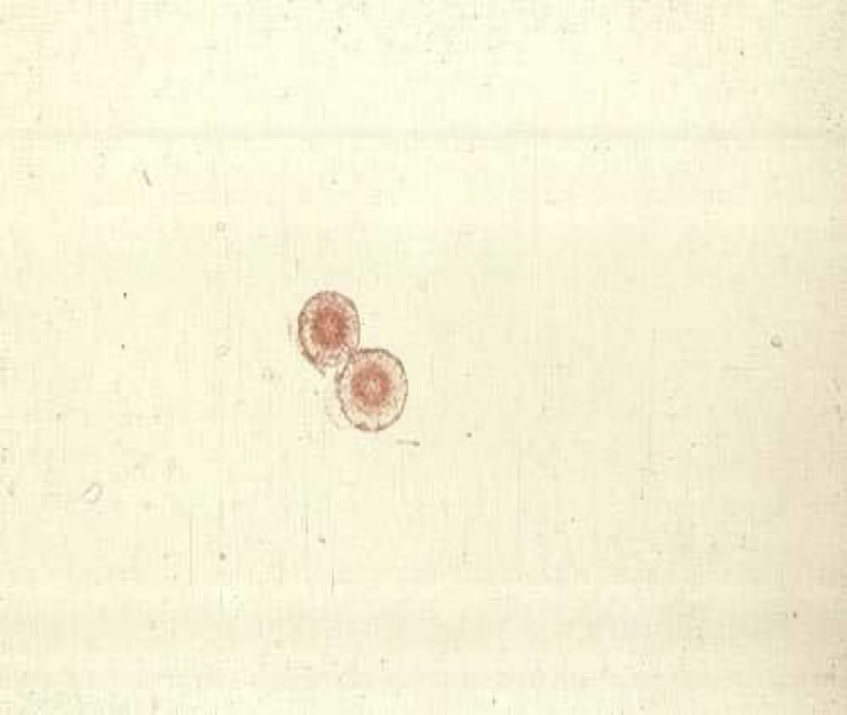
Typically seen in acidic urine.
Resemble a wheel or a “pin cushion”.
Can be seen in animals with liver disease.
Leucine crystals.
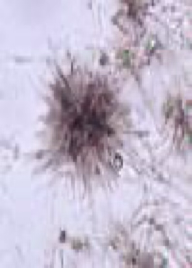
Typically seen in acidic urine.
Often seen in small clusters.
Not commonly found in dogs or cats but can be found in animals with liver disease.
Tyrosine crystals.
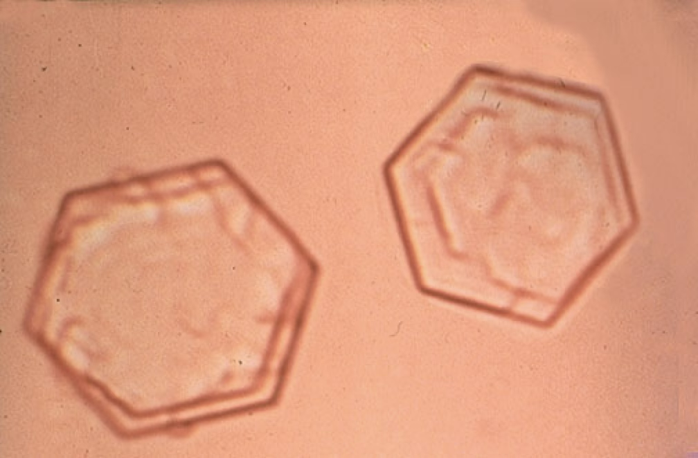
Typically seen in a cidic urine.
Appear flat and 6-sided (hexagonal).
Can be associated with renal tubular dysfunction or cystine urolithiasis.
Cystine crystals.
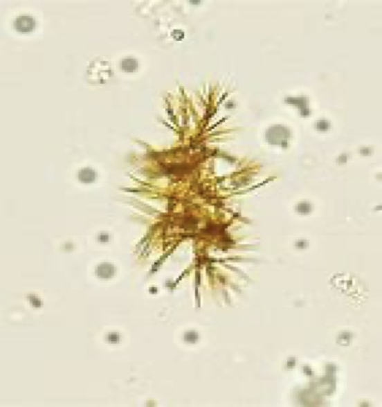
Typically seen in acidic urine
Appear as rhomboid plates, needles, or granules and are yellow, ruby red, or dark brown in color.
Commonly seen in conjunction with bilirubinuria and have little to no additional significance.
Bilirubin crystals.
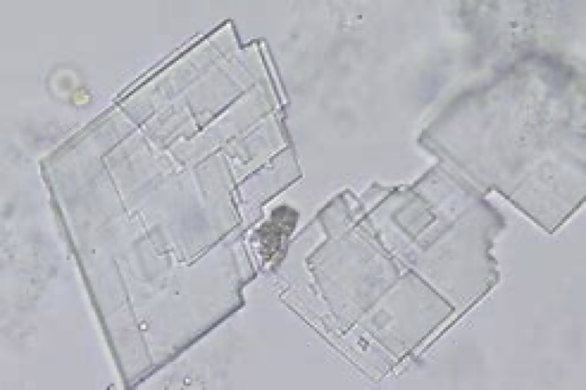
No clinical significance, normal.
Can be seen in normal urine samples of dogs.
Cholesterol crystals.
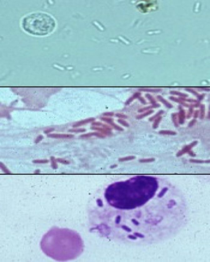
Can only be identified under the 40x high power objective.
May appear to be quivering- Brownian’s movement.
Large # accompanied by WBC’s can indicate infection/ inflammation of the urinary or genital tract.
Bacteria.
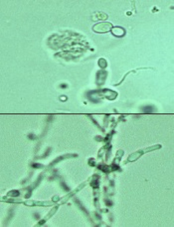
Often confused with RBC’s or fat droplets.
Peanut man/footprint shaped.
Usually considered contaminants when seen in urine sample because this type of infection is rare in animals.
Yeast.
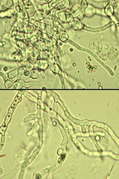
Filamentous and usually branching.
These kind of infections are rare, but considered quite serious when they do occur.
Fungi.
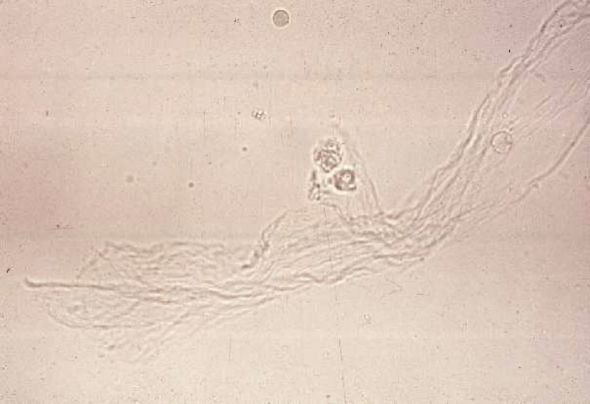
Often confused with casts.
Resemble a twisted ribbon.
A large amount is normal in equine.
In other animals, their presence indicates urethral irritation or contamination of the sample with genital secretions.
Mucus threads.
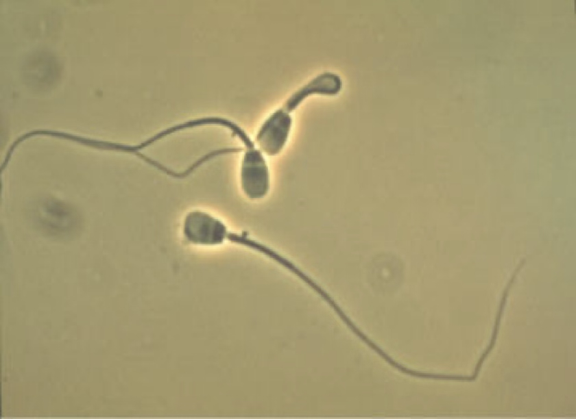
Occasionally seen in the sediment of intact male animals or recently bred females.
A large amount in urine can produce a false positive for protein.
Spermatazoa.
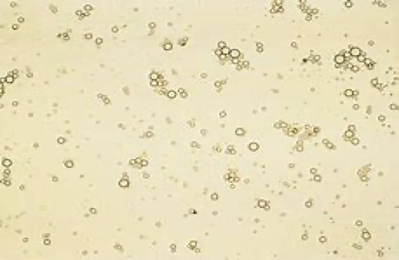
Seen to some degree in most cats.
If a prepared urine sediment on a microscope slide sits for a few minutes before being examined, this will rise to a plane of focus just beneath the coverslip.
Fat droplets.
Calculi/stones can cause severe, acute inability to what?
Urinate or remain in the bladder.
Can cause inflammation and bleeding.
Can have blood in their urine.
Most uroliths found in dogs and cats are usually ___________, but oxalate and cystine can also be seen
Struvite.
Removal of uroliths are typically done via a ____________ or urohydropropulsion.
Cystotomy.
How can you prevent uroliths from reoccurring?
abx therapy (for animals that also have a UTI)
Special diets
Increase water consumption
Possibly other medications to control urine pH.