Pathophysiology Module 9
1/36
There's no tags or description
Looks like no tags are added yet.
Name | Mastery | Learn | Test | Matching | Spaced | Call with Kai |
|---|
No analytics yet
Send a link to your students to track their progress
37 Terms
It is important for the nurse to understand that as intracranial pressure rises above normal limits and compensatory mechanisms fail
neurons can be injured and a change in level of consciousness occurs.
Brain herniation is the movement of the brain from a compartment of high pressure to a compartment of lower pressure. The movement of the brain
increases hypoxia and ischemic injury to brain tissue, reducing the level of consciousness.
Which parts of the brain and its structures contribute to the formation cranial pressure?
Blood, brain tissue, and cerebral spinal fluid
What does the Monro- Kellie hypothesis state?
Small increase in volume can be compensated for by one or both of the other components.
Trama in the Head
Trama in the head can cause head injury. When it happens, it is important to monitor intracranial pressure for disruption. Which can lead to additional brain injury.
What are the three components of intracranial pressure (ICP)?
Brain tissue, Cranial spinal fluid, cranial blood flow
Cerebral blood flow (3-10%)
The brain is able to utilize autoregulation to maintain cerebral perfusion despite changes in systemic blood pressure.
How does cerebral perfusion change in systemic blood pressure?
It does this by dilating or constricting the cerebral arteries depending on arterial pressure.
What is the normal range for ICP
0-15 mm Hg
Brain Tissue (80%)
The brain tissue is the last component to adjust to changes in ICP. The brain can compensate for small changes in ICP by partial collapse of the cisterns, ventricles, and vascular systems. When compensation is not sufficient, brain herniation occurs.
Cerebral Spinal Fluid (8-12%)
Cerebral spinal fluid (CSF) circulates in a closed system of the ventricles, subarachnoid space, and down and around the spinal column. The arachnoid villi absorb the cerebral spinal fluid. Any changes in the cycle of CSF production, circulation, and absorption can affect ICP.
Primary Brain Injuries
Caused by initial trauma
What are the results in immediate disruption for a primary brain injuries
Skull
Brain structures (meninges, blood vessels, brain tissue, neurons)
Brain functions (blood flow, oxygenation, cellular metabolism)
Secondary Brain Injuries Result in
Cerebral infarction
Coma
Increased cerebral edema
Secondary Brain Injuries that are caused by effects of primary injury
Uncontrolled ICP
Cerebral ischemia
Hypotension
Hypoxemia
Local or systemic infection
Intracerebral Pressure
Cerebral spinal fluid and cranial blood flow may be able to adjust to relieve ICP.
Signs of increased Intracerebral Pressure
Increased intracerebral pressure can progress quickly
Some signs of increased Intracerebral Pressure
Level of consciousness
Pupi size and reaction to light
Motor function
Vital signs
other signs
Early signs of increased Intracerebral Pressure
Consciousness: Decreased alertness to drowsiness
Pupil size and reaction to light: Small and sluggish reaction
Motor Function: Hemiparesis
Vital signs: No change
other signs: Hache, slurred speech
Late signs of increased Intracerebral Pressure
Consciousness: Stupor or coma
Pupil size and reaction to light: Large and nonreactive
Motor Function: Hemiplegia
Vital signs: Hypertension, widened pulse pressure, bradycardia, abnormal respiratory patterns
other signs: Vomiting
Cerebral edema
It is a secondary brain injury that occurs as a result of a primary brain injury.
Cerebral edema (swelling of brain tissue) results from abnormal water accumulation from intracellular fluid.
Cerebral edema Disorders
These disorders cause an expansion of brain tissue, and this expansion within the solid cranial vault causes decreased cerebral fluid circulation leading to hypoxia.
The first type of Cerebral edema
Cytotoxic - Results from intracellular sodium potassium pump failure
The second type of Cerebral edema
Vasogenic - Results from disruption to blood- brain barrier
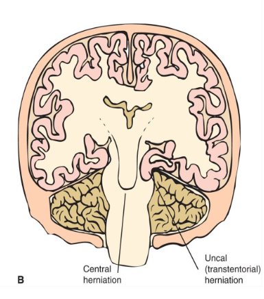
Brain herniation
Occurs when pressure within the cranium is greater than what the brain can compensate for. When pressure increase to this high of a level, it causes brain tissue to be displaced.
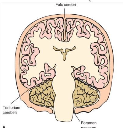
Normal Brain
Herniated Brain
Herniation associated with brainstem compression is called central herniation, whereas herniation associated with the supratentorial structures is called uncal (transtentorial) herniation.
Which key points would you include when helping a new nurse understand increased ICP?
ICP above 15 mm Hg is considered cerebral hypertension.
Nurses must carefully monitor signs and symptoms of increased ICP to prevent secondary brain injury.
Decreasing compliance of cranial components to maintain equilibrium results in increased ICP.