The Prime Factors
1/76
There's no tags or description
Looks like no tags are added yet.
Name | Mastery | Learn | Test | Matching | Spaced | Call with Kai |
|---|
No analytics yet
Send a link to your students to track their progress
77 Terms
Prime factors
Affect x-ray emission and are under DIRECT CONTROL OF RADIOGRAPHER
milliamperage-second (mAs)
kilovoltage (kV)
distance (d)
quantitative factors
-milliamperage-second (primary quantitative)
-kilovoltage
-distance (further away, radiation spreads out more)
-filtration (more stuff in between)
Qualitative factors
how strong it is
Kilovoltage
Filtration
mA
quantity of electrical current flowing through a circuit
how is mA described as
the rate representing the number of electrons passing down a wire per second
mA control of machine
selection which taps different series of resistors in filament circuit to control the intensity of the flow of electricity
when selecting higher mA
greater flow rate of electricity
more electrons in shorter amount of time
Intensity=
quantity
the intensity rate of x-ray beam is
directly controlled by the mA sstation set at console
Number of x-rays created is a product of
number of electrons crossing tube and amount of time electrons are allowed to cross (mAs)
change in mA or mAs
affects the number of electrons
BUT NOT the kinetic energy of electrons flowing from cathode to anode
mAs
product of mA and exposure time (s)
if either mA station or set exposure time is doubled
the total radiation exposure is doubled to both the detector and the patient
mA and exposure time are
inversely proportional
if mA is doubled
time is halved
mAs=
mAs
formula for mAs
mA1xs1=mA2xs2
mAs is the primary controller of
quantity/intensity of x-ray photons (directly proportional)
in film, mAs was
density (directly proportional)
mAs and exposure relationship
direct
if mAs goes up, detector exposure/exposure index goes up
Insufficient mAs produces
underexposed image
quantum mottle
excessive mAs produces
overexposed image
saturation
harder to discern in digital systems but will provide inappropriate EI numbers
kilovoltage (keV or kV)
measure of the electrical force or pressure behind a current of electricity, which causes it to flow
-measure of electrical energy
-greater the potential difference, the more pressur exerted
Due to AC electricity
kV is measured in terms of average value/peak value
(Kilovolt peak KVP)
X-ray beams with higher average energy
Capable of penetrating through different types of tissue
Most important function of kVp
to provide at least partial penetration through all tissues to be recorded
kVp is primary controller of
total quality of x-ray photons
kVp in film
contrast
kVp impacts this as well indirectly
quantity of photons due to more interactions by incident electrons in target material
UNDESIRED SIDE AFFECT
kVp is primary controller of differences in radiographic densities/IR exposures, known as
image contrast (brightness)
As we increase kVp
Increased penetrability, which will result in image with less contrast (think of an abdomen)
15% rule
an increase in kVp by 15% will cause a doubling in exposure'
a decrease by 15% will cause a halving of exposure
If maintaining a particular exposure is desired, but more penetration is needed
increase kVp by 15%
cut mAs in half
kVp adjustments should NOT
be used to control radiographic exposure
Do not oversimplify 15% rule to
10-kVp rule, stating that with every 10 kVp, changes in exposure occur by factor of 2
since kVp controls both quality and quantity of x-ray photons
there will be some increase in patient exposure
using 15% rule to increase kVp increasess
patient exposure by 1/3 (28-38% avg 33%)
however if using 15% rule and cutting mAs in half
then exposure is offset by this 50% cut in quantity of photonss
Using 15% kvp rule averages a
net savings in patient dose of about 1/3
net result for apply 15% rule is exposure averaging about 67% of the original
distance
x-ray photons from a point source begin to spread out or diverge with distance
-causes decrease in intensity
x-ray beam is
divergent
x-ray intensity is measured with a dosimeter
previously recorded in roentgens (R or mR)
now recorded in milliGray (mGy)
more distance
decrease in intensity
As SID is increased and collimators are closed further to maintain field size
Further cutting photons and decreasing intensity
Inverse square law
As distance increases, beam intensity decreases
Amount of exposure received is less
The intensity of radiation at a given distance from the point source iss
inversely proportional to the square of the distance
Inverse square law concerning distance and intensity formula
I1/I2=D2squared/D1squared
I1= original intensity (mR)
I2= new intensity (mR)
D1= original distance
D2= new distance
The concentration of radiation will be
inversely proportional to the square of the distance (at twice the distance, the radiation will be 1/2squared or 1/4 as intense)
Doubling the distance
allows the x-ray beam to spread out over a fourfold area, reducing exposure intensity to one quarter the original
isotropically- evenly all the way around
mAs and detector exposure relationship to distance
As distance increase
Intensity decreases
Decreases IR exposure
Direct Square Law
formula for mAs, detector exposure relationship, and distance
direct square law
-to compensate for SID changes
-determines amount of mAs necessary to provide enough photonss to create an image after SID changes
Direct relationship is necessary to
compensate for the changes in intensity and IR exposure
Direct square law is also known as
exposure maintenance formula
and density maintenance formula (for film)
Exposure maintenance formula
mAs1/mAs2=D1^2/D2^2
or
mAs2= mAs1 x D2^2/ D1^2
Exposure maintenance dissected
Direct square law also applies to each individual component of mAs- ma or s separately
mA1/mA2= D1^2/D2^2
T1/T2= D1^2/D2^2
Increasing SID
-reduces patient dose
-increases spatial resolution
-reduces magnification
-because of reduced magnification of anatomy within field, more of body part can be included with projection
Technique changes mobile radiography application
with digital imaging systems
the relationship of kvp being contrast and mAs being density has been decoupled
brightness and contrast are now controlled primarily through
post-processing
milliamperage
measures the rate of electricity flowing through the X-ray tube, and controls the x-rays emitted from it
mA stations at console
select from diffrent resistors to control amount of amperage flowing through the filament to maintain a steady space charge boiled off by thermionic emission
mAs overall
controls the totaly amount of x-rays delivered from x-ray tube during exposure
mAs is preferred controlling factor for
total exposure/intensity
overexposure from excessive mAs
is not apparent in digital images, and can only be monitored by checking the expossure indicator readout
mAs is not considered a factor in controlling
contrast
mAs has no direct relationship with recognizability factors such as
sharpness, magnificaation, distortion
however, shorter exposure times make unsharpness due to motion less likely to occur
the predominance of different tissues within a body part
determine minimum kVp that should be used
no amount of radiation intensity can ever compensate for
insufficient penetration of x-ray beam
a 15% change in kVp
alters intensity of radiation reaching detector by a factor of 2
optimum kVp
level well above the minimum needed for sufficient penetration, which strikes a balance between saving patient exposure and preventing excessive scatter
kvp has no direct impact on the geometrical aspects of image
sharpnesss, magnification, distortion
increasing SID
reduced exposure intensity at detector by inverse square of distance, because x-rays spread out isotropically
when should technique be adjusted to compensate SID
any change greater than 15%
adjustment for radiographic technique for changes in SID follows
direct square law
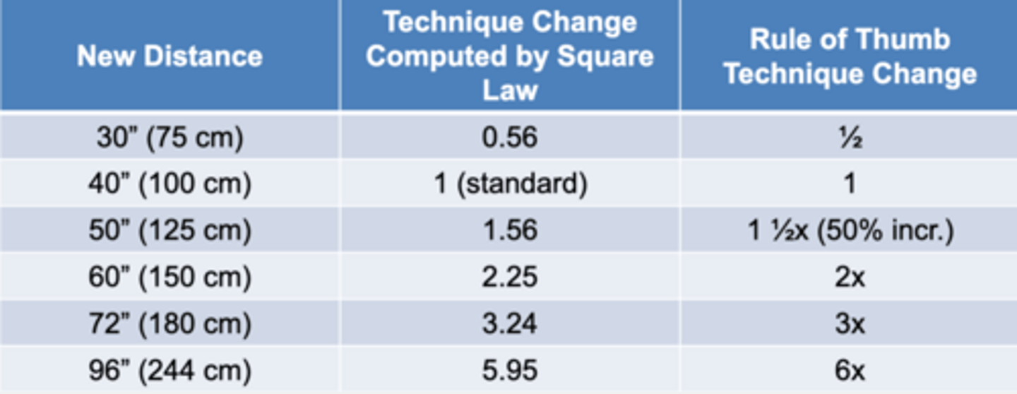
rule of thumb for distance changes from 40'' to 72''
adjust technique by 3x
increased SID
if increase in SSD
because source to skin distance changes by a greater ratio than SID
increased SID can be used to reduce patient skin dose