anat & phys unit 8
1/78
There's no tags or description
Looks like no tags are added yet.
Name | Mastery | Learn | Test | Matching | Spaced | Call with Kai |
|---|
No analytics yet
Send a link to your students to track their progress
79 Terms
What are the 4 functions of the muscular system?
Movement, maintain posture/position, stabilize joints, generate heat
Describe the structural organization of a muscle down to the sarcomere.
Muscle → Fascicle → Muscle fiber → Myofibril → Sarcomere
Describe what occurs at the neuromuscular junction. Include the role of Ca2+ and acetylcholine.
At the neuromuscular junction, extracellular Ca2+ is taken in by the neuron. This triggers the release of the neurotransmitter Acetylcholine into the synaptic cleft. Acetylcholine is taken up by the muscle which triggers depolarization of the muscle and begins the contraction process.
thin filaments
actin fibers
thick filaments
myosin fibers
z-disc
edges of the sarcomere, come closer together during contraction
a-band
denotes the length of the myosin fibers
h-zone
the space between actin fibers on each z-disc; shrinks during contraction
i-band
space between z-disc and start of myosin, never changes size

what is A?
z disc

what is B?
thick filament

what is C?
thin filament

what is D and F?
I band
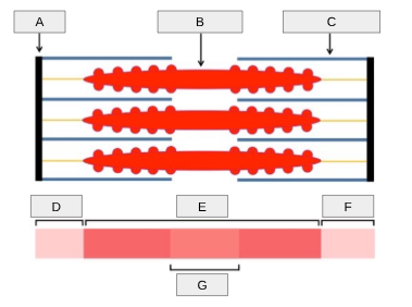
what is E?
A band

what is G?
H zone

what is happening in A?
The myosin head is bound to the actin filament following a previous powerstroke.
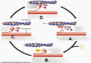
what is happening in B?
A new ATP molecule binds to the myosin head, releasing the crossbridge.

what is happening in C?
The ATP breaks down into ADP + Pi. This allows the myosin head to rebind to the actin filament.

what is happening in D?
The myosin head is bound to the actin and once the ADP + Pi releases from the myosin, the resulting conformational change is what causes the powerstroke to occur.
What is the role of Ca2+ in the sliding filament theory of muscle contraction?
Ca2+ is stored in the sarcoplasmic reticulum and released when depolarization occurs due to a signal from a neuron. The Ca2+ binds to troponin, a protein associated with the actin filament. This binding causes another protein associated with the actin filament (tropomyosin) to undergo a conformational change that exposes the myosin binding site.
what are isotonic contractions?
the myosin head is successful in performing a powerstroke and the sarcomere contracts
what are isometric contractions?
the myosin is unable to complete the powerstroke because it is unable overcome a counteracting force on the muscle
What are the 5 golden rules of skeletal muscle activity?
Skeletal muscles must cross at least one joint.
The bulk of the skeletal muscle lies proximal to the joint crossed.
Skeletal muscles attack the skeleton in at least 2 spots (origin & insertion)
Skeletal muscles can only pull; they never push.
During contraction the insertion of a muscle moves toward the origin.
flexion
decreases the angle of the joint, brings 2 bones closer together
extension
opposite of flexion, increases the angle of the joint
abduction
moving a limb away from the midline
adduction
moving a limb towards the midline
circumduction
moving a limb in a circular motion; proximal end is stationary while distal end moves in a cone shape
rotation
movement of a bone around its longitudinal axis
Dorsiflexion & Plantarflexion
Up and down movement of the foot at the ankle
dorsiflexion specifically
toes up
plantarflexion specifically
toes down
Inversion & Eversion
Rolling of the ankle
inversion specifically
roll foot inward
eversion specifically
roll foot outward
Supination & Pronation
Rotation of the hand at the wrist
supination specifically
rotating the palm upward
pronation specifically
rotating the palm downward
opposition
touching each of your fingers to your thumb
what is muscle tone and what is its purpose?
Muscle tone is the small continuous contractions of skeletal muscles even while at rest. This keeps the muscles firm and healthy
slow twitch muscle fibers
low force production, utilize oxygen and cell resp, high fatigue resistance, used for endurance
fast twitch muscle fibers
high force production, utilizes anaerobic respiration, low fatigue resistance, used for burst of speed

what is A?
frontalis

what is B?
orbicularis oculi

what is C?
orbicularis oris
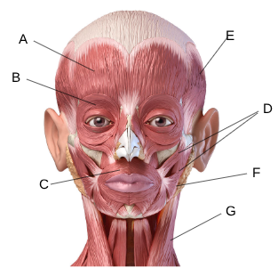
what is D?
zygomaticus

what is E?
temporalis

what is F?
masseter

what is G?
sternocleidomastoid

what is A?
trapezius

what is B?
deltoid

what is C?
pectoralis major

what is D?
rectus abdominis

what is E?
external oblique

what is F?
latissimus dorsi

what is G?
bicep brachii

what is H?
tricep brachii

what is I?
brachioradialis

what is J?
gluteus maximus

what is K?
hamstring group

what is L?
quadricep group
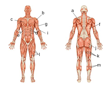
what is M?
gastrocnemius
What 3 muscles make up the hamstring group?
The bicep femoris, semitendinosus, semimembranosus
What 4 muscles make up the quadricep?
The rectus femoris, vastus lateralis, vastus medialis, vastus intermedius
step 1
Acetylcholine is released from a motor neuron at the synaptic cleft.
step 2
Acetylcholine binds to receptors on the muscle fiber.
step 3
Ca2+ ions are released from the sarcoplasmic reticulum.
step 4
Ca2+ binds to troponin.
step 5
A conformational change in troponin causes a conformational change in tropomyosin exposing the myosin bind site.
step 6
ATP binds to myosin head.
step 7
ATP breaks down into ADP + Pi.
step 8
Myosin binds to actin at the binding site forming a crossbridge.
step 9
ADP + Pi is released from the myosin.
step 10
Myosin pulls on actin resulting in a powerstroke.
step 11
The distance between the Z-lines in the sarcomere shortens.
step 12
A new ATP molecule binds to the myosin-actin complex.
step 13
The myosin releases from the actin filament.
step 14
The cycle repeats as long as there is adequate Ca2+ and ATP.
step 15
Fibers relax and Ca2+ is pumped back into the sarcoplastic reticulum.