ANAT W3 key words
1/114
Earn XP
Description and Tags
skeletal system, atricular system, muscle system
Name | Mastery | Learn | Test | Matching | Spaced | Call with Kai |
|---|
No analytics yet
Send a link to your students to track their progress
115 Terms
yellow bone marrow
triglyceride storage in bone
axial skeleton
head, neck, trunk
what tissue type is bone
connective tissue
tissue structure of bone?
ECM that surrounds widely spread out cells
what is the ECM in bone made out of and for what purpose
collagen that resists tension and mineral salts that resist compression
appendicular skeleton
pectoral girdle (including clavicle, scapula), pelvic girdle, and the arms and legs
compact bone
dense bone outside spongy bone
spongy bone
inside of the bone, filled with bone marrow
cortical
compact bone
trabecular, cancellous
spongy bone
trabeculae
column of connective tissue in spongy bone
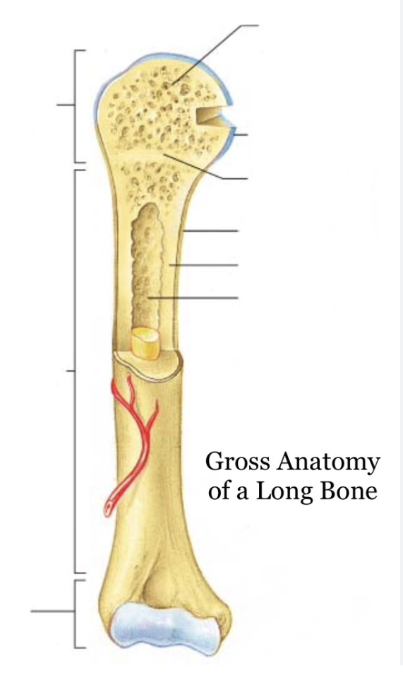
proximal epiphysis, diaphysis, distal ephysis, atricular cartilage, spongy bone, epiphyseal line, periosteum, medullary cavity, endosteum, compact bone, yellow bone marrow, nutrient artery, metaphysis
label this
long bones
longer than they are wide, weight bearing
short bones
similar width and length, stability and support
sesamoid bone
special type of short bone that forms in a tendon
flat bone
thin and flattened, can be curved, protect vital organs, large surface for muscle attatchments
irregular bones
bones in no other category
perioseal arteries
supply periosteum, outer compact bone, enter through many small canals
large nutrient arteries
enter through nutrient foramen at center of diaphysis, enters meduallary cavity and goes towards epiphysis
epiphyseal arteries
supply end of long bones especially epiphysis
metaphyseal arteries
supply end of long bones especially metaphysis
osteoblast
bone building cells, synthesise and secrete collagen and organic components to build ECM, intiate calcification
osteoclasts
break down bone ECM for resorption, release lysosmal enzymes and acids to digest protein + mineral of ECM
intramembranous ossification
development directly from mesenchyme, first ossification during fetal development
flat bones of skull, facial bones, mandible, parts of clavicle, fontanelles
bones that develop from intramembranous ossification
ossication center
where cells cluster due to signalling from osteoblasts during intramembranous ossification
chondrocytes
what mesenchyme cells condense into to form cartilage model
primary ossification center
initiated by blood bringing nutrients, in the middle of the bone where bone starts to replace cartilage in endochondral ossification
endochondral ossification
development of mesenchyme into cartilage during fetal stage then bone
secondary ossification center
in the epiphysis of the bone where the spongy starts replacing cartilage
25 years old
age when bone starts to only grow appositionally
interstitial growth
type of growh of cartilage on the epiphyseal side of the epiphyseal plate
zone of resting cartilage
anchor epiphyseal plate of the epiphysis bone
zone of proliferating cartilage
zone for interstial growth
zone of hypertonic cartilage
zone of mature chondrocytes
zone of calcified cartilage
the last zone where the cartilage becomes bone in longitudinal growth
endochondral ossification
replacement of cartilage on the diaphysis side of the epiphyseal plate for longitudinal growth
18 F and 21 M
the time when the epiphyseal plate closes forming the epiphyseal line
appositional bone growth
growth in the thickness of the bone
periosteum differentiate into osteoblasts, osteoblasts secrete collagen and other organic materials that make up the ECM of bone, osteoblasts surrounded by ECM develop into bone, old bone lining medullary cvity destroyed
mechanisms for appositional bone growth
bone remodelling
bones changing shape throughout life time
gluteal tuberosity on femur due to attatchment of gluentous maximus
example of bone marking for muscle attatchment
radius and ulna articular with humerous
bone marking forming joints
foramen magnum in the occipital lobe for the spinal cord
example of bone marking for vesseles and nerves
atricular system
system of joints jointed by ligament and accessory structures
cartilage
avascular, resilient, semirigid connective tissue
diffusion
how is cartilage nourished
atricular cartilage
smooth cartilagenous surface for joints
hyaline cartilage
most common cartilage, covers bone atriular surfaces, model for early skeleton, moderate amount of collagen
elastic cartilage
flexible, forming external ear, containing bundles of elastic
fibroncartialge
what is this cartilage’s name? specialised in joints eg. disc, mix of fibrous tissue and hyaline tissue, withstand prolonged pressure, substantial collagen
synovial joints
movable joints jointed by atricular cavity
fibrous joint
immovable joints, joined by connective tissue, movement depends on length of fibres
cartilaginous joint
minimal movement, bones jointed by hyaline of fibrocartilage, strength and shock absorption, no joint capsule
sutures
fibrous joint: connective joints in the skull
sutural ligament
special material that connects sutures in the skull
gomophosis
fibres between root of tooth and bony socket
syndesmosis
ligament joining 2 bones eg. radius and ulna, length of ligament determines movement
interosseous membrane
connective material in syndesmosis
synchondrosis
a bar or plate of hyaline cartilage between 2 ossification of developing bone
primary cartilagenous
synchondrosis -
symphysis
fibrocartilage connecting 2 seperate bones
secondary cartilagenous
symphysis
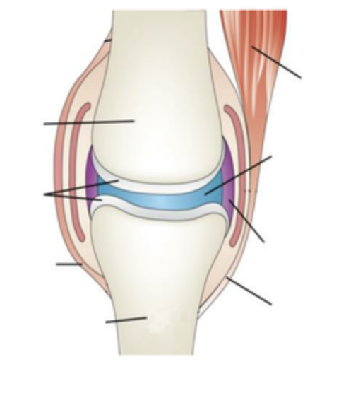
atricular cartilage, joint capsule, inner synovial membrane, outer fibrous membrane, accessory structures
label the synovial joint
plane joint
non axial movement, bones sliding past each other
hinge joint
uniaxial, movement at right angles to the joint, flexion and extension, surfaces one rounded and one curves around
pivot joint
unaxial, movement in line with the longitudinal axis of the bone, rotational movement, sleeve of bone + ligament and the axle bone
condyloid/ellipsoid joint
biaxial joint, movement around 2 axis at right angles, one indented and one oval surface, flexion, extension, abduction, adduction. eg. the knuckle joint
saddle joint
biaxial joint, movement around 2 axes at right angles, flexion, extension, adduction, abduction eg. the carpometacarpal joint of the thumb
ball andd socket joint
multiaxial, bones are shaped like ball and socket, flexion, extension, adduction, abduction, circumduction and rotation eg. hip and shoulder
range of motion
the range in degrees through which bones can be moved
atricular discs
binds strongly to fibrous membrane of joint capsule, seperates bone so more movement and seperate movement at same time can occur
fibrocartilage, extending inwards from the atricular capsule and partially or completely dividing it, absorb compression force + distribute weight
atricular menisci
only in the knee, C shaped
fibrocartilage, extending inwards from the atricular capsule and partially or completely dividing it, absorb compression force + distribute weight
labrum
wedge shaped, outer margin of the ball and socket joint, made out of firbocartilage, deepens the socket, increasing SA
fat pads
space fillers, intracapsular but extrasynovial, spreads the synovial fluid
bursa
bags of synovial fluid that reduce friction, wrap around tendons that pass bones, numerous in joints that have increased mobility, extracapsular
tendon sheath
tube like synovial structure that wraps around a tendon, smooth gliding over long surfaces
thermogenesis
muscle generating heat as they contract
skeletal muscle
attatch to skeleton, voluntary for some, straited
cardiac muscle
only in the heart and some great vessels, involuntary (without nerve system), branching chain of cells
smooth mucle
not straited, uninucleated, in the walls of hollow organs, involuntary (through autonomic nervous system)
endomysium
wraps individual muscle fibres
perimysium
wraps fascicles
epimysium
wraps the entire muscle including muscle and nerves, blood vessles
circular muscles
fascicles in concentric rings, contraction closes
sphincters
external body openings where ciruclar muscles are present
convergent
broad origin by fascicles converge towards single tendon of insertion, fan shaped, strongest contraction eg. pectoralis major
pectoralis major
example of convergent muscle
parallel
lengt of fasciles parallel to long axis of muscle, strap like eg. sartorious or fusiform eg. bicep
sartorious
strap like muscle
fusiform
belly of the muscle protruding
pennate
fascicles are short and attatch obliquely to the tendon
unipennate
fascicles insert onto one side of the tendon
bipennate
fascicles insert onto opposite sides of the tendon
multipennate
fascicles insert onto the tendon from many directions
deltoid
example of multipennate mucle
rectus femoris
example of bipennate muscle
extensor digitorus longus
example of unipennate muscle
ligament
joining bone to bone, blend with periosteum at joint
tendon
joining muscle to bone