Parasitology Lab Exam 2
1/35
There's no tags or description
Looks like no tags are added yet.
Name | Mastery | Learn | Test | Matching | Spaced | Call with Kai |
|---|
No analytics yet
Send a link to your students to track their progress
36 Terms
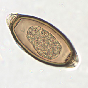
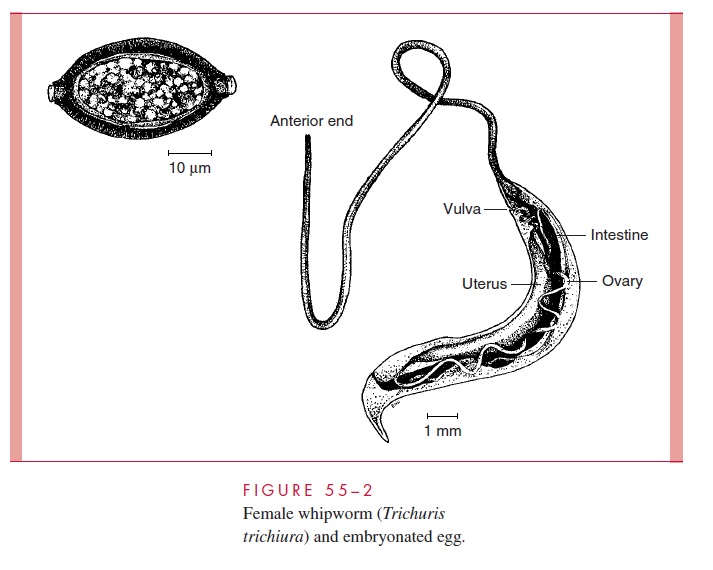
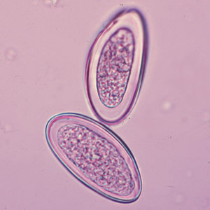
Trichuris trichiura Life Cycle
Whipworm
Phylum - Nematoda
Eggs are barrel-shaped and have polar “plugs” at each end
Unembryonated eggs are passed in the stool
In the soil, eggs develop into a two-cell stage
An advanced cleavage stage takes place
Embryonated eggs are ingested (contaminated soil or food)
The eggs hatch in the small intestine and release larvae
The larvae mature and establish as adults in the colon
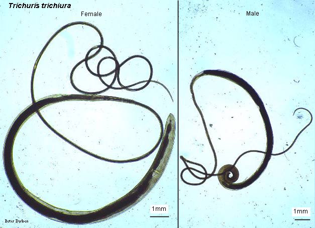
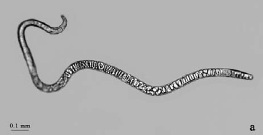
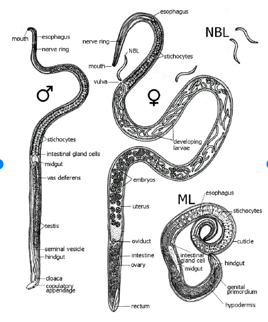
Trichuris trichiura Female Anatomy
Sexual dimorphism, females are larger
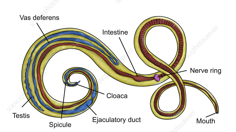
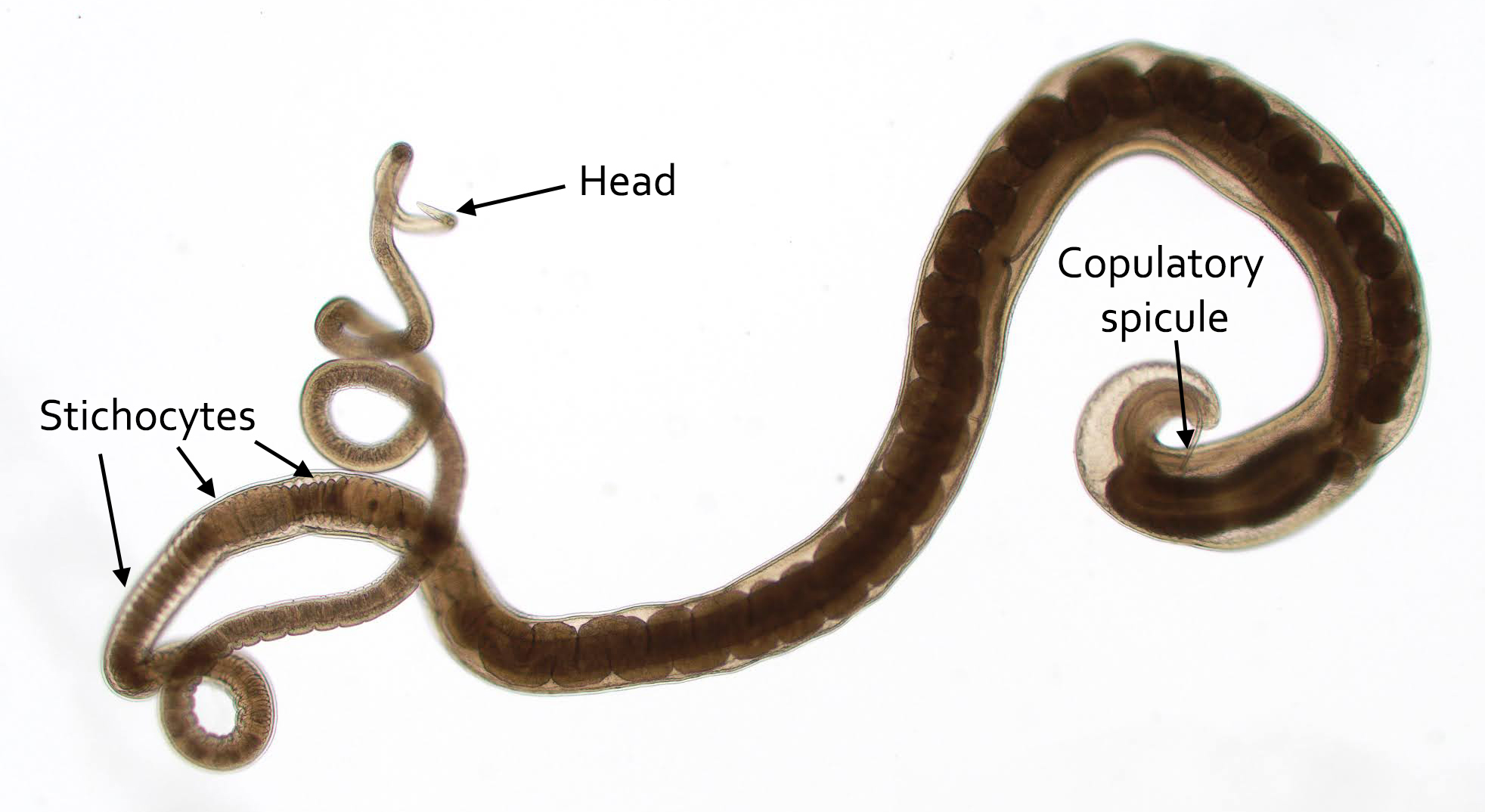
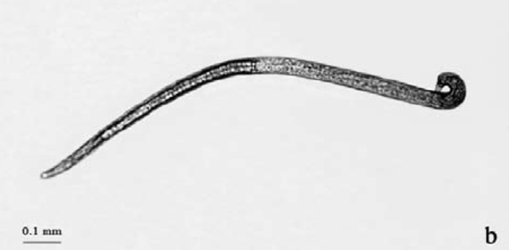
Trichuris trichiura Male Anatomy
Males are smaller than females
Have a curved tail
The curved tail has a copulatory spicule with a retractable sheath
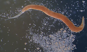
Trichinella spiralis Life Cycle
Contracted via undercooked pork containing encysted larvae
Two host life cycle
Larvae are released from cysts after exposure to gastric acids
Larvae invade the small bowel mucosa and develop into adult worms
Females release larvae that migrate to striated muscles where they encyst

Trichinella spiralis Female Anatomy
Sexual dimorphism, females are larger
Trichinella spiralis Male Anatomy

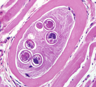
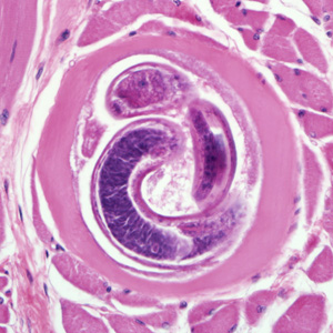
Trichinella spiralis Juvenile in Muscle
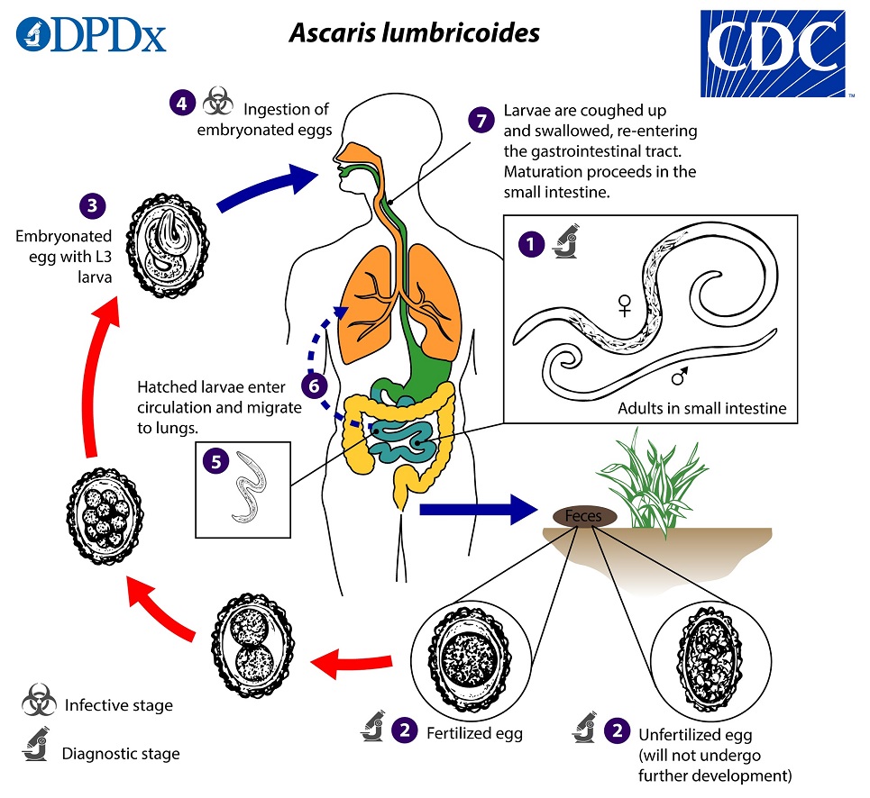
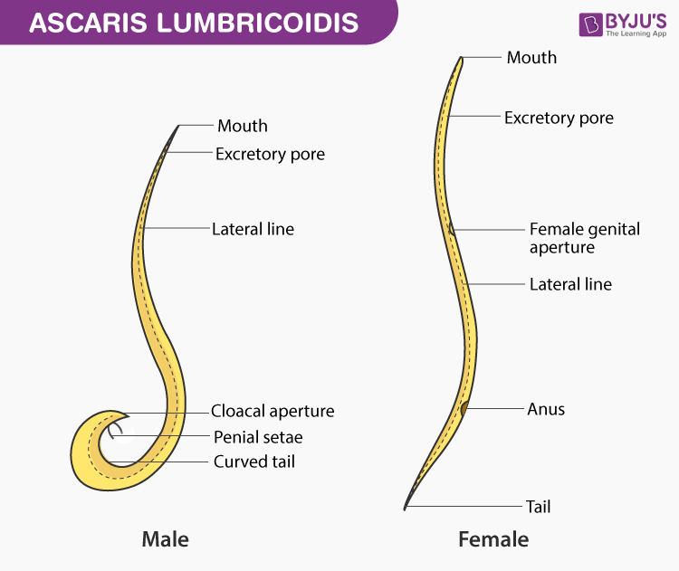
Ascaris lumbricoides Life Cycle
Unfertilized eggs are passed in stool
Eggs become fertilized in the environment (ideal conditions: moist, warm, shaded soil)
Infective eggs are swallowed
The larvae hatch and infect intestinal mucosa
Larvae are carried to the lungs (10 to 14 days)
Crawl up the throat and are swallowed
Reach the small intestine and mature into adults
Eggs have a lipid coating and can persist in the environment
Humans and swine are the major hosts, dogs may also be infected
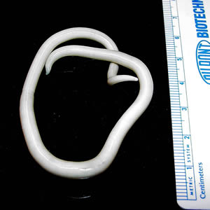
Ascaris lumbricoides Female Anatomy
Sexual dimorphism, females are larger
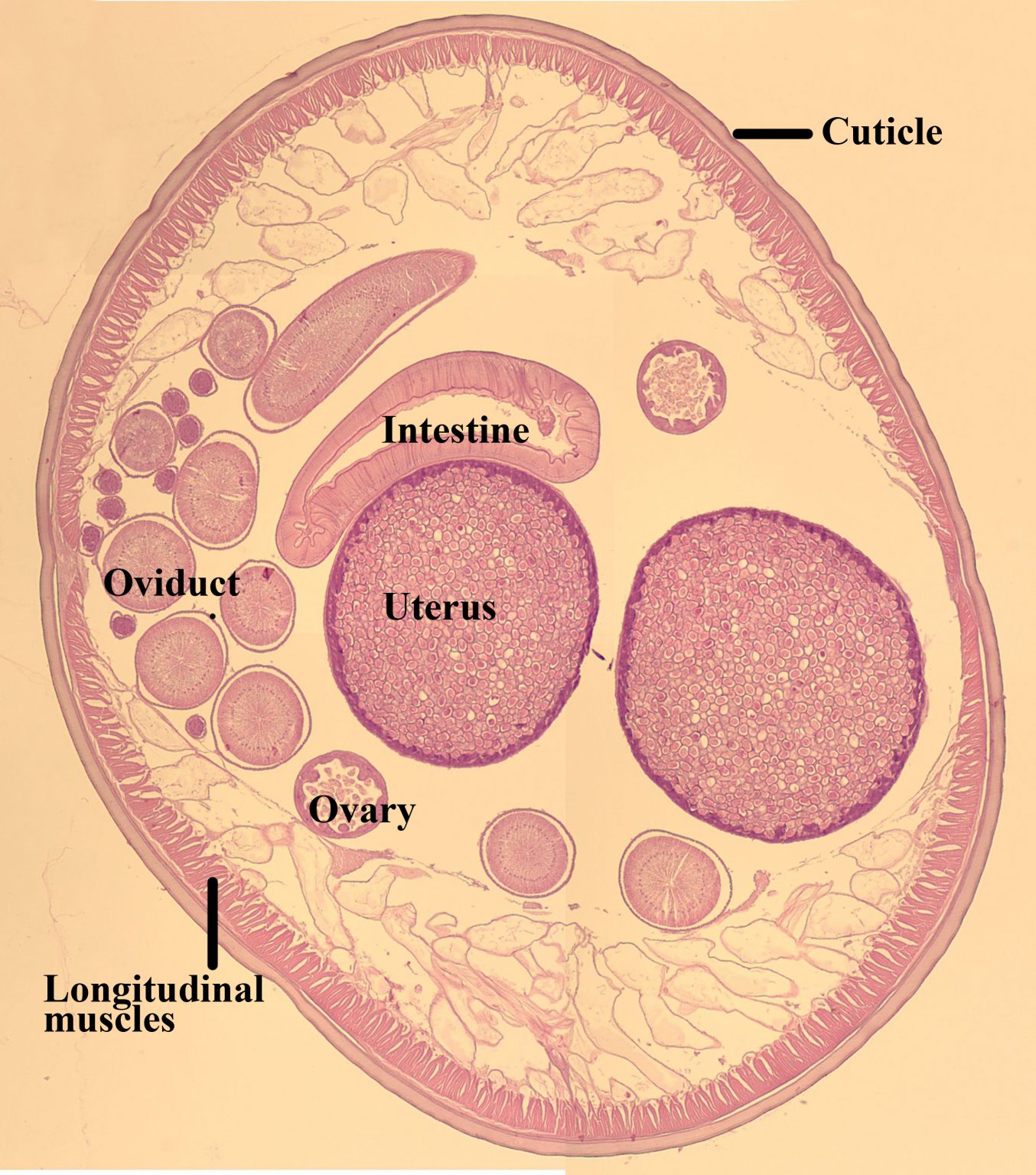
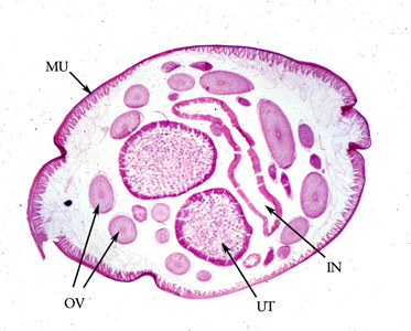
Ascaris lumbricoides Female Cross-section
Gravid uterus (UT)
Intestine (IN)
Coiled ovary (OV)

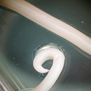
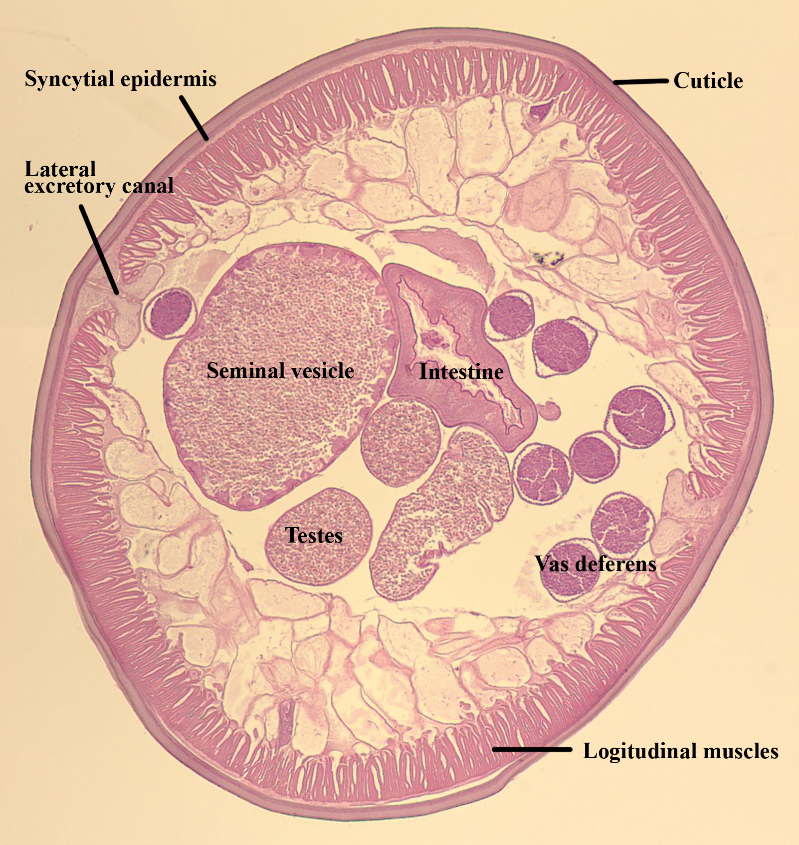
Ascaris lumbricoides Male Anatomy
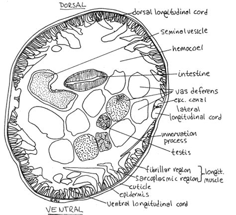
Ascaris lumbricoides Male Cross-section
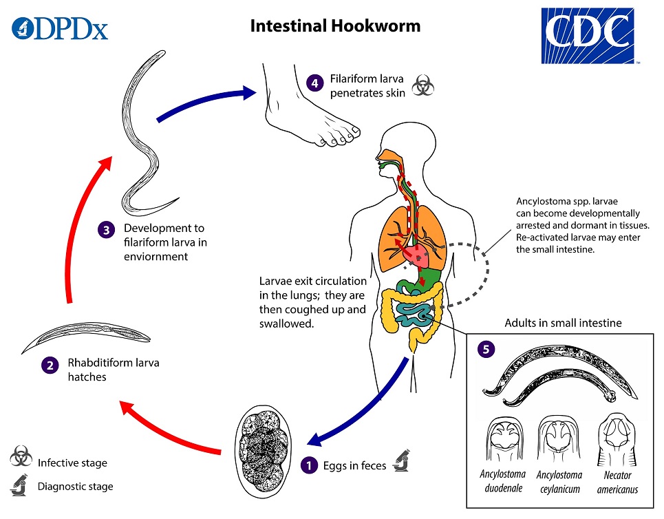
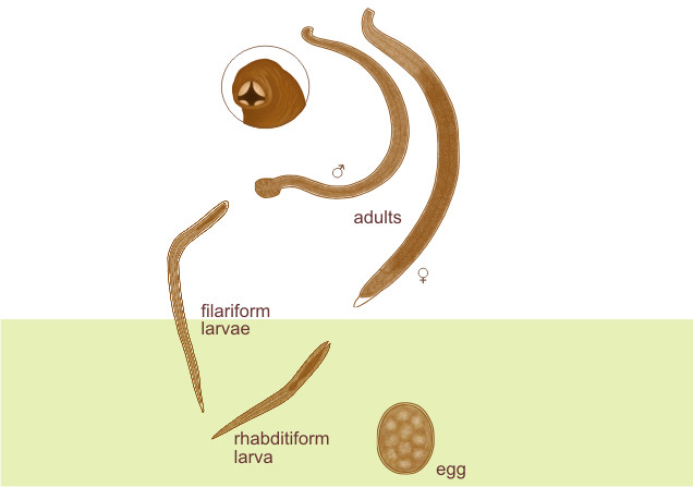
Hookworm Life Cycle
Eggs are passed in stool
Larvae grow in feces and/or soil
Become a filariform larvae after two molts
Larvae penetrate the skin and are carried through the blood to the heart and the lungs
Crawl to the throat, are swallowed, and reach the small intestine and mature into adults
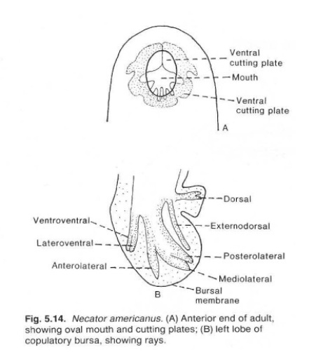
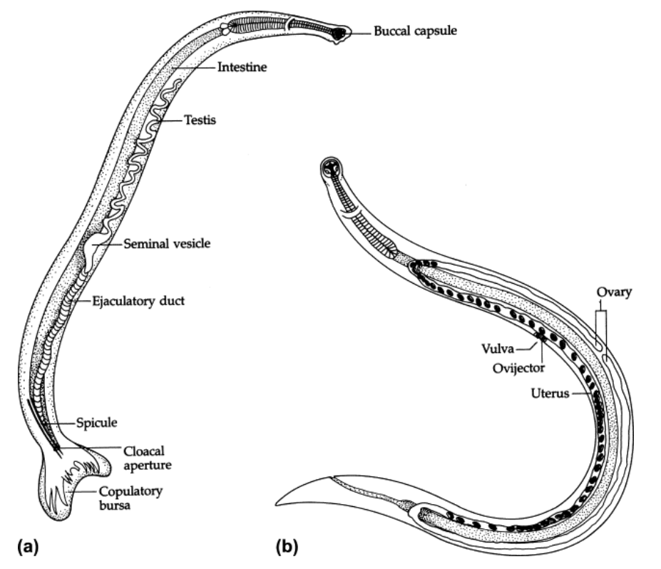
Necator americanus Adult Anatomy
Copulatory bursa at posterior of male
Two spicules
Fleshy rays support lateral and dorsal lobes
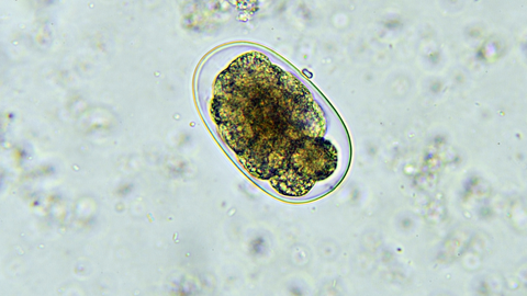
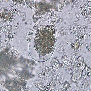
Necator americanus Eggs
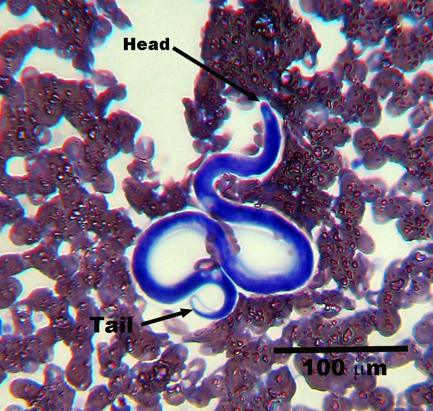
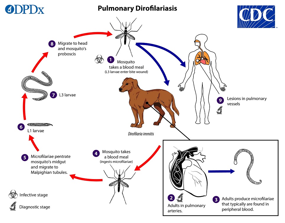
Dirofilaria immitis Life Cycle
A mosquito takes a blood meal and L3 larva enter the bite wound
In the definitive host L3 larvae undergo two more molts into L4 and adults
Adults reside in the pulmonary arteries
Female worms produce microfilariae which circulate in peripheral blood
A mosquito ingests the microfilariae, which migrate to the Malpighian tubes in the abdomen
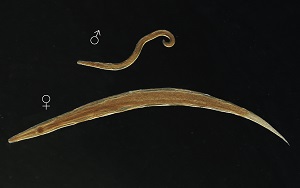
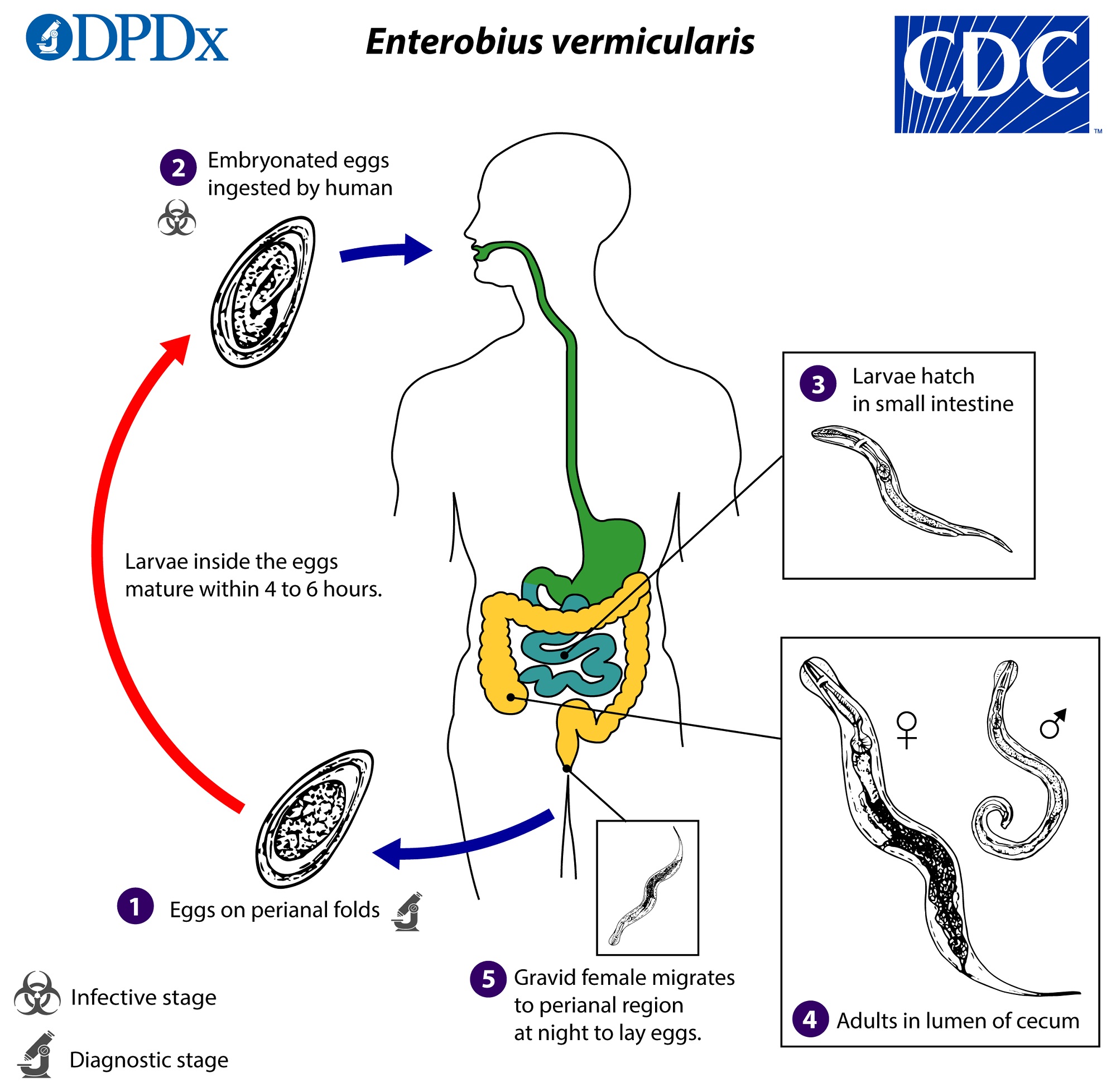
Enterobius vermicularis Life Cycle
Gravid females deposit eggs on perianal folds
Self infection occurs from the transfer of eggs from the perianal region to the mouth
Larvae hatch in the small intestine
Adults establish themself in the colon
Gravid females migrate nocturnally outside the anus and deposit eggs
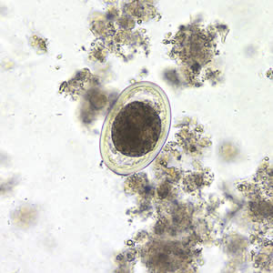
Enterobius vermicularis Egg
Class Insecta Characteristics
Body segmentation (head, thorax, and abdomen)
Parasitic adaptations include attachment organs, flattened bodies, and loss of wings
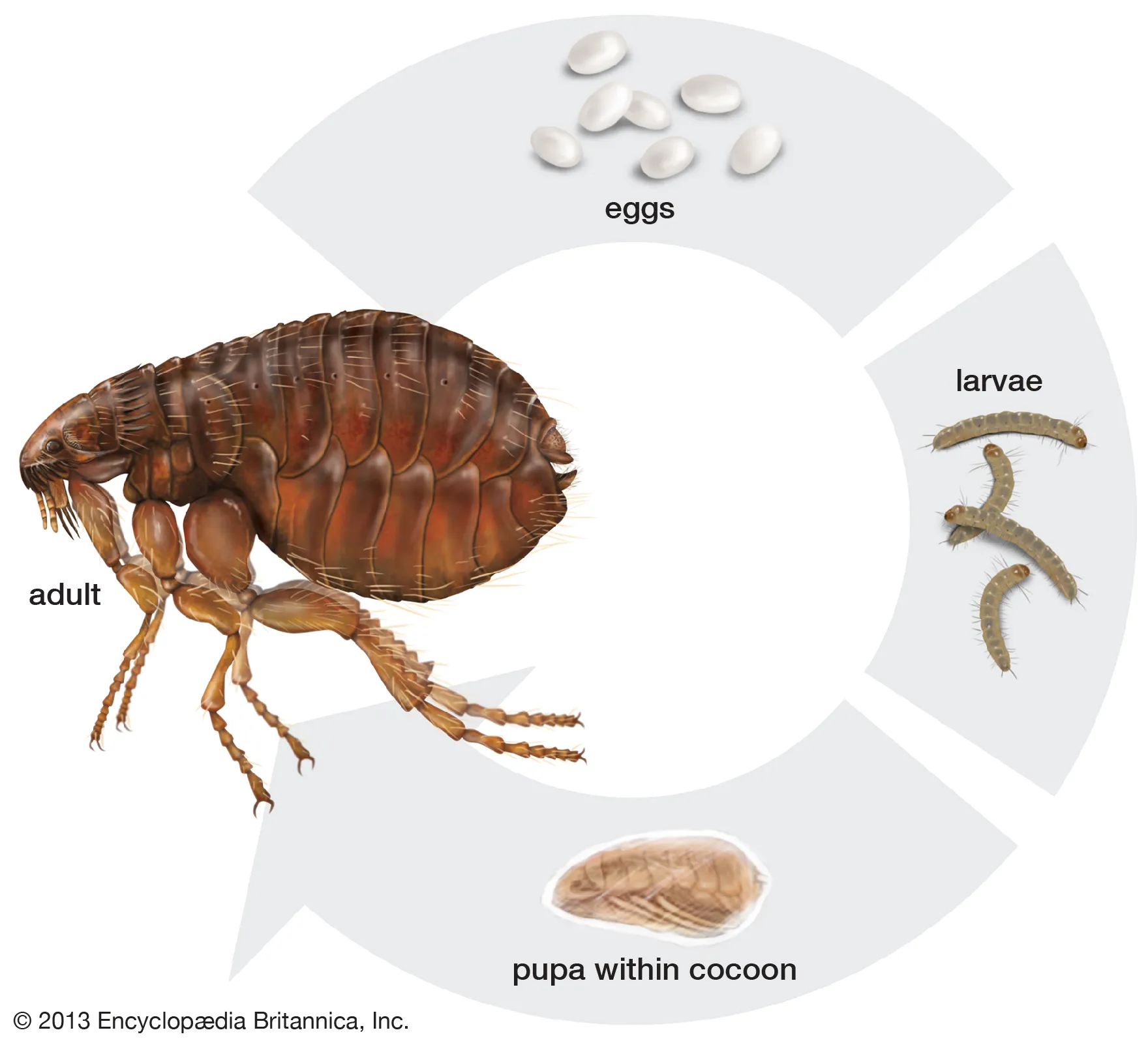
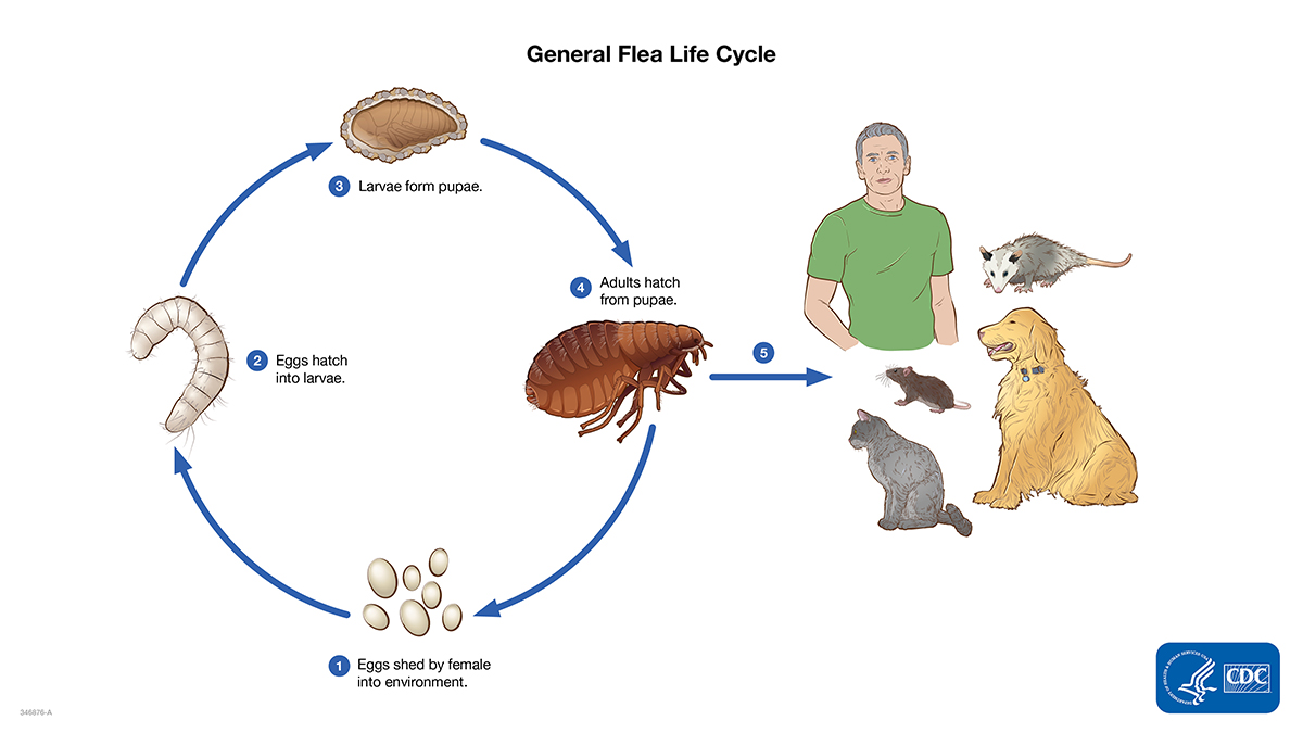
Flea (Siphonaptera) Life Cycle
After taking a blood meal, fleas mate and begin laying eggs
After hatching, fleas enter the larval stage
Larvae spin a cocoon and enter the pupa stage
Adult fleas emerge from the cocoon when there is the presence of a host
Phylum - Arthropoda
Class - Insecta
Order - Siphonaptera
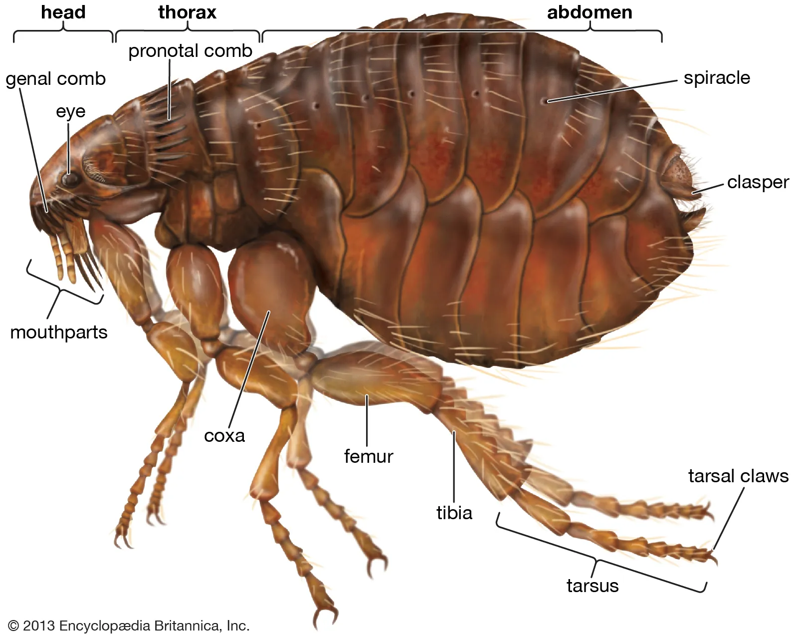
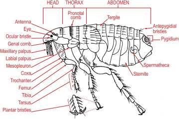
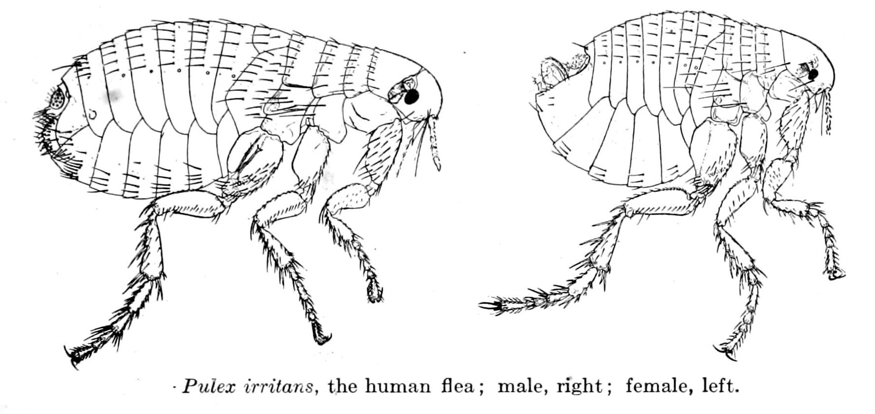
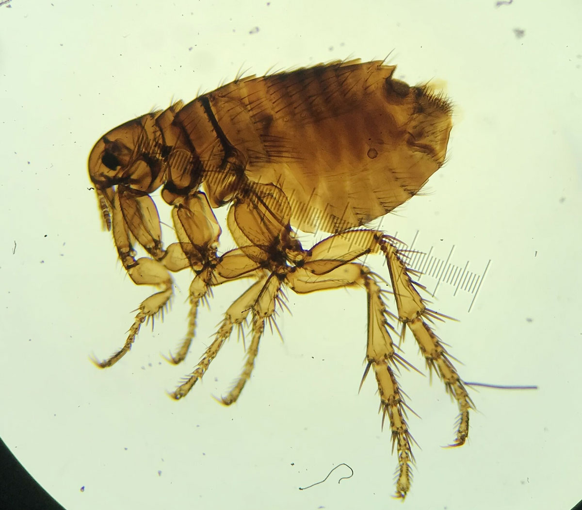
Flea Morphology
Identify:
Antenna
Compound eyes - reduced eye structure due to parasitic lifestyle
Genal ctenidia - anchoring mechanism, backward-facing spines lock onto fur, makes it difficult for hosts to remove the flea
Pronotal ctenidia - anchor the flea in place, assist in locomotion through the host’s hair
Pygidium - detect air movements and vibrations, allow the flea to sense potential hosts and threats
Spiracle - regulate respiration, minimize water loss, allow for air exchange
Spermatheca (females) - stores, nourishes, and protects sperm for an extended period of time
Copulatory apparatus (males) - locks securely onto the female and ensures sperm transfer
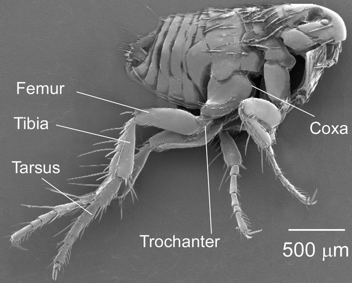
Flea Leg Morphology
Coxa - the base of the jumping mechanism, enables movement through the host’s hair
Trochanter - acts as a lever to release jumping power, transfers stored energy from the resilin protein
Femur - activates the internal spring system (resilin), compress the elastic protein before releasing it
Tibia - allows for long distance leaps, is pressed against the the ground with the trochanter to build energy, backward facing spines help to grip the substrate
Tarsus - anchor point for jumping, equipped with claws that grip the host’s hair and allow the friction to push off the ground

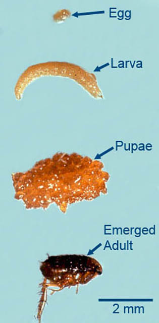
Ctenocephalides Larvae/Adult
The cat flea, primarily impacts mammal animal hosts
Four-stage, 30-75 day life-cycle (egg, larvae, pupa, adult)
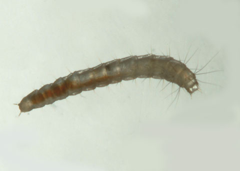
Pulex irritans Larvae/Adult
Human flea or house flea
Wide host spectrum
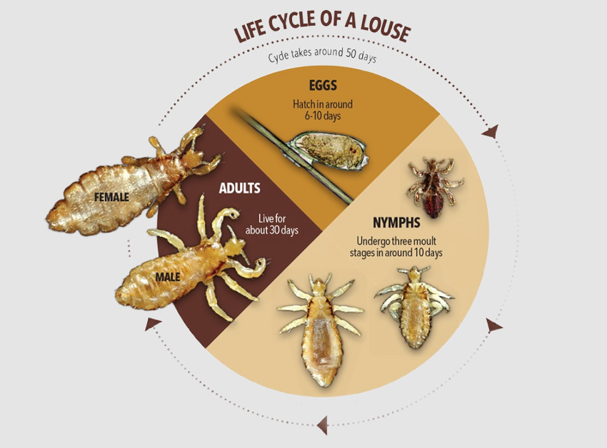
Lice (Phthiraptera) Life Cycle
Nits, or eggs, are laid by the adult female and are cemented at the base of the hair shaft near the scalp
The egg hatches and releases a nymph, nymphs mature after 3 molts
Nymphs become adults after 7 days after hatching
Adult lice are the size of a sesame seed and have six legs
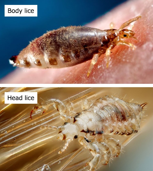
Body lice - body lice reside and lay their eggs on clothing and migrate to the human body to feed
Phylum - Arthropoda
Class - Insecta
Order - Phthiraptera
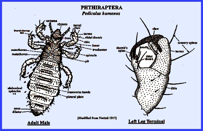
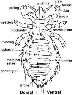
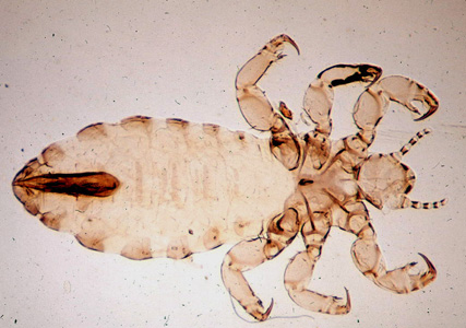
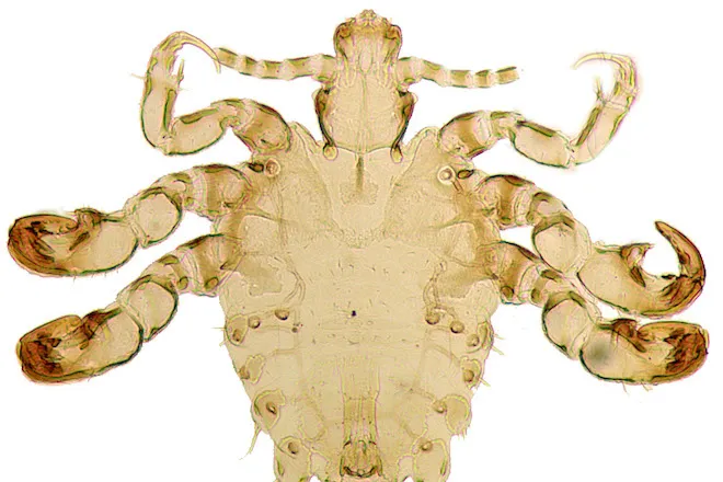
Lice Anatomy
Antennae
Compound eye
Coxa - helps to anchor the large muscles
Trochanter - provides flexibility and allows the rotational movement of the leg
Femur
Tibia - used to anchor the louse, a tibial thumb works in opposition to the tarsal claw
Tarsus - grasps human hair and clothing fibers, allows a secure hold on the hair during movement
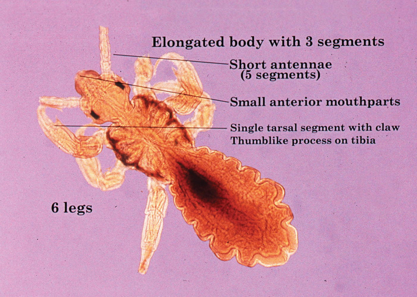
Pediculus humanus humanus (body louse)
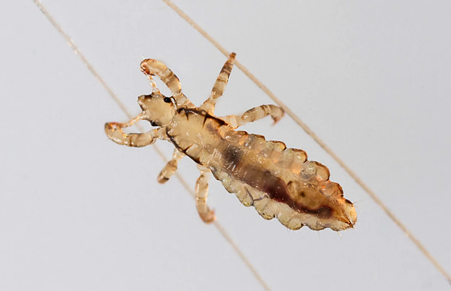
Pediculus humanus capitalis (head louse)
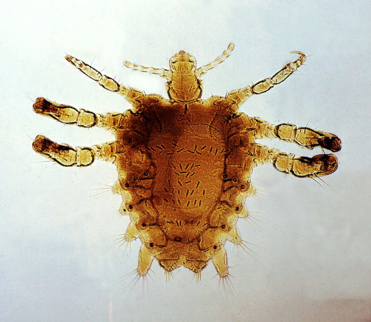
Phthirus pubis
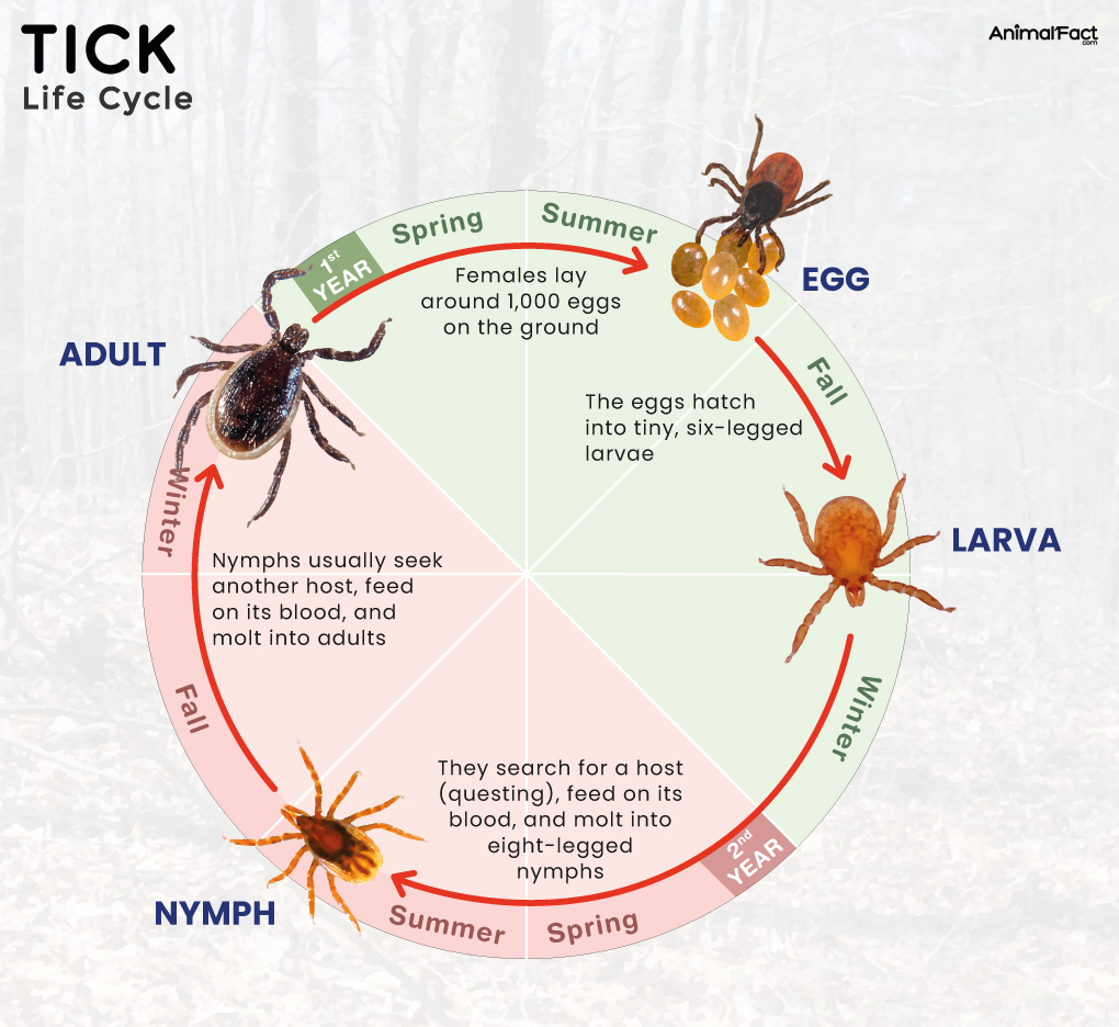
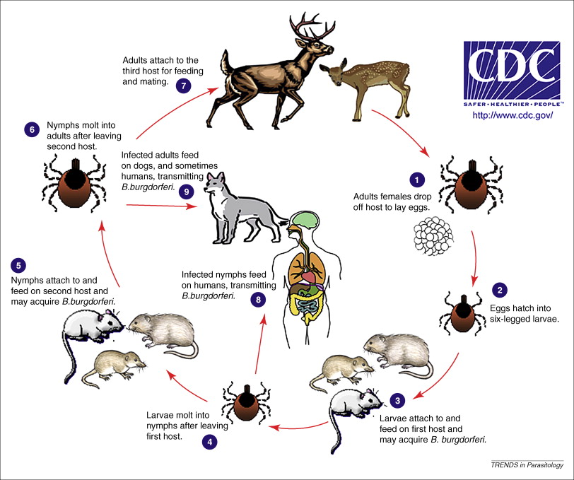
Tick Life Cycle
Four life stages: egg, larva, nymph, adult
A tick needs a blood meal at each stage to survive
Spring - eggs are laid
Summer - hungry 6-legged larva hatch
Next spring - 8-legged nymphs look for their next blood meal, can now transmit pathogens
Fall or winter - nymphs transition into adults, females seek out another blood meal to reproduce
Phylum - Arthropoda
Class - Arachnida
Subclass - Acari
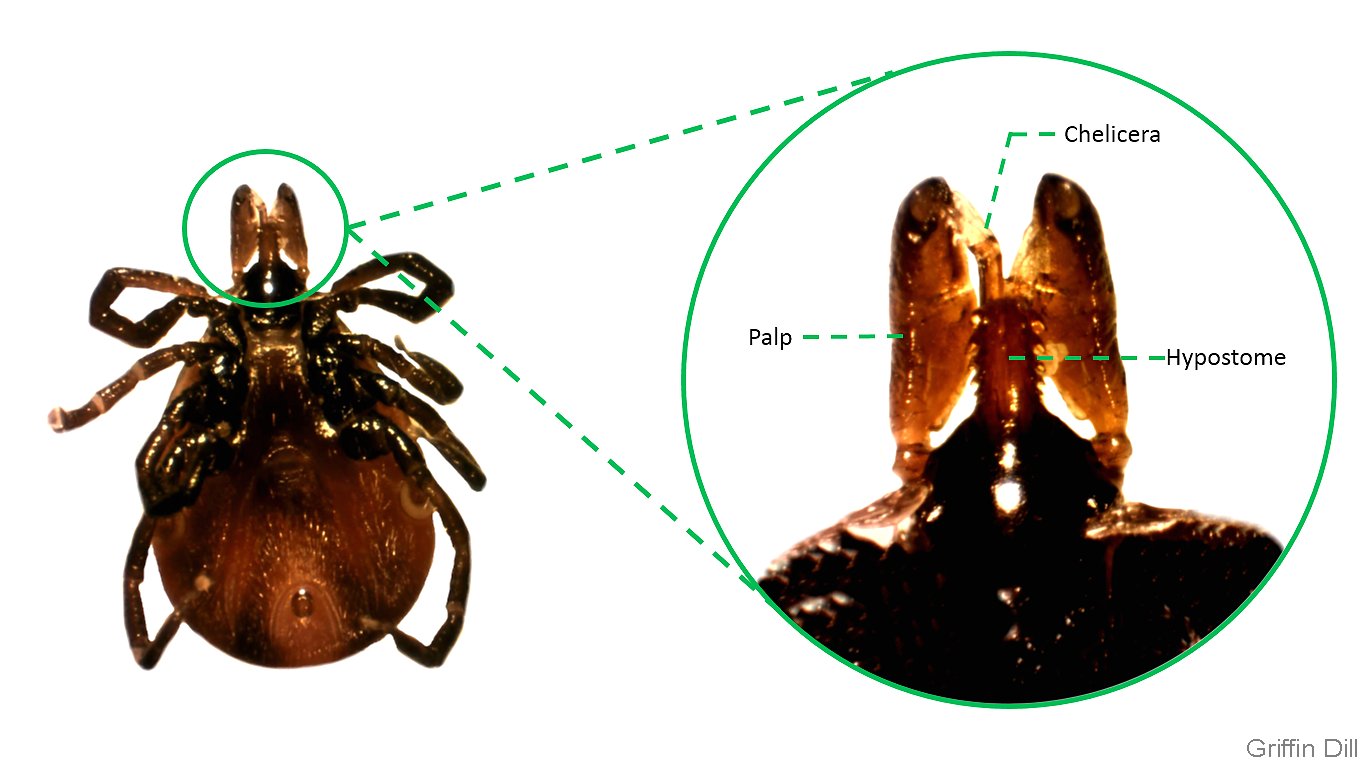
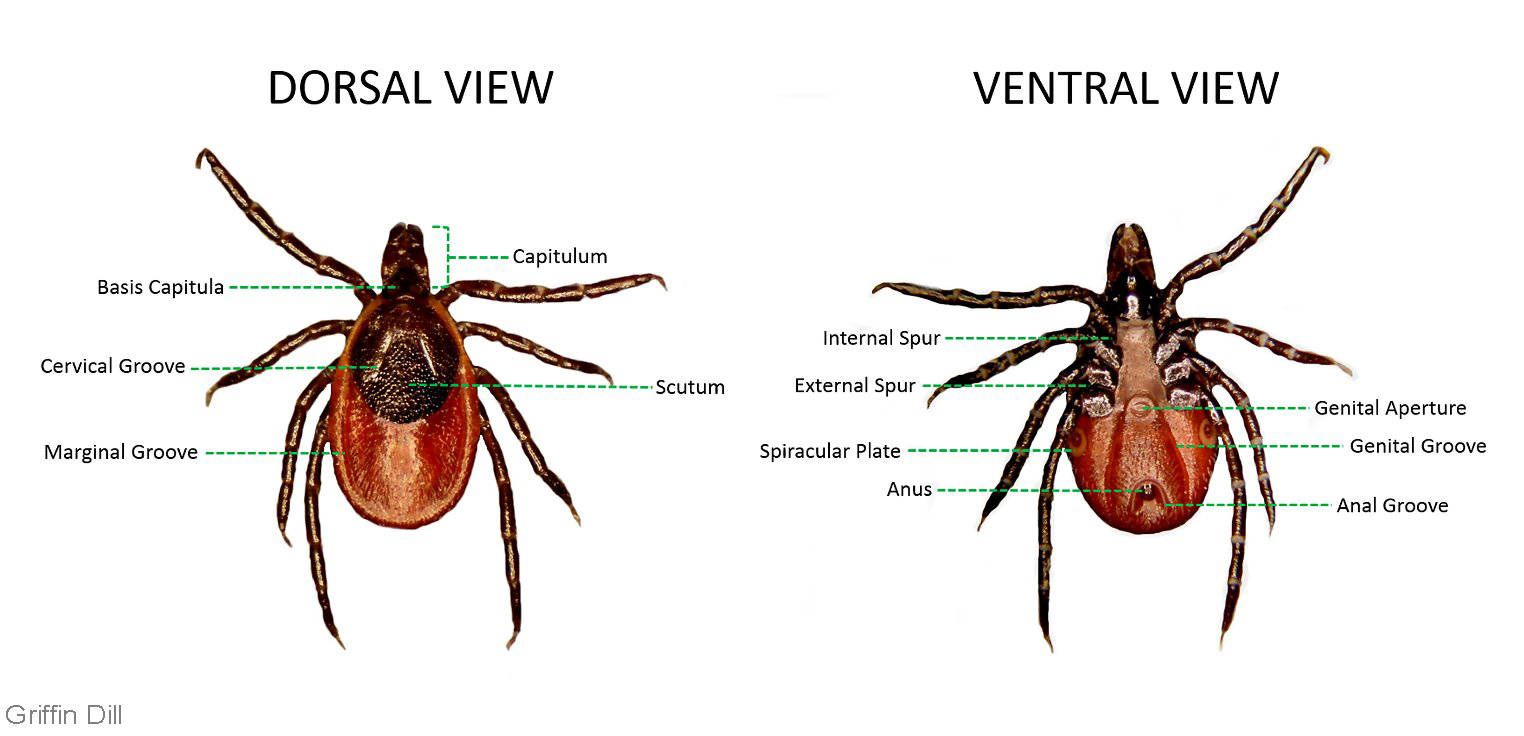
Tick Anatomy
Anterior - gnathosoma
Posterior - idiosoma
Hypostome - embeds in skin
Scutum - provides structural support and protection
Spiracular plate - allow oxygen to enter and carbon dioxide to exit, manages water loss
Festoon - allow for expansion of the body with blood
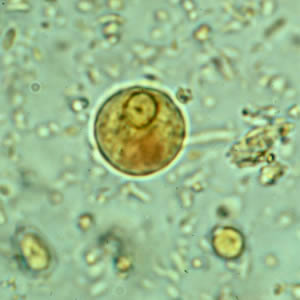
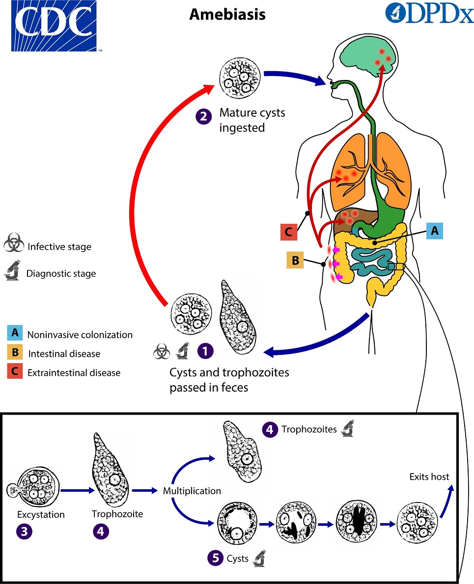
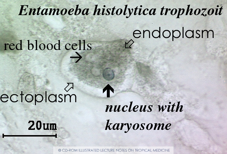
Entamoeba histolytica Life Cycle
Cysts and trophozoites are passed in feces
Mature cysts are ingested
Excystation occurs in the small intestine
Trophozoites can invade the intestinal mucosa and other sites like the blood, liver, and brain
Phylum - Amoebozoa
Class - Lobosea
Order - Amoebida
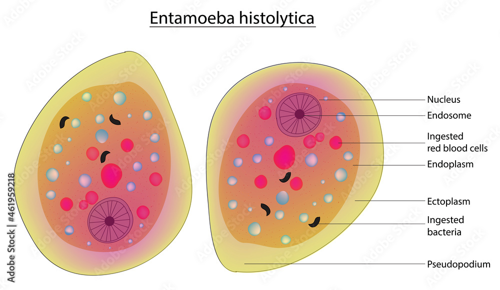
Entamoeba histolytica Anatomy
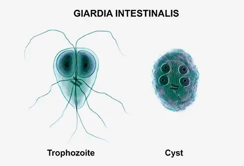
Giardia lamblia Life Cycle
Phylum: Metamonada
Class: Trepomonadea
Order: Distomatida
Genus: Giardia
Cysts or trophozoites are found in feces
Ingestion of cysts
Trophozoites multiply in the small bowel
Encystation occurs and the parasites travel towards the colon
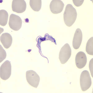
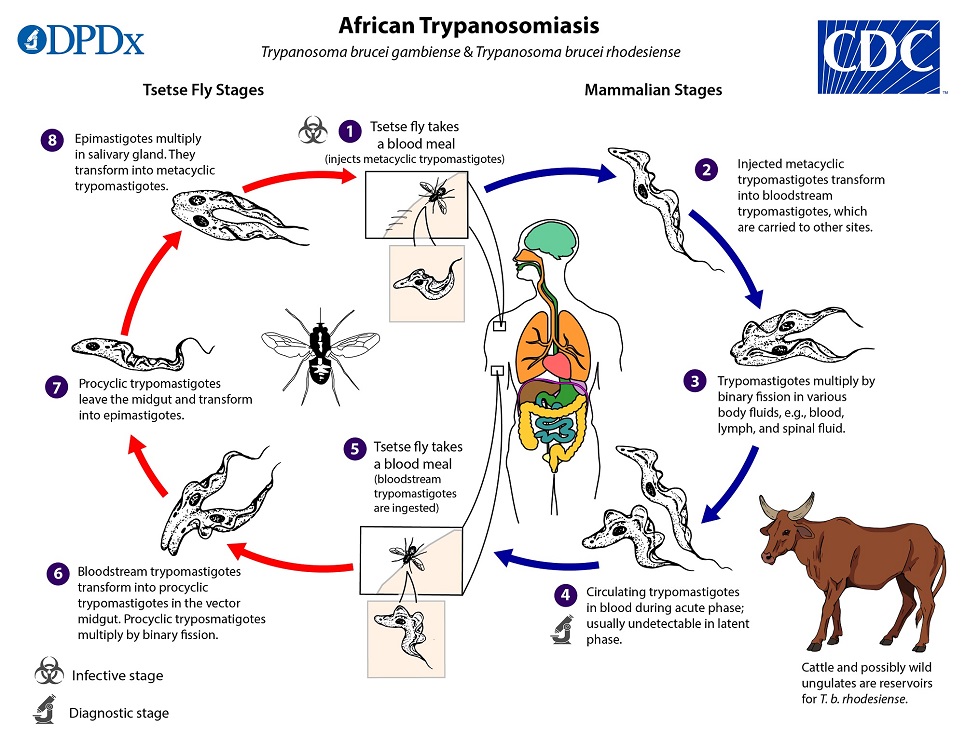
Trypanosome brucei
Phylum: Euglenozoa
Class: Kinetoplastea
Order: Trypanosomatida
Genus: Trypanosoma
A tsetse fly takes a blood meal
Parasites enter the blood stream
Transform into trypomastigotes, are carried to other sites in the body
A fly takes a blood meal and becomes infected
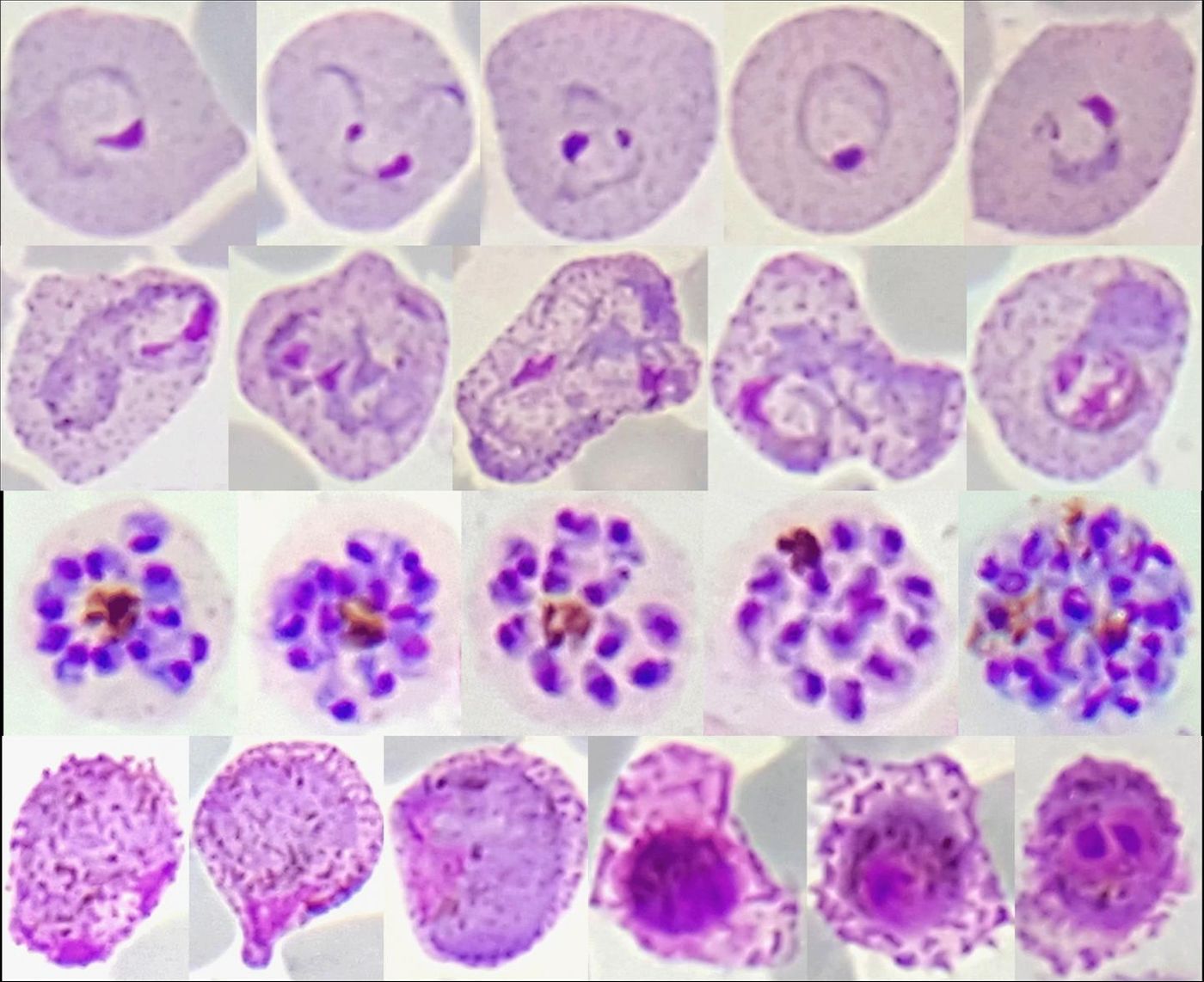
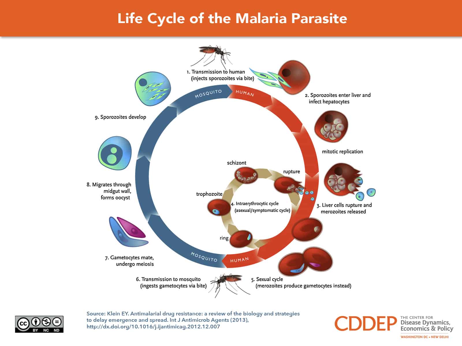
Plasmodium spp.
A mosquito ingests gametocytes that mate within the gut of the mosquito
Sporozoites are injected into a human during the next blood meal
Sporozoites travel to the liver, infect hepatocytes, and begin asexually replicating
Merozoites infect the red blood cells, the cell then ruptures
The cycle repeats